Introduction
Prenatal treatment with glucocorticoids induces maturational changes in fetal organ systems, some of which can be beneficial, such as promotion of lung maturation, leading to great therapeutic benefits for babies born preterm.Reference Bishop 1 , Reference Ward 2 However, prenatal glucocorticoids treatment can also predispose the fetus to cardiovascular and metabolic diseases in adult life, as demonstrated in animal models such as sheep and rats.Reference Dodic, Abouantoun, O’Connor, Wintour and Moritz 3 – Reference Drake, Tang and Nyirenda 6
In terms of reproductive function, most disorders in sperm production appear to originate during fetal life, thus any disruption of the process of fetal testis development will likely have long term impacts on function.Reference Sharpe and Franks 7 In this regard, adult men with congenital adrenal hyperplasia who have been exposed to excess adrenal-derived testosterone during development, show low sperm counts and reduced fertility.Reference Recabarren, Rojas-García and Recabarren 8 One disruptive factor is glucocorticoid exposure. We have previously shown that exogenous glucocorticoids during fetal and postnatal development affect the apoptotic and proliferative pathways in testicular tissueReference Pedrana, Viotti and Souza 9 which, in addition to issues with fertility, could have broader health implications because abnormal sperm counts, observed in 6–8% of adult men,Reference Sharpe and Skakkebaek 10 are associated with a 20-fold higher incidence of testicular cancer.Reference Raman, Nobert and Goldstein 11
The timing of glucocorticoids treatment during fetal testis development is critical. In all eutherian mammals studied to date, two or three populations of Leydig cells arise in sequence, where interstitial cells gradually differentiate into the first fetal population that is the predominant source of circulating androgens in the fetus. After birth, this population is replaced by an adult population that produces the pubertal surge in testosterone.Reference O’Shaughnessy and Fowler 12 The number of adult Leydig cells and their steroidogenic capacity determine the circulating levels of testosterone.Reference Hardy, Gao and Dong 13 These two populations of Leydig cells arise separately and appear to be distinct, both functionally and morphologically, although a connection between their precursor stem cells is possible.Reference Griswold and Oatley 14 A third neonatal population of Leydig cells has been suggested for a number of species, including humans, on the basis of a post-natal rise in androgens.Reference Prince 15
Evidence is accumulating for the direct effects of glucocorticoids on the development of Leydig cells. It was noted long ago that, in adult male rats, dexamethasone treatment reduced testicular LH receptor numbersReference Bambino and Hsueh 16 and testosterone production.Reference Welsh, Bambino and Hsueh 17 In addition, exposure to corticosterone increases apoptosis in Leydig cells.Reference Gao, Tong and Hu 18 Many of these effects are mediated by changes in the activity of 3β-hydroxysteroid dehydrogenase (3β-HSD), a dimeric bifunctional steroidogenic enzyme that is essential for the biosynthesis of all classes of steroid hormones, including androgens. According to uniprot databases (http://www.uniprot.org/uniprot/P14060#similar_proteins) and the literature, 3β-HSD is a single-pass membrane protein located in both the endoplasmic reticulum and the mitochondria. In the rat testis, 3β-HSD is restricted to the mitochondriaReference Pelletier, Dupont and Simard 19 where it catalyzes the sequential 3-hydroxysteroid dehydrogenation and Δ5- and Δ4-isomerization of the 5-steroid precursors.Reference Samuels, Helmreich, Lasater and Reich 20 , Reference Simard, Durocher and Mébarki 21 In male rats born to mothers that had been stressed during delivery, steroidogenesis is inhibited, with a decrease in the activities of 17α-hydroxylase and 3β-HSD in Leydig cells.Reference Orr and Mann 22 For sheep, 3β-HSD is first detected in Leydig cells on Day 30, and then progressively develops over days 40, 75 and 130 of gestation.Reference Sweeney, Saunders, Millar and Brooks 23 At 30–75 days of gestation (DG), 3β-HSD mRNA is abundant throughout the testis, particularly in somatic cells outside the seminiferous tubules.Reference Quirke, Juengel, Tisdall, Lun, Heath and McNatty 24
In searching for an understanding of the processes that mediate the responses of the developing testis to glucocorticoids, we need to consider the paracrine factors that play roles in the control of cellular proliferation and differentiation. Most important are the members of the transforming growth factor-β family,Reference Feige and Chambaz 25 such as the inhibins; a group of dimeric signalling peptides that exert paracrine effects on cells surrounding their site of production in the testis.Reference Anderson and Cambray 26 In the sheep testis, inhibins are involved in the regulation of the functional development of Leydig cells, including steroidogenesis.Reference Jarred, Cancilla and Richards 27
Therefore, in the present study, we aimed to test whether maternal administration of glucocorticoids to sheep in late pregnancy would affect the expression of 3β-HSD in Leydig cells, and expression of inhibin-α in testicular parenchyma, during fetal and postnatal development.
Methods
Animals and prenatal treatments
All animal procedures and experimental design were approved by the Animal Experimentation Ethics Committee of the University of Western Australia and/or the Western Australian Department of Agriculture. The experiments were performed at Allandale experimental station of the University of Western Australia. Pregnant Merino sheep (n=42) bearing a singleton fetus were randomly allocated to receive intramuscular injections of 0.5 mg/kg, betamethasone as previously describedReference Sloboda, Newnham and Challis 28 , Reference Sloboda, Moss and Li 29 (Chronodose Celestone, Schering Plough, Baulkham Hills, NSW, Australia) or saline (control) at 104, 111 and 118DG. Saline injections were of a comparable volume (5–6 ml). At 100DG, all animals were intramuscularly injected with 150 mg medroxyprogesterone acetate (Depo Provera, Upjohn, Rydalmere, NSW, Australia) to reduce pregnancy losses because of subsequent glucocorticoid treatment, as previously described.Reference Sloboda, Moss, Gurrin, Newnham and Challis 5 The dose of betamethasone was chosen after previous studies in sheep in which the importance of dose and time of treatment were assessed.Reference Sloboda, Newnham and Challis 30 The total dose of betamethasone, correlates with clinical doses used for fetal lung maturation in women at risk of early preterm birth.Reference Liggins and Howie 31 Pregnant ewes were killed by captive bolt at 121DG (five per treatment) and 132DG (treated n=5; control, n=6) and their fetuses were immediately delivered by caesarean section and killed by decapitation. Male lambs delivered spontaneously at 150DG were kept with their mothers on pasture supplemented with cereal grain and lupin seed. Lambs were weighed and then anaesthetized with ketamine (15 mg/kg) and xylazine (0.1 mg/kg, Troy Laboratories, Smithfield, NSW, Australia) and killed by decapitation at postnatal days (PD) 45 (treated, n=5; control, n=6) and 90 (treated, n=5; control, n=5).
Tissue processing
Fetal and post-natal testes were dissected, weighed, cut longitudinally and immersed in Bouin’s fixative solution (75% picric acid saturated, 20% formaldehyde 40, 5% glacial acetic acid) for 24 h at room temperature (20°C). Subsamples were immersed in increasing concentrations of ethanol (70, 95, 100%) and chloroform, and then embedded in paraffin wax. Sections (5 μm) were cut by microtome (Leica Reichert Jung Biocut 2030, Wetzlar, Germany), placed on glass slides and dried in an oven at 60°C for immunohistochemistry.
Immunohistochemistry
We used the streptavidin-biotin-peroxidase technique. Tissue sections were de-paraffinized and rehydrated, and heat-induced epitope retrieval (HIER) was performed by incubating the sections in 0.01 M citrate buffer (pH 6.0) with Tween 20 for 5 min in a microwave (set on ‘high’) and then washed with distilled water and phosphate-buffered saline (PBS, pH 7.4). Endogenous peroxidases were deactivated with 3% hydrogen peroxide for 20 min, followed by washing in PBS and incubation with normal serum sheep to block non-specific binding proteins. The sections were incubated in a humidified chamber at 4°C for 18 h with two primary antibodies, Western blots were done to confirm the proteins using the same antibody in testis. The proteins of interest 3β-hydroxysteroid dehydrogenase and inhibin-α (Ovis aries) had been already demonstrated in ovine testis. independently on separate slides: mouse monoclonal anti-3β1 HSD at 1 mg/ml, diluted 1:100 (ab 55268, Abcam, Cambridge, MA, USA), and mouse monoclonal anti-inhibin-α, diluted 1:100 (ab105927, Abcam). Subsequently, the sections were washed with PBS and incubated for 30 min with biotinylated anti-mouse secondary antibody (ab 64259 Abcam). The sections were washed with PBS and incubated for 30 min with streptavidin-peroxidase enzyme complex (HRP/DAB detection kit, ab 64259, Abcam). They were then washed again with PBS, followed by incubation for 5 min with diaminobenzidine (DAB; ab64259, Abcam). Sections were counterstained with Mayer’s hematoxylin, dehydrated, and mounted in Biopack (Argentina).
Image analysis
Digital images were retrieved with DinoLite Capture 2.0 software (AnMo Electronics Corporation, Taiwan) and a digital camera (Dino-Eyepiece, AM-423X, AnMo Electronics Corporation) connected to a microscope (Premiere Professional Binocular, Model MRP-5000, Manassas, USA) at 400×magnification. The quantitative freehand selection tool from the image analysis software (ImageJ 1.43 m, Wayne Rasband, National Institutes of Health, USA; http://rsb.info.nih.gov/ij.) was applied to 50 fields to select and measure the DAB-positive (brown) immunostained area and to determine the intensity of staining in Leydig cells. Images were evaluated through colour segmentation analysis in which all objects of a specific brown threshold were selected from RGB images and converted to binary images. The threshold values were verified and normalized with controls carried across several runs for calibration. This process provided quantitative values for the percentage of area immunostained and for the intensity of immunostaining in the Leydig cells and sex cords. A macro was used to automate the process.
Statistical analysis
The continuous variables analysed were immunostained area for inhibin-α in testis parenchyma, and the proportion of immunostained area and staining intensity for 3β-HSD in Leydig cells and sex cords/seminiferous tubules. All values were expressed as means (±s.e.m.) and compared using SAS (v 9.1, SAS Institute Inc, Cary, NC, USA), considering in the model the main effects of day (gestation, postnatal) and betamethasone treatment, and the interaction between these effects. As an alternative to the transformation of percentages to normalize the data, generalized linear models (GLM) were used that allow percentages to be treated as normally distributed variables. Post hoc differences between groups were assessed with Tukey’s tests. In all cases, the level of significance was P<0.05.
Results
Inhibin-α in Sertoli and Leydig cells
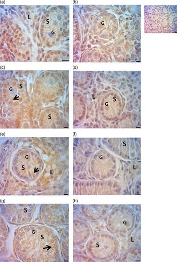
Immunostaining for inhibin-α was primarily localized to Sertoli and Leydig cells. Within the sex cords during the prenatal period, the most intense staining was observed in Sertoli cells, with less detected in gonocytes. In the postnatal period, this situation remained the same but inhibin-α was also detectable in Leydig cells on both 45PD and 90PD (Fig. 1).
Immunostaining for inhibin-α in testicular parenchyma at 121 days of gestation (DG) (a, b); 132DG (c, d); 45PD (e, f) and 90PD (g, h) following prenatal treatment with betamethasone or saline. Magnification 600. Scale bar: 20 μm. Top right: negative control for immunohistochemistry. Note the strong positive immunostaining for inhibin-α (black arrows) in Sertoli cells (S) cytoplasm; gonocytes (G) showed negative or slight staining; Leydig cell (L) staining varied from positive to slightly positive.

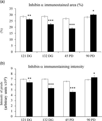
The percentage immunostained area did not change as development progressed in the control group (Fig. 2a). However, in the treated group, the values decreased progressively from 121DG to 45PD (P<0.0001), before increasing again between 45PD and 90PD (P<0.0001). As a consequence, a smaller proportion of testicular tissue was stained in betamethasone-treated than in control tissues at 121DG (P=0.005), 132DG (P<0.001) and 45PD (P<0.0001). By 90PD, the recovery in the treated group was such that the values exceeded those for the control group (P=0.03).
Effects of prenatal treatment with betamethasone (black bars) or saline (white bars) on inhibin-α expression in testis parenchyma at 121 days of gestation (DG) and 132 DG, 45PD and 90PD. Intensity of immunostaining is measured as the mean grey value of pixels. Asterisks indicate differences between groups (*P<0.05; **P<0.01; ***P<0.001).

3β-HSD in Leydig cells
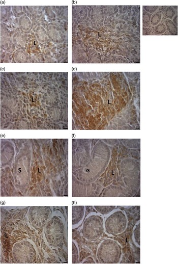
The enzyme 3β-HSD was localized in Leydig cell cytoplasm during fetal and post-natal development. Leydig cells from fetal to prepubertal testis were isolated, or distributed in groups, surrounding lymphatic vessels and blood vessels around at the interstitial tissue between the sex cords (Fig. 3).
Immunostaining for 3β-HSD in testicular parenchyma at 121 days of gestation (DG) (a, b); 132DG (c, d); 45PD (e, f) and 90PD (g, h) following prenatal treatment with betamethasone or saline. Magnification 600. Scale bar: 20 μm. Top right: negative control for immunohistochemistry. Note the strong positive immunostaining for 3β-HSD in Leydig cells (L) varies from positive to slightly positive in Sertoli cells (S); gonocytes (G) showed negative or slight immunostaining.

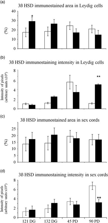
The percentage of immunostained area did not change from pre- to post-natal development in the control group (Fig. 4). However, in the betamethasone-treated group, 29.2±4.6% of testicular tissue was stained at 121DG (compared with 17.2±4.6% in the control group; P=0.098), and this proportions became smaller as the experiment progressed so, by 90PD, it was smaller than at 121DG (P=0.046). With respect to immunostaining intensity, the values for the control group did not change significantly as development progressed (Fig. 4). However, by 90PD, the intensity had increased in the betamethasone-treated group to be significantly more intense than the control (P=0.033).
Effects of prenatal treatment with betamethasone (black bars) or saline (white bars) on 3β-HSD expression in testis parenchyma at 121 days of gestation (DG) and 132 DG, 45 postnatal days (PD) and 90PD. (a, b) % and intensity of immunostaining in Leydig cells. (c, d) % and intensity of immunostaining in sex cords (121DG, 132 DG) seminiferous tubules (45PD, 90PD). Intensity of immunostaining is measured as the mean grey value of pixels in arbitrary units. Asterisks indicate differences between groups (*P<0.05; **P<0.01; ***P<0.001).

3β-HSD in sex cords and seminiferous tubules
Staining for 3β-HSD was less intense in the sex cords and seminiferous tubules than in the Leydig cells. In gonocytes, 3β-HSD was not detectable at any stage of development. In Sertoli cells, the enzyme was again located in the cytoplasm, at all pre-and post-natal stages. No differences in percentage immunostained area were observed between the control and betamethasone-treated groups at any stage of development (Fig. 4c and 4d). In the treated group, the percentage immunostained area remained constant but, in the control group, it tended to be higher at 90PD than DG 121 (P=0.07). The intensity of staining increased continually as development progressed in the controls (P=0.0001 for 90PD v. both 121DG and 132DG), with an especially marked increase between 45PD and 90PD (P=0.002; Fig. 4d). By contrast, in the betamethasone-treated group, there was no significant change in the intensity of staining, apparently because in the normal pattern of developmental increase had stopped by 132DG. As a consequence, by 90PD, the intensity was significantly lower in the betamethasone-treated group than in the control group (P=0.006).
Discussion
Prenatal betamethasone administration during the last third of pregnancy produced biphasic responses in the sheep developing testis, for the expression of both inhibin-α and 3β-HSD. Inhibin-α expression was initially downregulated during the prenatal period and then upregulated at the onset of spermatogenesis (90 days after birth). By contrast, 3β-HSD expression was initially upregulated during prenatal testicular development but then decreased between birth and puberty. We have previously shown biphasic protein expression patterns in response to exogenous glucocorticoid in placental lactogen in male sheep treated with dexamethasone administration during early pregnancy,Reference Braun, Meng and Shang 32 and other show biphasic responses to GH-releasing hormone in rat pituitary cells.Reference Ceda, Davis and Hoffman 33 It has been shown that elevated levels of endogenous glucocorticoid are usually associated with stress, and stress can reduce and then enhance reproductive function through an inhibition-rebound effect in sheep.Reference Tilbrook, Canny, Serapiglia, Ambrose and Clarke 34 A sequence of positive and negative outcomes for reproductive function might also be related to opposite biphasic patterns elicited by prenatal betamethasone in the expression of 3β-HSD and inhibin.
The effects of glucocorticoid treatment on inhibin-α expression are likely to affect the development of reproductive capability because inhibin is a powerful inhibitor of FSH secretion in ram lambs from as early as 1 month of age, with maximal responses established by the onset of puberty.Reference Tilbrook, De Kretser and Clarke 35 Puberty in male sheep is reached at 120PD, as evidenced by histological observation of spermatozoa in the seminiferous tubules,Reference Schanbacher, Gomes and VanDemark 36 and between 185 and 213 days of age, as evidenced by the appearance of spermatozoa in the ejaculate in Merino-type breeds (review:Reference Skinner and Rowson 37 ), although this measure varies with genotype.Reference Skinner and Rowson 37 , Reference Castrillejo, Moraña and Bielli 38 For example, compared with normal testis, the staining intensity for inhibin-α in Sertoli cells is much stronger in cases of impaired spermatogenesis, including Sertoli-cell-only syndrome.Reference Bergh and Cajander 39 Clearly, this hypothesis needs to be tested directly by assessing the effects of prenatal glucocorticoids on the onset of reproductive life. We would therefore expect that changes in inhibin production elicited by betamethasone treatment would suppress FSH secretion with consequences for spermatogenesis.
The progressive increase in 3β-HSD activity during development of the sex cords and seminiferous tubules has been reported previouslyReference Walsh, McCormick, Martin and Stocco 40 , Reference Stocco and McPhaul 41 and shows that Sertoli cells can synthesize steroid hormones as they go through morphological and functional differentiation. The effects of betamethasone on 3β-HSD expression in Leydig and Sertoli cells are therefore also likely to affect fetal and postnatal testicular development. Betamethasone could act directly on testicular tissue where in rat Leydig cells in vitro, dexamethasone inhibited 3β-HSD activity.Reference Agular, Vinggaard and Vind 42 The initial prenatal increase in expression is consistent with a previous study using a model of maternal stress from 14 to 21DG. High concentrations of maternal stress-induced corticosterone were associated with an increase in fetal testosterone concentrations at fetal day 17 followed by a decrease in testosterone concentration and the absence of the testosterone peak normal seen between days 17 and 19.Reference Ward and Weisz 43 Our observed betamethasone induced biphasic pattern of an increase followed by a decrease in immunostaining of Leydig cells is consistent with a compensatory mechanism for testosterone production mediated by the enzymatic machinery in the few cells that are expressing the enzyme.
Conclusion
In conclusion, the present study is the first quantitate the dynamic changes in the expression of the enzyme 3β-HSD in Leydig and Sertoli cells after glucocorticoid administration. The outcomes suggest that prenatal glucocorticoid therapy might compromise the production of paracrine factors and hormones that are involved in the functional development of the testis, perhaps leading to long-term consequences for male offspring phenotype, fertility and health.
Acknowledgements
Authors thank Dr Alejandro Bielli, Head of Department of Morphology and Development, where laboratory procedures were done.
Financial Support
This work was supported by the Comisión Sectorial de Investigación Científica (CSIC), Universidad de la República, and Comisión de Investigación y Desarrollo Científico (CIDEC), Veterinary Faculty, Universidad de la República (Uruguay) and the University of Western Australia.
Conflicts of Interest
None.
Ethical Standards
The authors assert that all procedures contributing to this work comply with the ethical standards of the relevant national guides on the care and use of laboratory animals of the Western Australian Department of Agriculture and has been approved by the institutional committee Animal Experimentation Ethics Committee of the University of Western Australia.