The pulmonary valve is usually easy to assess in a standard two-dimensional transthoracic echocardiography study; however, an approach that defines its cross-sectional morphology accurately is lacking. The short-axis view of the pulmonary valve would show a similar image to the one of the aortic valve from the parasternal short-axis view, but the particular anatomical position of the pulmonary valve in standard echocardiographic views makes this difficult to assess.Reference Lemler and Ramaciotti 1 Although there are a few cases reported in the literature where right-sided heart disease results in modification of the anatomy of the right heart that allows a short-axis view of the pulmonary valve to be obtained from a parasternal window,Reference McAleer, Kort and Rosenzweig 2 , Reference Fernández-Armenta, Villagómez, Fernández-Vivancos, Vázquez and Pastor 3 this situation is changing with the gradual introduction into our clinical practice of new imaging techniques such as three-dimensional echocardiography and cardiac MRI that are able to identify such anomalies of the pulmonary valve.Reference Cho, Kim and Kim 4 , Reference Vendanthan, Sanz and Halperin 5
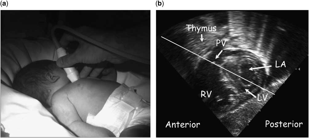
Neonates and small infants have unique characteristics that make it possible to obtain echocardiographic views that are inaccessible in older patients. In the early stages of life, the sternum is predominantly cartilaginous, and therefore allows ultrasound transmission. In addition, the thymus gland is located in the anterior mediastinum and has a homogeneous echotexture that facilitates visualisation of cardiovascular structures. Finally, the normal right ventricular dominance in term newborns contributes to the different anatomical position of the pulmonary annulus at this age, making the transsternal approach an excellent way to obtain a cross-sectional view of the pulmonary valve (Fig 1a). Thus, and only in this age group, we have noticed during routine clinical practice that we can easily obtain a short-axis view of the pulmonary valve from a high transsternal approach. It also has allowed us to obtain very good pictures of the pulmonary arteries and the pulmonary venous drainage.
( a ) High transsternal view – transducer position. ( b ) Subcostal short-axis view: the transducer is placed on the manubrium sternum and then gradually tilted until the cross-section of the pulmonary valve is imaged (white line). LA=left atrium; LV=left ventricle; RV=right ventricle; PV=pulmonary valve.

The high transsternal view is performed with the patient in the supine position, ideally with the head turned towards the left, because this slightly left lateral decubitus position facilitates the approximation of the heart to the chest wall. The transducer must be positioned on the manubrium sternum with its major axis parallel to the intermammillary line (Fig 1b). The cardiovascular structure that is often initially identified is the pulmonary artery bifurcation, and this should be taken as a marker for identifying the pulmonary valve itself. Subsequently, from this position, the transducer is tilted slightly anteriorly and inferiorly to follow the pulmonary trunk in order to display the cross-section of the pulmonary valve, as shown in the Supplementary video.
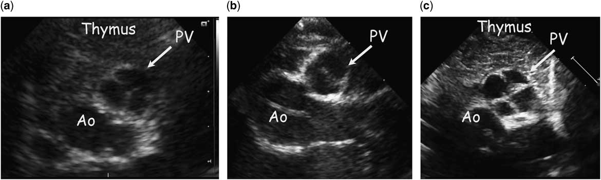
The transsternal view is routinely used by paediatric radiologists to study infant mediastinal structures;Reference Gassner and Geley 6 however, after reviewing the literature and specific paediatric echocardiography texts, we have found no reported use of this view as part of a systematic echocardiographic study of the heart in neonates and young infants. After including this approach in the daily echocardiographic assessment of our patients, we can conclude that this high transsternal view allows the sonographer to easily define the pulmonary valve anatomy of infants with different underlying pathologies, even in patients undergoing mechanical ventilation, accurately identifying congenital anomalies when present (Fig 2).
( a ) High transsternal view of an infant with an innocent murmur – normal pulmonary valve. ( b ) High transsternal view of an infant with prenatal diagnosis of pulmonary valve stenosis – bicuspid pulmonary valve. ( c ) High transsternal view of an infant with prenatal diagnosis of coarctation of the aorta not confirmed postnatally – quadricuspid pulmonary valve. Ao=aorta; PV=pulmonary valve.

The systematic study of the pulmonary valve morphology in neonates and young infants can provide information about the real incidence of the anomalies of this usually hidden valve. We believe that the high transsternal view should be included as part of routine protocols for echocardiographic examinations performed in this age group. In order to provide objective support for this belief, we have recently launched a prospective study to further define the characteristics of this approach in children and assess its utility when imaging the congenitally malformed heart.
Acknowledgement
None.
Financial Support
This research received no specific grant from any funding agency, commercial, or not-for-profit sectors.
Conflicts of Interest
None.
Supplementary Material
To view supplementary material for this article, please visit http://dx.doi.org/10.1017/S1047951115001845