Background
Even a “perfect Fontan” deteriorates over time, as the own author stated. Attrition due to multiple reasons shows survival rates of 86% at 5 years and 74% up to 15 years. Reference Fontan, Kirklin and Fernández1 Refinement in surgical techniques made the results to improve, lowering morbi-mortality and slowing the progression to congestive heart failure. Although late death remains an issue after Fontan completion, which is regarded as a definite palliation for univentricular heart patients. Reference Gentles, Mayer and Gauvreau2–Reference Iyengar, Winlaw and Galati6 As a result, “failed Fontan” patients turn out to become candidates for heart transplants. Reference Michielon, Parisi and di carlo7–Reference Mauchley and Mitchell14
From a surgical perspective, former procedures render a new scenario in which arteries and veins connections to the heart look totally different, so that alternative technical skills are requested to match graft and recipient. Many records about the topic can be found Reference Doty, Renlund, Caputo, Burton and Jones15–Reference Gil-Jaurena, Pérez-Caballero and Pita21 and, nowadays, nearly any distortion from previous surgery can be successfully approached when facing a transplant.
Our aim in this paper is to show a series of patients with Fontan physiology who were eventually transplanted. Literature is reviewed, technical hints are underlined and a sequential analysis of the different anastomoses is depicted in a sort of algorithmic approach (Fig 1).
Sequential analysis for the five anastomoses in transplant surgery after Fontan. Top row shows the five structures after the recipient’s heart removal. Middle row depicts the commonest modifications performed (in their respective columns). Bottom row adds ancillary procedures. Circ. arrest stands for brief periods of deep hypothermia plus circulatory arrest. SVC stands for superior vena cava.

Methods
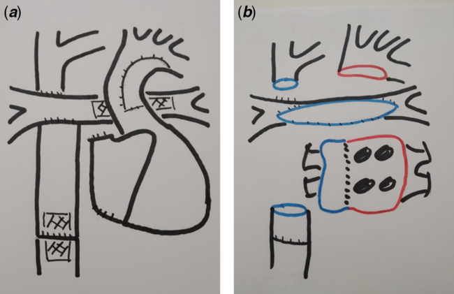
The records of 20 patients who were transplanted between January, 2013 and December, 2019 after Fontan completion are collected (Table 1). Our sample includes all consecutive patients with Fontan physiology transplanted in the authors’ centre. Institutional review board approval to collect and reprint the clinical data was obtained. Median age was 13 years (range 7–49) with 7 patients older than 18 years. Median weight was 37 kg. (range 17–61). Hypoplastic left heart syndrome (HLHS) was the main diagnosis in 13 patients. Extracardiac Fontan (Fig 2a) was performed in 15 cases (18–20 mm. diameter), atrio-pulmonary connection in 4, and intracardiac lateral tunnel in 1. Along the same 7-year period, 52 congenital non-Fontan transplants and 66 Fontan completion were undertaken. The comparison between transplant in Fontan (20) and non-Fontan (52) patients cumulative survival was performed with log-rank (Mantel–Cox) and χ2.
Demographics

IVC=inferior vena cava; LPA=left pulmonary artery; PLE=protein-losing enteropathy; SVC=superior vena cava
Scheme of pre- and post-cardiectomy in extracardiac Fontan (HLHS). ( a ) Bilateral Glenn and PTFE extracardiac conduit. Stents in LPA and IVC. ( b ) Surgical field after stents retrieval and pulmonary branches through patching. PTFE sleeve in situ (IVC). Ascending aorta removed for hemi-arch suture. Right-side structures in blue. Left-side structures in red.

Interval between Fontan and transplant surgery was 7 years as a median, ranging from 3 months to 37 years, being the gap lower than 1 year in four cases. The Fontan was taken down in two patients during the first 48 hours after its completion. Protein-losing enteropathy (PLE) was found in 6 patients. Seventeen stents had been previously deployed in 12 patients: left pulmonary artery (10), superior vena cavae (3), and inferior vena cavae (4). One patient had been assisted with a centrifugal pump (right atrium and ascending aorta cannulation) for 3 weeks.
Peripheral vessel patency was checked in previous cath lab studies and eco-Doppler when requested, in order to schedule anaesthetic monitoring and feasibility of femoral cannulation for cardiopulmonary bypass. Hepatic profiles were routinely assessed in the adult patients to rule out associated liver disease. MRI was particularly useful to depict pathways and anatomical relationships, as well as to define adherences between the sternum and underlying structures (right atrium and ventricle, aorta, innominate vein, and atrio-pulmonary connections).
Heart arrest was achieved with 20 ml/kg. of Crystalloid cardioplegic solution (Celsior; Genzyme Corp, Boston, MA, USA), plus an extra dose on arrival if overall ischaemia was expected to be longer than 4 hours. Our routine approach for graft procurement includes extra donor tissue. This regards aorta with aortic arch (beyond left subclavian artery), superior vena cavae with innominate vein, and a patch of pericardium. Pulmonary branches are hardly available because of the increased number of simultaneous lung harvesting. Patent formate ovale (PFO) is routinely closed and the left atrial appendage is checked for leaking (if used for decompression) upon arrival.
Results
Transplants were carried out with the five anastomoses technique, rather than the classical Shumway approach, provided that both vena cavae were no longer connected to the right atrium (in the extracardiac Fontan). In fact, this is our current strategy for most congenital heart transplants. Peripheral cannulation (vein, artery, or both) was undertaken in seven patients, whilst the central approach for extracorporeal circulation (ECC) was mainly performed. Patients were cooled down to 25 °C for better controlling blood return due to collateral circulation. Mean ischaemia time was 261.15 ± 58.56 minutes (140–370). Deep hypothermia (18 °C) with short periods of circulatory arrest was requested in 11 cases for manipulation of profound and fragile structures (inferior vena cava and pulmonary branches) and unpredicted aortic arch surgery. Inferior and superior vena cavae anastomoses were routinely performed after cross-clamp removal, unless unexpected issues.
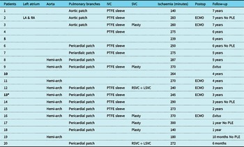
The five anastomoses underwent technical changes as outlined (Table 2, Fig 2):
-
1. Left atrium (1): size mismatch between donor and recipient; a small left cuff in the recipient was managed by keeping the perimeter of both atria (after septum removal) to match the anastomoses with the left atrium graft.
-
2. Aorta (9): hemi-arch anastomoses in short and distorted aorta (particularly for HLHS patients, with a bizarre amalgamation of aorta, pulmonary root, and patch augmentation tissue). Selective cannulation in the first supra-aortic vessel was electively accomplished when planned (in the same way as to neonatal arch strategy), so as to spare a circulatory arrest period.
-
3. Pulmonary arteries (15): after Glenn and extracardiac conduit previous sutures detachment, right and left branches are splint opened. If present, stents are removed (10) and donor aortic wall (3) or pericardium (12) is used as a patch from hilum to hilum. A customised slot in the patch hosts the graft pulmonary trunk.
-
4. Inferior vena cavae (12): a short sleeve (1–2 cm) from the extracardiac conduit was left in place to accommodate the graft. Stents were previously removed (4).
-
5. Superior vena cavae (7): direct anastomosis to innominate vein was accomplished in two patients because of former stenosis (1) and thrombosis (1). Beveled anastomoses were carried out in three patients for size mismatch. A persistent left superior vena cava (dual SVC) was approached in two patients with extra tissue from the donor (innominate vein). Stents were removed in three cases.
Anastomoses variations and follow-up. Patients No. 5 and No. 10 had no modifications. Patient No. 13* was on an assist device

LA=left atrium; LSVC=left superior vena cava; PLE=protein-losing enteropathy; PTFE=polytetrafluoroethylene graft; RA=right atrium; RSVC=right superior vena cava
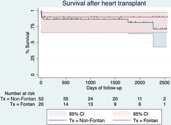
Follow-up has been complete with a median of 42 months (range 6–84). One patient died in the operation theatre due to massive bleeding (case number 9). The second patient died in the ICU because of a brain haemorrhage on postop day 3 (No. 16). Six patients required ECMO after coming off bypass because of right ventricle dysfunction (progressive ventricle dilatation plus atrial pressure raising above 20 mmHg). Likely causes are as follows: suspected pulmonary hypertension, graft ischaemia, volume overload for bleeding, etc. ECMO was discontinued before 72 hours in all cases. Bleeding re-exploration was performed in four patients. Collateral circulation closure in the cath lab was carried out in four patients in the early postop. Two patients had their aortic and superior vena cava anastomosis stented, respectively, in the first month of post-transplant. PLE resolved in all but one patient (theatre death) in their first 6 months. Cumulative survival was 90%, compared to 83% in the non-Fontan congenital transplant group (9/52 mortality rate), as shown in Fig 3. No statistical significance was found between them (p = 0.551). The overall survival was 85% for the whole group (72 patients).
Kaplan–Meier survival curve. Ninety percentage in the Fontan group (red line) compared to 83% in non-Fontan congenital transplants (blue line).

Discussion
Provided that a bi-ventricular physiology is not achievable, Fontan surgery is regarded as a palliative strategy in certain uni-ventricular conditions. Even though surgical techniques have evolved since the original reports, attrition was close to 26% at 5 years in Fontan’s own words. Reference Fontan, Kirklin and Fernández1 Whether the causes of eventual deterioration are due to left or right single ventricle anatomy, or ventricle dysfunction versus Fontan circuit failure are beyond the scope of this paper. Reference Backer, Russell and Pahl12,Reference Michielon, vanMelle and Wolff13 The authors focus their attention in the surgical strategy.
Transplant after Fontan completion is actually a surgical challenge. Besides the hurdles of a re-do surgery (fourth sternotomy in HLHS patients), uni-ventricular physiology adds unique requirements. The lack of a sub-pulmonary pump makes both systemic and pulmonary circuits to be in serial connection. Preload, chronically diminished, enhances diastolic dysfunction. Afterload changes, either pulmonary or systemic, drive to systolic dysfunction. Non-modifiable causes such as ventricle morphology, heterotaxy, PLE, or plastic bronchitis (PB) increase morbidity. Adult Fontan patients need to have their liver and kidney function assessed Reference Mauchley and Mitchell14,Reference Rychik, Atz and Celermajer22 to check a Fontan-associated-liver disease (FALD) either to contraindicate the heart transplant or to indicate a combined liver-heart transplant.
Before evaluating transplant candidacy in Fontan patients, catheter-based and surgical options must be optimised. Aorta and pulmonary branches angioplasty (or stenting) to release afterload, valvular regurgitation repair (or replacement), anti-arrhythmia measures (including pacemaker and resyncrhonisation), extracardiac Fontan conversion, Reference Mavroudis, Backer, Deal, Johnsrude and Strasburger23 etc., should be scrutinised. In comparison to bi-ventricular hearts, uni-ventricular patients are considered as suboptimal candidates for mechanical circulatory support. A review shows more than twofold mortality (57.7 versus 24.4%) in uni- as opposed to bi-ventricular patients, Reference Home, Conway, Rebeyka and Buchholz24 inasmuch several improvements are in progress. Reference Adachi, Burki and Fraser25
Pre-transplant assessment is paramount. This cohort of patients have undergone many catheter-based and surgical procedures, including transfusions, and lymphocytic reactivity must be checked. Neck and groin vessels might not be patent any longer. Pulmonary arteries pressure and resistance should be considered as slightly elevated, and it proves that a reliable method to assess it has not been yet defined. Reference Mitchell, Campbell and Ivy26
Several papers report a comprehensive approach to these complex operations. Reference Mauchley and Mitchell14,Reference Gil-Jaurena, Pérez-Caballero and Pita21,Reference Hosseinpour, González-Calle and Adsuar27,Reference Iyengar, Sharma and Weintraub28 Close coordination between procurement and transplant teams are key, in order to keep at a minimum ischaemia (in a scenario where a spare hour is needed between cardiectomy and proper graft stitching). As previously stated, extra tissue from the donor is harvested to patch the recipient with “autologous” material. The rationale is trying to avoid foreign pieces, which might be more prone to endocarditis in the setting of a immunodeficiency post-transplant. We routinely evaluate the “5 sutures” to be carried out, in a sort of algorithm (Fig 1), and anticipate any modifications. Peripheral vessel patency beforehand information allows quick and safe anaesthetic monitoring and groin preparedness for cannulation. The more information and setup, the less improvisation at transplant time.
Table 2 displays the changes carried out for the 5 anastomoses in the 20 patients enrolled. Every structure has been modified at least once. Although previously reported on isolation, Reference Doty, Renlund, Caputo, Burton and Jones15–Reference Gil-Jaurena, Pérez-Caballero and Pita21 few papers actually consider it a strategy (Fig 2b) facing complex procedures. Reference Mauchley and Mitchell14,Reference Gil-Jaurena, Pérez-Caballero and Pita21,Reference Hosseinpour, González-Calle and Adsuar27,Reference Iyengar, Sharma and Weintraub28
-
1. The size mismatch between left atria was sorted out leaving a “bi-atrial” cuff in the recipient (No. 2).
-
2. Our aortic anastomoses have evolved since one of our early patients had his/her anastomoses stented in the postop (No. 3). Currently, we advocate for a more aggressive “hemi-arch” procedure when a size mismatch is anticipated, particularly in HLHS patients with a short and bizarre new aorta. Reference Kanter, Mahle, Vincent and Berg29 Thus, we have recently cannulated the brachiocephalic artery interposing a PTFE conduit to avoid deep hypothermia and circulatory arrest. As an alternative, axillary cannulation can be applied (not used yet in our experience). Nevertheless, unexpected findings can lead us to cool down and arrest the pump to fix the aortic anastomoses. That was the case of a patient whose (fragile) aorta torn apart when trying to release a scar in a former Waterston shunt (No. 19).
-
3. Pulmonary arteries plasty has been already reported, Reference Mauchley and Mitchell14 choosing synthetic material. Reference Iyengar, Sharma and Weintraub28 As abovementioned, an autologous (recipient/donor) patch is preferred when available. Donor aorta was selected in our first patient, but we then switched to donor pericardium in order to spare the graft aorta (arch) for hemi-arch techniques. Previously implanted stents are completely removed, Reference Gil-Jaurena, Zunzunegui and Pérez-Caballero30 mainly in the left branch, before patching from hilum to hilum.
-
4. A short sleeve is left in situ for the suture of the inferior vena cavae after removing the extracardiac conduit. We found this maneuver helpful in avoiding tearing and deep cannulation close to the diaphragm. Again, stents were removed Reference Gil-Jaurena, Zunzunegui and Pérez-Caballero30 in 4 patients in a low flow setting (2) or femoral cannulation and circulatory arrest (2).
-
5. Superior vena cavae suture changes were anticipated in the preoperative screening. Cannulation sites were modified and suturing was carried out under cross-clamp, instead of on a beating-heart as routinely.
Generally speaking, short period of deep hypothermia and circulatory arrest were scheduled as we earned experience, particularly for profound structures (left pulmonary artery, inferior vena cava) and when stents were deemed to be removed. Interestingly, some patients underwent three intermittent periods of circulatory arrest at 18 °C to address LPA, IVC, and aortic arch. We have learned to cool the patients down slowly, anticipating any such low-flow (or even arrest) safe periods. Owing to the complexity and changes in the five anastomoses, plus the average long-distance procurement, ischaemia was longer than 4 hours in 15 patients (Table 2). Not surprisingly, on average one hour is necessary to “prepare” the field after cardiectomy and before graft stitching begins (Fig 2). Only two patients in our series (No. 5 and No. 10) did not undergo any changes in their sutures: both were atrio-pulmonary Fontan-type (assuming that detachment of atrium from the pulmonary artery is not a modification).
Post-operative complications are to be expected in this subset of patients. Four patients returned to theatre because early postop bleeding. Re-do surgery and plenty of collateral vessels have been blamed for that. Reference Iyengar, Winlaw and Galati6,Reference Kanter11,Reference Backer, Russell and Pahl12,Reference Mauchley and Mitchell14 Even worse, one of the patients died in the operation theatre because of massive and uncontrollable bleeding, as reported elsewhere Reference Michielon, vanMelle and Wolff13 particularly in caquectic and coagulopathic patients. Right ventricular dysfunction, likely caused by subtle pulmonary hypertension, lead to ECMO in six patients (successfully removed before 72 hours). Interestingly, patient No. 13 was on a systemic assist device for 3 weeks before transplant and on a sub-pulmonary assist device (ECMO) after transplant for 2 days. Not surprisingly, the right atrial appendage and aorta were cannulated in both instances (same structures, different ventricles). Collateral vessels required closure in the cath lab in four patients. Step up on inotropes with good bi-ventricular function should give the clue in such circumstances. Reference Krishnan, Lamour, Hsu, Kichuk, Donnelly and Addonizio31 PLE resolved in five out of the six patients (sixth one died in theatre), as has been reported. Reference Backer, Russell and Pahl12 Whether PLE is a predisposing factor for post-operative infection in a immunodepression setting Reference Michielon, vanMelle and Wolff13 cannot be ascertained in our short experience.
Patients over 18 years deserve further attention. Reference Murtuza, Hermuzi and Crossland32,Reference Hernández, Lemor and Clark33 Most of them were morphologically left single-ventricle and atrio-pulmonary Fontan type connection. “Old-fashioned” techniques like thoracic and Waterston shunts were found in this subgroup. One has to be prepared for more surprises (if any) amongst older patients who encompass the evolution of Fontan-type surgical techniques. On the contrary, most young single-ventricle transplant candidates belong to the HLHS category and have been stepwise and uniformly operated on.
Bernstein et al Reference Bernstein, Naftel and Chin10 published in 2006 the first multicentric study about transplant in failed Fontan. Since several groups have reported papers with series including more than 10 patients. Reference Backer, Russell and Pahl12–Reference Mauchley and Mitchell14 They all agree that surgical mortality is far worse than in other-than-Fontan transplants, approaching 20%. Interestingly, the group in Emory Reference Kanter11 (Atlanta, United States of America) shows astonishing low rates such as 4%. A European multicentric study in 2015 gathers 61 patients from 11 institutions along 20 years, Reference Michielon, vanMelle and Wolff13 mirroring the scattering of data and the hurdles on accumulating experience on Fontan-transplanted patients in any given centre. In this paper, Reference Michielon, vanMelle and Wolff13 26% of the patients come from HLHS morphology, whilst 72% are extracardiac connection type. Most of the authors underline pre-operative assessment of utmost importance, including vessel patency and image techniques (MRI) to precisely define adherence to the sternum. Reference Mauchley and Mitchell14 Conventional interventionism and surgery to delay or improve pre-transplant status are strongly advised. Reference Michielon, vanMelle and Wolff13 Amongst these procedures are: collaterals closure/embolisation, vessel angioplasty/stenting, valve repair/replacement, Fontan conversion plus Maze procedure, etc. Data collection will provide insight into Fontan failure mechanisms such as: single-ventricle dysfunction or sub-pulmonary failure (even both), right versus left morphological single ventricle, late versus early failure. Backer et al Reference Backer, Russell and Pahl12 stated that late failure (ventricle dysfunction) yields better prognosis (82% survival at 5 years post-transplant) compared to early deterioration for Fontan sub-pulmonary circuit failure – with good ventricle function – (33% survival at 5 years). PLE has been recently accepted as an indication for transplant listing, Reference Michielon, vanMelle and Wolff13,Reference Mauchley and Mitchell14 provided it is a surrogate of Fontan circuit failure. Accordingly, Fontan takedown is considered a truly early Fontan failure, with a 25% mortality rate after transplant in the European study. Reference Michielon, vanMelle and Wolff13
From a surgical standpoint, most papers agree that transplant after Fontan is actually a challenge. Re-do surgery, collateral circulation, coagulation disorders, caquexia, etc., increase complexity. Venous system (both and, occasionally, three cavae), pulmonary branches (from hilum to hilum), and aorta (aortic arch) reconstruction are routine in this setting (Fig 2). Exquisite coordination between procurement and implanting teams is of paramount importance, foreseeing 3–4-hour headstart prior to graft arrival. Extra donor tissue is suggested. Bypass and ischaemia times are longer than usual. Deep hypothermia plus circulatory arrest (intermittent periods) are becoming familiar. Altogether, these are ingredients for a morbidity and mortality higher than average. Bleeding and post-operative right ventricular failure (ECMO included) are to be expected in the postop, as well as a hyperdynamic state caused by residual collateral circulation (needing cath lab transfer for vessels closure Reference Mauchley and Mitchell14,Reference Krishnan, Lamour, Hsu, Kichuk, Donnelly and Addonizio31 ).
Our series enrolled 20 consecutive transplants (kids and adults) in a 7-year lapse, performed by the same team of congenital surgeons, with a follow-up of nearly 4 years. 13 patients were under 18 years (mostly HLHS, extracardiac Fontan) and 7 of them, over 18 years (mainly morphologically left single ventricle and mixture of Fontan-type procedures). A sequential analysis focused on the five proposed anastomoses is carried out, planning in advance any change pertaining to them all (Fig 1). This rationale includes lowering temperature and pump flows (anticipating arrest periods), as well as stents removal. The goal is to avoid improvisation as much as possible. Morbidity and mortality results are similar to other groups, Reference Bernstein, Naftel and Chin10 assuming our low figures and follow-up as a limitation. Interestingly, mortality rate is not worse than non-Fontan transplants in our own Institution.
Refinements in surgical technique and post-operative care yield good results nowadays, allowing more children to reach adulthood. Conversely, more palliated patients (e.g., Fontan) are likely to become transplant candidates according to their attrition rate. Michielon et al Reference Michielon, Carotti, Pongiglione, Cogo and Marino34 foresee that 70% of congenital heart transplants will belong to uni-ventricular conditions in the future. A likely mismatch between the increase of candidates and donors enhances us either to exhaust conventional approaches, either to develop surgical expertise in the transplant. Reference McCormick and Schumacher35 Encouraging reports with good results should consider allocation Reference Rychik, Atz and Celermajer22,Reference Murtuza, Hermuzi and Crossland32 and resources for this emerging and demanding cohort of patients.
Conclusions
Transplant surgery after Fontan poses a surgical challenge in a high-risk setting. Careful preoperative assessment and a sequential analysis focused on the five anastomoses and their likely modifications (including hypothermia and stents removal) have paved the way for improving results. Extra donor tissue allows for autologous reconstruction, prior to graft implantation. ECMO for right ventricular dysfunction was needed in nearly one-third of the cases, prompting us to lower our threshold for such an assistance. On gaining experience, overall results can match that of non-Fontan transplant patients.
Acknowledgements
To Dr. Reza Hosseinpour (Sevilla, Spain) for his kind revisions of the manuscript.
Financial support
This research received no specific grant from any funding agency, commercial, or not-for-profit sectors.
Conflicts of interest
None.