Despite the remarkable, intrinsic ability of bone to regenerate without scarring, there are a number of clinical conditions where healing is impaired. In approximately 10% of fractures, for example, bone healing is either very slow (delayed union) or the bone does not heal at all (nonunion). The reasons for this are unclear, although it is more common in smokers (Ref. Reference Kwiatkowski, Hanley and Ramp1), alcohol abusers (Ref. Reference Chakkalakal2) and diabetics (Ref. Reference Gandhi3). Bone healing is also an issue for the elderly (Ref. Reference Gruber4) and patients with osteoporosis (Ref. Reference Giannoudis5). Healing is also difficult under conditions where a large segment of bone is lost because of injury, infection or tumour resection; indeed, segmental defects beyond a certain ‘critical size’ never heal spontaneously, even in young, healthy individuals (Ref. Reference McKee6). Problems in bone healing also arise for iatrogenic reasons; for instance, when the surgeon needs to fuse two vertebrae together as a way of treating back pain (Ref. Reference Daniels7). An improved ability to form bone would also be useful for the treatment of osteonecrosis and for increasing bone stock around prosthetic joints, with the possibility of preventing or treating aseptic loosening. Moreover, there are numerous potential applications in the craniofacial, orthodontic and dental areas.
The most reliable way to form bone under these conditions is by the surgical implantation of some of the patient's own living bone (autograft). This is usually harvested surgically from the iliac crest of the pelvis. Although this method has a high success rate, its usefulness is restricted by the limited amounts of bone available for autografting, and side effects, especially pain, at the harvest site (Ref. Reference Kim8). As an alternative or supplement to autograft bone, it is possible to use bone recovered from cadavers (allograft) (Ref. Reference Delloye9). Although allograft bone is available in almost unlimited quantities, there are concerns about disease transmission, and because processing kills endogenous cells, it is essentially dead bone. Therefore, unlike autograft, it is unable to participate actively in the formation of new bone and thus serves as an inert filler. If the allograft is implanted into areas where it needs to bear load, it frequently fails, because unlike living bone, it cannot remodel.
In recent decades, the search for new ways to promote bone healing has increasingly turned to biology (Ref. Reference Watson10). In general, there are two routes through which the body forms bone: endochondral and intramembranous (Ref. Reference Shapiro11). The former process involves the local differentiation of progenitor cells into chondrocytes that lay down a cartilaginous matrix. The cartilage is then replaced by bone through a process known as endochondral ossification, during which the cartilaginous matrix is degraded, blood vessels invade the cartilage and chondrocytes are replaced by osteoblasts. The intramembranous formation of bone involves the differentiation of progenitor cells directly into osteoblasts without a cartilaginous intermediate. Both the endochondral and intramembranous routes lead to the formation of immature, woven bone that undergoes remodeling into mature, lamellar bone (Ref. Reference Shapiro11).
Study of osteogenesis has identified a number of growth factors with the ability to stimulate one or more of the steps involved in endochondral and intramembranous bone formation (Refs Reference Simpson, Mills and Noble12, Reference Lieberman, Daluiski and Einhorn13). The best characterised of these factors are the bone morphogenetic proteins (BMPs). Several members of this large family, including BMP-2, BMP-4, BMP-6, BMP-7 and BMP-9, are able to induce the formation of bone when implanted experimentally into the muscles of laboratory animals (Ref. Reference Tsumaki and Yoshikawa14). Recombinant, human BMP-2 and BMP-7 are available clinically as the active components of the products Infuse® and OP-1® (osteogenic protein-1), respectively. Despite the powerful osteogenic properties of these proteins in laboratory animals, their clinical performance is mixed (Ref. Reference Kwong and Harris15). For instance, they appear to be much more effective for spinal fusion than for long-bone healing, and need to be administered at extremely high doses of several milligrammes – many orders of magnitude greater than the levels at which they occur naturally in bone. The use of such high doses not only raises safety concerns, but also enormously increases the cost. In the USA, for example, a single dose of Infuse® can cost around $5000. There is considerable optimism that gene-therapy approaches can be harnessed to improve the effectiveness of osteogenic factors, such as BMP-2, while lowering both costs and the potential for side effects.
As its name suggests, gene therapy involves the transfer of genes, or more usually cDNA (complementary DNA), to patients for therapeutic purposes. Gene therapy was originally developed as a means of curing genetic diseases, but in recent years its potential use in treating nongenetic disorders has become increasingly appreciated and a range of nonmendelian diseases, including various cancers (Ref. Reference Gillet16), arthritis (Ref. Reference Evans17) and Parkinson disease (Ref. Reference Fiandaca, Forsayeth and Bankiewicz18), have been the subject of clinical trials. In the context of bone healing, the aim is to deliver to the fracture site cDNAs encoding osteogenic proteins, such as BMPs. Successful gene delivery and expression leads to the continuous, local, focal synthesis of the osteogenic protein, which is likely to have undergone authentic post-translational modification and, unlike the recombinant product, to be uncontaminated by inactive, misfolded products that can trigger immune responses. Safety is further enhanced by gene transfer because the local production of osteogenic proteins at approximately physiological concentrations is less likely than the bolus application of a large amount of recombinant protein to lead to the systemic spread of these proteins to ectopic sites.
As with most other applications of gene therapy, the key questions for bone healing are: which gene(s) to transfer? Where to transfer them? How to get them there? Does it work? Is it safe? Much of the remainder of this article addresses these questions, bearing in mind that different clinical applications, such as fracture nonunions versus segmental defects, and different parts of the skeleton, such as maxillofacial bones versus long bones, might require different approaches. Gene therapy for bone healing has been recently reviewed by Carofino and Lieberman (Ref. Reference Carofino and Lieberman19)
Which genes to transfer?
There are a large number of growth factors known to stimulate one or more of the processes involved in osteogenesis and bone healing. Most of these either stimulate the differentiation of progenitor cells into chondrocytes or osteoblasts, or enhance the bone-forming activities of mature osteoblasts. Because bone is highly vascularised, vascular endothelial growth factor (VEGF) and other angiogenic factors are also important, especially for intramembranous bone formation; chondrogenesis, the first step in endochondral ossification, does not require a blood supply. The importance of angiogenesis in bone healing has been nicely demonstrated by Peng and colleagues (Ref. Reference Peng20), who used gene transfer to show that VEGF enhanced, and a VEGF antagonist (sFlt1) inhibited, repair of cranial defects in mice. Because individual growth factors act at different stages of osteogenesis, combinations of different factors should promote bone healing more potently than a single factor alone. This has been confirmed in animal models using gene delivery of BMP-2 and BMP-7 (Refs Reference Koh21, Reference Zhu22); BMP-4 and VEGF (Ref. Reference Peng20); BMP-4 and transforming growth factor-β (TGF-β) (Ref. Reference Li23).
An alternative approach to the delivery of cDNAs encoding growth factors involves the delivery of transcription factors associated with osteogenesis, such as RUNX2 (Ref. Reference Zhao24) and osterix (Transcription factor Sp7) (Ref. Reference Tu25). Because these are intracellular proteins, they are not easily delivered by traditional methods of protein delivery, but are well suited to gene transfer. Their safety might be greater than that of growth factors, because they are not secreted and do not circulate. This is a disadvantage in terms of efficiency, however, because gene transfer would have to be very effective to modify most of the progenitor cells in the vicinity of the fracture. Timing of gene transfer would then be important, to synchronise with the entry of progenitor cells into the defect. In the case of a secreted gene product, by contrast, there is only the need to modify adequate numbers of cells to create a sufficiently high local concentration of the secreted factor for a sufficient period of time. LIM mineralisation protein-1 (LMP-1) is another intracellular, osteogenic molecule of interest. It was discovered during a screen of transcripts that are induced during osteogenesis, and its function is still unclear (Ref. Reference Boden26). However, some of its activity may be explained by its ability to prevent the degradation of Smad signalling molecules (Ref. Reference Sangadala27). Remarkably potent and dramatic preclinical results in models of spinal fusion have been reported using plasmid DNA-liposome as the vector to deliver LMP-1 in rats (Ref. Reference Boden28) and adenovirus vectors in rabbits (Ref. Reference Viggeswarapu29). Vectors expressing a related protein, LMP-3, are also osteogenic in animal models (Refs Reference Lattanzi30, Reference Parrilla31, Reference Pola32)
A slightly different approach is indicated by data which show that the knockdown of the BMP inhibitors noggin (Ref. Reference Wan33) and chordin (Ref. Reference Kwong, Richardson and Evans34) also stimulates osteogenesis. The delivery of siRNA molecules is possible by RNA transfection or the delivery of a vector that encodes the inhibitory RNA molecule. There do not appear to have been any experimental attempts to enhance fracture repair by inhibition of osteoclast function using gene transfer, although this might be a promising short-term approach to increase the net accumulation of bone.
Where to transfer genes?
Although it is possible to deliver osteogenic genes to sites where secreted gene products are able to circulate systemically, such strategies are considered only for conditions such as osteoporosis, where there is disseminated loss of bone (Refs Reference Baltzer35, Reference Bolon36, Reference Kostenuik37). There is some evidence that mesenchymal stem cells (MSCs) can home to fracture sites and deliver a genetic payload (Ref. Reference Shen38), but this has not been widely studied. Instead, because fractures occur locally, gene-therapy strategies are based upon the direct, local delivery of genes to cells within or around the fracture site. This has the advantage of achieving high local concentrations of the gene product, with minimal exposure of other organs. As noted earlier, this introduces an important safety component.
How to transfer genes to fractures?
Basic principles
A variety of different vectors have been developed for gene transfer. These can be divided into nonviral and viral gene delivery; nonviral gene transfer is known as transfection, and viral gene transfer is known as transduction. Each has advantages and disadvantages. Viral vectors are much more complicated to manufacture and they raise greater safety concerns, but continue to be used for the majority of human gene-therapy trials because they are so much more efficient than nonviral vectors. For most gene-transfer applications, viruses are engineered to reduce any pathogenicity they might have had, while retaining their ability to infect target cells. For nearly all purposes, the viruses are attenuated, so that after they have delivered their genetic payload to the cells they infect, they cannot replicate. A variety of different viruses have been engineered in this way, and a number have found use in human clinical trials (www.wiley.co.uk/genetherapy/clinical/). There is not space here to describe them all in detail (for review see Ref. Reference Young39), but some general comments can be made about the vectors used in studies of bone healing.
Oncoretroviruses (usually referred to as retroviruses) and lentiviruses (also members of the retrovirus family) are RNA viruses that integrate their genetic material into the chromosomal DNA of the cells they infect, thus providing the potential for long-term transgene expression. Although this is highly desirable when treating a genetic disease for the lifetime of the patient, it is probably not necessary for healing a fracture, which might be expected to take 6–8 weeks. The continued expression of a transgene beyond this time would probably be counterproductive. Another disadvantage of integrating retrovirus and lentivirus vectors is their potential for causing insertional mutagenesis. This has led to leukaemia in children being treated for severe combined immunodeficiency disease (SCID) (Ref. Reference Kohn, Sadelain and Glorioso40). Retrovirus, but not lentivirus, has the additional disadvantage of requiring host-cell division for successful transduction (Ref. Reference Kurian, Watson and Wyllie41). Lentivirus vectors are being engineered to prevent integration (Ref. Reference Sarkis42), but it still remains doubtful that such viruses could be used in humans to treat a non-life-threatening condition, such as an osseous nonunion, especially as the most commonly used lentiviral vectors come with the ‘psychological baggage’ of having been derived from HIV.
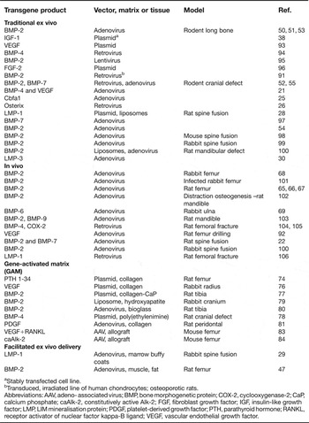
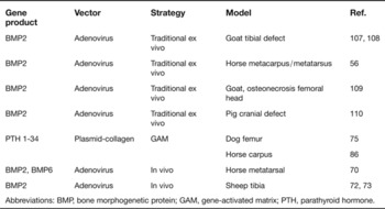
Adenovirus and recombinant (unlike wild-type) adenoassociated virus (AAV) are DNA viruses whose genomes do not integrate with high frequency into host-cell DNA. Nevertheless, they are capable of maintaining transgene expression for lengths of time compatible with the needs of bone healing. Among the advantages of adenovirus is the relative ease with which recombinant vector can be produced at high titre, and its high infectivity towards many cell types, regardless of whether they are dividing or not (Ref. Reference Douglas43). As reflected in Tables 1 and 2, they have been the most widely used vectors for studies of gene therapy for bone healing. A perceived disadvantage of adenovirus is its high antigenicity (Ref. Reference Douglas43). Most of the population have circulating, neutralising antibodies to adenovirus serotype 5, the serotype most commonly used for gene transfer, which might reduce the effectiveness of adenovirus-based gene therapy. Moreover, the inflammatory response to adenovirus may be detrimental to healing.
Use of gene therapy to treat osseous defects in small-animal models

aStably transfected cell line.
bTransduced, irradiated line of human chondrocytes; osteoporotic rats.
Abbreviations: AAV, adeno- associated virus; BMP, bone morphogenetic protein; COX-2, cyclooxygenase-2; CaP, calcium phosphate; caAlk-2, constitutively active Alk-2; FGF, fibroblast growth factor; IGF, insulin-like growth factor; LMP, LIM mineralisation protein; PDGF, platelet-derived growth factor; PTH, parathyroid hormone; RANKL, receptor activator of nuclear factor kappa-B ligand; VEGF, vascular endothelial growth factor.
Use of gene therapy to treat osseous defects in large-animal models

Abbreviations: BMP, bone morphogenetic protein; GAM, gene-activated matrix; PTH, parathyroid hormone.
AAV is perceived to be safe, and the death in 2007 of a subject receiving intra-articular injections of recombinant AAV in an arthritis gene-therapy trial was not attributed to the vector (Ref. Reference Frank44). Although it has a small carrying capacity, most of the growth factors of interest have small cDNAs that fit comfortably into the recombinant AAV genome. The single-stranded DNA genome of AAV was an earlier limitation for many purposes, because transgene expression required the host cell to synthesise the second strand of DNA and this was sometimes very inefficient. This limitation was overcome by the development of recombinant, selfcomplementing AAV with double-stranded DNA genomes (Ref. Reference McCarty, Monahan and Samulski45) that give much higher levels of transgene expression much more quickly and reliably. Use of AAV remains constrained by the technological challenges of producing vector in large quantities and the need for relatively high multiplicities of infection.
Nonviral vectors (Ref. Reference Glover, Lipps and Jans46) can be as simple as naked plasmid DNA. The efficiency of gene transfer can be increased by associating the DNA with a carrier, such as a liposome, polymer or nanoparticle, or by using a physical stimulus such as electroporation or sonication.
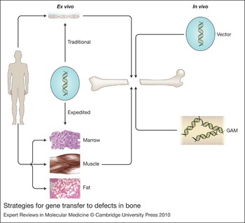
Regardless of the vector, there are two basic strategies for gene transfer: ex vivo and in vivo (Fig. 1). For ex vivo delivery, cells are removed from the host, genetically modified outside the body and then reimplanted. This was very common in the early days of gene therapy, because the first useful vectors were retroviruses that require cell division for effective transduction. Usually the cells are expanded in tissue culture, which, for human application, is very cumbersome and expensive. As described later, when host-cell division is not required, cell culture may not be necessary and tissue can be removed, genetically modified and reimplanted intraoperatively in an expedited ex vivo fashion (Ref. Reference Evans47). For in vivo delivery, the vector is introduced directly into the host, often by injection or by implantation in association with a matrix (Fig. 1).
Strategies for gene transfer to defects in bone. There are two general strategies: in vivo (right-hand side) and ex vivo (left-hand side). For in vivo gene delivery, the vector is introduced directly into the site of the osseous lesion, either as a free suspension (top right) or incorporated into a gene-activated matrix (GAM) (bottom right). For ex vivo delivery, vectors are not introduced directly into the defect. Instead they are used for the genetic modification of cells, which are subsequently implanted. Traditional ex vivo methods (top left) usually involve the establishment of cell cultures, which are genetically modified in vitro. The modified cells are then introduced into the lesion, often after seeding onto an appropriate scaffold. Expedited ex vivo methods (bottom left) avoid the need for cell culture and scaffolds by genetically modifying tissues such as marrow, muscle and fat, intraoperatively and inserting them into the defect during a single operative session.

For certain applications, such as when treating a disease such as rheumatoid arthritis that has flares and remissions, it might be desirable to regulate the level of transgene expression. There are a number of strategies for this, most of which involve the use of inducible promoters. This has not been widely investigated in the context of bone healing because, as far as we know, the levels of the most commonly used growth factors do not need to be closely regulated, and many of the commonly used vectors express for suitable lengths of time without the need for modification. Nevertheless, it has been confirmed that BMP-2 expression and consequent osteogenesis can be regulated in vivo using a TetON inducible-expression system (Ref. Reference Gafni48).
Applications in bone healing
Four strategies have emerged for gene transfer to osseous lesions; two of them are in vivo and two are ex vivo (Fig. 1). All have shown promise, and it is not yet clear which of them will find eventual clinical use in human or veterinary medicine.
Ex vivo strategies
Traditional ex vivo gene transfer
As noted above, ex vivo gene transfer to culture-expanded cells was the first gene-therapy strategy, and the first to be used in human clinical trials (Ref. Reference Rosenberg49). It was also among the first experimental approaches to be used to enhance bone healing in experimental animal models. Lieberman and co-workers (Refs Reference Lieberman50, Reference Lieberman51) used a recombinant adenovirus vector carrying the cDNA for human BMP2 (Ad.BMP-2) to transduce cultures of bone marrow stromal cells. The transduced cells were seeded onto collagen scaffolds and implanted into critical-sized defects in the femora of rats. All defects healed within 12 weeks of implanting the genetically modified cells, and showed better histological integrity than control defects treated with recombinant human BMP-2. Because rat marrow stromal cells differentiate into osteoblasts in response to BMP-2, the transduced cells have the potential to serve both as a provider of BMP-2 and also as a source of osteoprogenitors, thereby enhancing the reparative response. Subsequent investigators have confirmed the utility of this strategy in experimental animals using osteoprogenitor cells derived from additional sources, such as periosteum (Ref. Reference Breitbart52), fat (Refs Reference Peterson53, Reference Hsu54) and muscle (Ref. Reference Lee55). Despite the success of these methods, it is still unclear whether the use of osteoprogenitor cells as vehicles for ex vivo gene delivery confers much of an advantage. It has been difficult to identify donor cells in the healed bone (Ref. Reference Lee55), and some investigators report success with skin (Refs Reference Lattanzi30, Reference Ishihara56) and gingival (Ref. Reference Shin57) fibroblasts, which are not noted for their osteogenic potential. In agreement with this, the abilities of different cell types to promote the formation of heterotopic bone intramuscularly in mice was compared, but it was concluded that the cell type was not important (Ref. Reference Gugala58).
Although most investigators use adenovirus vectors for experiments of this kind, gene expression usually lasts for only 2–3 weeks in vivo. Virk and colleagues (Ref. Reference Virk59) compared lentivirus, which expresses BMP-2 within osseous lesions for 8 weeks in vivo, with adenovirus. Although both vectors achieved bony union in the femoral critical-size-defect model, the repaired bone formed in response to the lentivirus vector had better mechanical and histological properties. In contrast to this, Gazit and colleagues (Ref. Reference Gazit60) have achieved remarkable success in murine models of bone healing using nonviral delivery of plasmids containing a human BMP2 cDNA. Under these conditions, BMP-2 expression is low and transient. There has been very little research into species differences when using gene-transfer approaches, and it is possible that the murine model is especially responsive to human BMP-2.
Expedited ex vivo gene transfer
Ex vivo strategies have the safety advantage that vectors are not introduced directly into the body. However, there are disadvantages to the traditional approaches described above: they require two invasive procedures, one to harvest cells and one to implant them, and they incur the high costs of expanding cells under GMP (good manufacturing practice) conditions. To obviate these problems, expedited ex vivo methods are being developed in which tissues are removed from the body, genetically modified and reimplanted within a single operative period (Ref. Reference Evans61). The first use of this approach in bone healing was in a rabbit model of spinal fusion (Ref. Reference Viggeswarapu29). Marrow was withdrawn and the buffy coats isolated, transduced with a recombinant adenovirus vector encoding LMP-1 and implanted locally during a single operative session. Full spinal fusion occurred in each rabbit receiving the transgene in this fashion.
Skeletal muscle and fat have also been evaluated as vehicles for an expedited, ex vivo gene-based technology, known as ‘facilitated endogenous repair’, for the healing of lesions in bone, cartilage and other connective tissues (Refs Reference Evans47, Reference Evans61). Muscle (Ref. Reference Bosch62) and fat (Ref. Reference Dragoo63) were selected because they contain osteoprogenitor cells, can serve as space-filling scaffolds, and they can be harvested, genetically modified and reimplanted in a single sitting. Using these methods in conjunction with Ad.BMP-2, it was possible to heal rat critical-size femoral defects more rapidly and reliably than with recombinant human BMP-2 (Ref. Reference Evans47). Implantation of genetically modified muscle led to the rapid formation of cartilage within the lesion, presumably by the efficient differentiation of progenitor cells within the implant into chondrocytes, which underwent efficient endochondral ossification. Tracking of the donor cells confirmed that at least some of the newly formed bone was derived from the implanted muscle (Ref. Reference Evans47). The expedited ex vivo approach also eliminates the humoral response to the adenovirus vector, which is an advantage for safety, efficacy and redosing.
In vivo strategies
In vivo strategies include direct injection of genes and gene-activated matrices and have been comprehensively reviewed recently (Ref. Reference Pelled64).
Direct injection of vector
Among the simplest of all gene-delivery strategies is to inject the vector directly into the osseous defect; this can often be performed percutaneously. Adenovirus vectors have been widely used for this purpose, and success has been reported in rats (Refs Reference Betz65, Reference Betz66, Reference Betz67), rabbits (Refs Reference Baltzer68, Reference Bertone69) and horses (Ref. Reference Ishihara70) using BMP-2 (Refs Reference Betz65, Reference Betz66, Reference Betz67, Reference Baltzer68, Reference Ishihara70) and BMP-6 (Refs Reference Bertone69, Reference Ishihara70) as transgenes. Bone healing in a rat femoral defect model was improved by delaying the administration of the virus for 5–10 days after creating the defect (Ref. Reference Betz65). This could reflect the time it takes for osteoprogenitor cells to enter the defect, and also the possibility that the coxsackievirus adenovirus receptor (CAR), through which adenovirus enters cells, is maximally induced at the lesion site at this time (Ref. Reference Ito71).
There also appear to be species differences in the osteogenic response to the direct injection of adenovirus. As noted above, very promising results have been reported in rabbits, rats and horses. In the sheep, the results have been quite disappointing. First experiments were conducted with a tibial osteotomy model (Ref. Reference Egermann72). This lesion spontaneously heals, and the administration of Ad.BMP-2 delayed, rather than accelerated, healing. Antibodies against both adenovirus and human BMP-2 were detected in the recipient sheep (Ref. Reference Egermann72), suggesting that immune reactions were responsible for the poor healing response. In agreement with this conclusion, the administration of Ad.BMP-2 to sheep that had been treated with steroids enhanced the healing of the tibial osteotomy (Ref. Reference Egermann73). It is not known whether the sheep immune system is especially sensitive to adenovirus, or whether the sheep, which spend much of their lives outdoors on a farm, had been previously exposed to adenovirus. Regardless of the explanation, it highlights again the importance of considering species differences and the immune system in studies of this sort.
Gene-activated matrices
Gene-activated matrices (GAMs) were developed to provide an off-the-shelf, nonviral, gene-based method for healing bone. In the original GAM formulation, plasmid DNA was incorporated into a collagen sponge and fitted into an osseous defect. The concept was to transfect in situ host reparative cells that infiltrated the GAM after implantation. The transfected cells would then secrete the transgene locally, triggering reparative responses. Using parathyroid hormone (PTH) 1–34 as the transgene, impressive results were reported in large segmental defects in rats (Ref. Reference Fang74) and dogs (Ref. Reference Bonadio75). However, with the exception of a GAM containing DNA encoding VEGF, which showed activity in a rabbit segmental-defect model (Ref. Reference Geiger76), no further progress has been reported using plasmid DNA and a collagen matrix.
Some investigators have attempted to improve the efficiency of GAMs by using different scaffolds (Refs Reference Endo77, Reference Huang78, Reference Ono79, Reference Valimaki80) or by using GAMs containing viral vectors. Once such GAM containing recombinant adenovirus carrying a cDNA encoding platelet-derived growth factor (PDGF) B has shown preliminary success in clinical trials for healing diabetic skin ulcers (www.t-r-co.com), and has shown promise in growing periodontal bone in animal models (Ref. Reference Chang81). The fact that is already in clinical trials for wound healing will facilitate its adaptation for bone healing in humans. There is evidence that a collagen scaffold reduces the immune response to adenovirus (Ref. Reference Sonobe82).
Schwarz and colleagues have developed a modified GAM process in which allograft bone serves as the scaffold and AAV as the vector (Ref. Reference Ito83). Efficacy was first demonstrated in a murine segmental-defect model. Allograft bone which, as described earlier in this article, contains no living cells, was ‘revitalised’ by coating it with two different AAV vectors carrying receptor activator of NF-κB ligand (RANKL), to promote osteoclastogenesis, and VEGF, to promote angiogenesis. When the construct was implanted into the mouse femoral defect, AAV transduced the host cells surrounding the implant, leading to the remodelling of the allograft and its replacement with host bone (Ref. Reference Ito83). Similar responses were noted using a constitutively active ALK2 cDNA (Ref. Reference Koefoed84). This encodes a type I BMP receptor bearing the same mutation as found in the heterotopic bone-deposition disease fibrodysplasia ossificans progressiva (Ref. Reference Shore85). Because the AAV-coated construct is relatively stable, it can form the basis of an off-the-shelf material for bone healing.
Does it work?
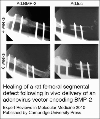
Table 1 lists the extensive literature confirming the ability of gene-transfer approaches to heal osseous defects in small laboratory animals, and Figure 2 shows one example of successful bone healing in a rat model. Far fewer studies have been performed in large-animal models (Table 2), but the results are encouraging, especially when using the traditional ex vivo approach in horses (Ref. Reference Ishihara56), goats (Refs Reference Rundle104, Reference Rundle105, Reference Strohbach106) and pigs (Ref. Reference Dai107). Results using GAM containing PTH 1-34 DNA were more equivocal (Refs Reference Bonadio75, Reference Backstrom86). Data from the in vivo delivery of Ad.BMP-2 and Ad.BMP-6 to osseous defects in horses were encouraging (Ref. Reference Ishihara70), whereas similar experiments sheep gave disappointing results (Refs Reference Egermann72, Reference Egermann73). Further research is clearly indicated.
Healing of a rat femoral segmental defect following in vivo delivery of an adenovirus vector encoding BMP-2. Representative radiographic images of segmental bone defects after direct injection of adenoviral vectors encoding human BMP-2 (Ad.BMP-2) or luciferase cDNA (Ad.luc). Most defects that had been treated with Ad.BMP-2 displayed bone formation within the defect by 4 weeks (top left) and complete union by 8 weeks (bottom left). Control defects treated with Ad.luc (and untreated defects) did not display appreciable signs of healing within this time (right panels). Images reproduced from Ref. Reference Betz66. © JBJS 2006.

Is it safe?
Over 1500 human gene-therapy clinical studies have taken place in the 20 years since the first properly authorised clinical gene-transfer study (Ref. Reference Rosenberg49) (www.wiley.co.uk/genetherapy/clinical/). Despite a common perception that gene therapy is unsafe, only two human deaths have been attributed to gene transfer (Ref. Reference Evans, Ghivizzani and Robbins87,). Nevertheless, safety issues continue to burden the clinical development of gene therapy. The 1999 death of Jesse Gelsinger (Ref. Reference Raper88) was widely reported and halted what had been a decade of spectacular growth in the number of clinical gene-therapy trials. His death resulted from an uncontrollable activation of the innate immune system following the in vivo infusion of a very high dose of recombinant adenovirus vectors into the hepatic portal vein. The second death occurred in Paris, where haematopoietic stem cells of children with SCID were treated ex vivo with a retrovirus vector. Several subjects developed leukaemia because of insertional mutagenesis, when the retroviral genome inserted adjacent to the LMO oncogene (Ref. Reference Kohn, Sadelain and Glorioso40). One of the affected individuals died, but the leukaemia was successfully treated in the other cases. The 2007 death of a subject who received an AAV vector by intraarticular injection in an arthritis gene-therapy trial was not attributed to gene transfer (Refs Reference Frank44, Reference Evans, Ghivizzani and Robbins87).
Safety issues are of particular concern when gene therapy is used for a nonlethal condition, such as bone healing, especially if alternative, nongenetic treatments are available. For this reason, vectors based upon integrating viruses, such as retrovirus and lentivirus, are unlikely to see early clinical application. Because the efficacy of nonviral delivery systems remains in doubt, adenovirus and AAV emerge as the vectors most likely to be used in the first clinical trials. Although recombinant adenovirus vectors were responsible for the death of Jesse Gelsinger, any application in bone healing will use far less virus and the vector will be delivered locally into an osseous lesion, rather than systemically into a major vessel. Moreover, it is likely that adenovirus will be used in an ex vivo fashion, which means that no virions enter the patient and the immune response to the virus is attenuated.
Progress towards clinical trials
Bringing gene-therapy protocols into the clinic is a lengthy and very expensive process. Most of the experimental data concerning bone healing, although very promising, have been generated with rodents and rabbits (Table 1). Efficient and reliable bone healing would need to be established in large-animal models (Table 2). Detailed pharmacological and toxicological studies are also mandated. These include, among other things, extensive biodistribution studies using very sensitive PCR-based methods, conducted under GMP conditions, to determine the possible spread of vector genomes throughout the body and their persistence. Demonstration of safety and efficacy in animal models is a necessary prelude to Phase I (safety, small number), Phase II (efficacy, small number) and Phase III (safety and efficacy, large number) studies in humans. Only if these are successful can the therapeutic enter clinical use. The total cost of such investigations is enormous. Because they lie beyond the typical means of an academic investigator, the clinical development of gene therapies for bone healing will almost certainly require the sustained interest of a commercial entity. Whether this occurs could determine the fate of the field. The other dominating factor is the ambient regulatory environment, which invariably tightens after a serious adverse event in a human gene-therapy trial. This makes a gene-therapy programme vulnerable to events occurring elsewhere, over which the investigator has absolutely no control. Nevertheless, GAMs containing adenovirus carrying PDGF cDNA are already undergoing clinical trials for wound healing and are thus favourably positioned to enter trials for bone healing.
In addition to human medicine, there are many veterinary uses for a gene-based approach to bone healing, and the route to veterinary application is less burdensome from the regulatory point of view. Demonstration of safety and efficacy in veterinary medicine should facilitate the application of a gene therapeutic for bone healing in humans.
Research in progress and outstanding research questions
There is a compelling logic to the strategy of healing osseous defects by delivering osteogenic cDNAs locally to sites of injury. However, further research is needed to determine which transgene and which gene-delivery system are the most appropriate for taking forward into advanced preclinical studies. One advantage of using transgenes encoding BMP-2, BMP-7 and PDGF, which are already used clinically as recombinant proteins, is the large body of accumulated data concerning the efficacy, pharmacology and toxicology of these substances in humans. This considerably smoothes the regulatory pathway to clinical application. The use of cDNAs encoding novel or esoteric osteogenic products, by contrast, although scientifically more exciting, will have a much more tortuous path to the clinic. The same is true for the simultaneous use of multiple transgenes. Thus, much of the current research uses cDNAs encoding well-studied proteins.
Similar considerations influence the choice of gene-delivery strategy and vector. Ex vivo gene delivery has many attractions, especially related to safety, and considerable research activity surrounds the evaluation of different types of cells for their suitability as vehicles for gene delivery. However, the traditional route of using monolayers of cells that have been serially expanded is very expensive, especially if the cells need to be autologous. The use of allografted cells would reduce the cost and complexity of this type of ex vivo gene therapy, and there is much interest in the possibility that MSCs can be successfully allografted because they produce immunosuppressive factors (Ref. Reference Xu89). However, femoral defects in rats could only be healed using allografted MSCs if the animals were immunosuppressed (Ref. Reference Tsuchida90). Nevertheless, the successful healing of fractures in immunocompetent, osteoporotic rats using xenografts of retrovirally transduced, irradiated, human chondrocytes expressing BMP-2 has been reported (Ref. Reference Yi91). The ability of MSCs to differentiate into osteoblasts promises to be an advantage, but, as noted earlier, it is still not clear to what degree transplanted MSCs become osteoblasts in animal models of bone healing. Moreover, other easily obtainable cells of mesenchymal origin, such as skin fibroblasts, seem able to differentiate into osteoblasts. MSCs are of additional interest because of their purported ability to home to sites of osseous damage, which, if true, would considerably simplify delivery.
Related to the choice of cell is the choice of scaffold upon which to seed the genetically modified cells. Discussion of scaffolds is beyond the scope of this review, but they are key adjuncts to traditional ex vivo methods of gene delivery to bone and the subject of much research. Expedited ex vivo strategies obviate the need to identify suitable cells types and scaffolds. Instead, the main research focus is to establish whether marrow, muscle, fat or some other tissue is the most appropriate one to use.
There is still no consensus concerning the most appropriate vectors to develop for clinical use. Although retrovirus and lentivirus have shown utility for demonstrating proof of principle, safety concerns will make them difficult to take forward into the clinic. Among the viral vectors, adenovirus and AAV seem to hold the most promise, because they are effective and the safety issues are manageable. They are thus the focus of much current investigation. The development of effective nonviral vectors would greatly accelerate the clinical development of the field, but these have not proved very effective. Nevertheless, there is much research in this area.
Finally, there is the need to move beyond the small-animal models that most of us use for our research. Because most of the skeleton bears load, the scale-up from a rodent to a large animal has important biomechanical consequences, superimposed upon which are species differences in responses to osteogenic stimuli. Although large-animal studies are expensive and slow, they need to be done if the field is to continue to move forward towards clinical application. Development of translational outcome measures of bone healing is central to the design of feasible gene-therapy studies in large animals and clinical trials. Novel, noninvasive techniques are being developed to determine mechanical strength and bone quality. Such techniques include cone-beam computed tomography, positron emission spectroscopy, dynamic-contrast-enhanced magnetic-reasonance imaging, union ratio, connectivity and bone mineral density.
Acknowledgments
The author's work in this area has been supported by The Orthopaedic Trauma Association, NIH grant R01 AR 050243 from NIAMS and the AO Foundation. I thank Elisabeth Ferreira, PhD, for reviewing an earlier draft of this manuscript. The suggestions of the four anonymous reviewers are also appreciated.