Introduction
Antiphospholipid syndrome (Hughes syndrome) is a systemic autoimmune condition which was first recognised in patients with systemic lupus erythematosus. The main feature is a tendency to thrombosis in both the arterial and venous systems, and the most frequent presentations are with the signs and symptoms of ischaemic events. These can affect any organ. Commonly seen features include: multiple fetal losses (particularly in the late stages of pregnancy); deep venous thrombosis; and the effects of thrombosis on the brain (including epilepsy, cerebrovascular accident, migraine, transient ischaemic attacks and multiple sclerosis like symptoms). These are accompanied by elevated serum levels of antiphospholipid antibodies, namely lupus anticoagulant and/or anticardiolipin antibodies. Due to its tendency to affect small and large vessels, the condition can manifest in many organ systems,Reference Cervera, Piette, Font, Khamashta, Shoenfeld and Camps1 including those managed by ENT surgeons. The impact of antiphospholipid syndrome on ENT practice is presented.
Case report
A young woman initially presented to the otorhinolaryngology department at the age of 19 years, with a one-year history of headaches, nasal obstruction and post-nasal drip. Examination revealed septal deflection to the left. Audiometric testing was normal.
As a child, the patient had attended the paediatric, orthopaedic and neurology departments. She had been diagnosed with hypercholesterolaemia at the age of six years and with osteochondritis at seven years, with subsequent development of reflex sympathetic dystrophy. She had no significant obstetric history.
At the age of 15 years, she had been diagnosed with migraine, following repeated episodes of transient headaches, dysphasia and hemiplegia. At this stage, platelet antibody and anticardiolipin antibody testing were negative. Magnetic resonance imaging (MRI) of the brain had demonstrated multiple, scattered foci of altered signal throughout both cerebral hemispheres, of unknown significance.
The patient underwent septoplasty and reduction of the inferior turbinates, with some initial resolution of her nasal symptoms.
However, six months after surgery, the patient's nasal obstruction recurred, with additional nasal crusting. Examination revealed a nasal septal perforation and atrophic rhinitis.
Despite regular review, intranasal medication and douching, the patient developed intranasal adhesions between the septum and the inferior and middle turbinates. These adhesions were divided surgically and a septal button inserted. This was not tolerated well. Therefore, open rhinoplasty was performed, with repair of the septal defect with conchal cartilage graft and augmentation of the nasal tip. Although the nasal tip remained elevated, the septal perforation recurred.
During the period she was under the care of the ENT department, the patient was also admitted to hospital on nine occasions with episodes of swelling of the knees, calves, feet and wrist. She was investigated for thrombophilia, including anticardiolipin antibody serology, which was normal at this time.
Approximately five years after her initial ENT consultation, the patient presented with transient ischaemia of the left foot and right hand. Peripheral arteriography found no abnormality of the upper limb but occlusion of the left dorsalis pedis artery. It was also noted at this time that she had extensive livedo reticularis over her limbs, with Raynaud's phenomenon of the hands and toes and possible early sclerodactyly. Anticardiolipin antibody levels were as follows: immunoglobulin IgG 2.9 GPL (IgG Phospholipid units, normal range, <20.1), IgM 9.5 MPL (IgM Phospholipid units, normal range, <11.0) and IgA 2.7 APL (IgA Phospholipid units, normal range, <7.0). The dilute Russell viper venom test was found to be outside the normal range, at 1.29 (normal range 0.84–1.16) with a correction of 1.13. This was thought to be consistent with lupus anticoagulant. In light of this finding, the dilute Russell viper venom test and anticardiolipin antibody levels were repeated after six weeks. On this occasion, the dilute Russell viper venom test result was 1.22, and the IgG and IgA anticardiolipin antibodies were within the normal range (2.9 GPL U/ml and 5.0 APL U/ml, respectively). However, the IgM anticardiolipin antibody level was 11.8 APL U/ml.
At the age of 24 years, the patient was referred to a national lupus unit for assessment with respect to possible antiphospholipid syndrome. At this stage, tests for antinuclear antibody, anti-deoxyribonucleic acid antibody, antinuclear cytoplasmic antibodies and rheumatoid factor were all negative. Complement testing revealed normal C3 levels but mildly elevated C4 levels. Echocardiography and pulmonary function tests were both normal.
Repeated peripheral arteriography confirmed an abrupt occlusion of the left dorsalis pedis artery. Computerised tomography (CT) of the paranasal sinuses and nose demonstrated the nasal septal perforation but no evidence of sinus disease. Magnetic resonance imaging of the brain demonstrated multiple, scattered areas of altered signal in both cerebral hemispheres, as previously described.
At the lupus unit, rheumatological opinion was that the patient's history, clinical findings and investigations were consistent with a diagnosis of antiphospholipid syndrome.
The patient was commenced on low molecular weight heparin, with a view to warfarin therapy for long term management. At the time of writing, the patient remained well on this therapeutic regimen, with no further apparent episodes of small or large vessel occlusion.
Due to the patient's tendency to thrombosis, no further attempt at septal perforation repair was made.
Method
In July 2005, we searched the Cochrane and Medline databases, using the term ‘antiphospholipid syndrome’, mapped to the Medical Subject Headings ‘complications’, ‘hearing loss’, ‘nasal septum’ and ‘jugular veins’.
Results
We identified 1768 citations. Of these, 22 were identified as relevant to otorhinolaryngology. These comprised case series and case reports.
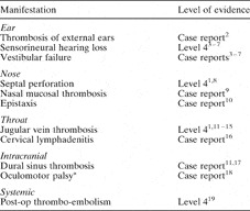
Table I summarises the findings of our literature search and the level of evidence of each study identified. It is apparent that present evidence is based on case series and individual case reports.
ENT associations of antiphospholipid syndrome

* Due to midbrain infarction. Post-op = post-operative
Discussion
In 2002, Cervera et al. Reference Cervera, Piette, Font, Khamashta, Shoenfeld and Camps1 reported a cohort of 1000 patients with antiphospholipid syndrome. Primary antiphospholipid syndrome (without any other underlying systemic autoimmune disease) occurred in 53.1 per cent of patients. Antiphospholipid syndrome was associated with systemic lupus erythematosus in 36.2 per cent; with lupus-like syndrome in 5.0 per cent and with other diseases in 5.9 per cent. The clinical features of antiphospholipid syndrome included deep vein thrombosis, thrombocytopenia, livedo reticularis, cerebrovascular accident, superficial thrombophlebitis, pulmonary embolus, recurrent fetal loss and haemolytic anaemia.
A subset of patients suffering a condition termed ‘catastrophic antiphospholipid syndrome’ was described in 1992.Reference Asherson20 This is a life-threatening condition in which patients who are antiphospholipid antibody positive develop thrombosis in three organs in a period of days to weeks, with a mortality rate of 50 per cent.Reference Asherson, Cervera, Piette, Sheonfeld, Espinosa and Petri11, Reference Erkan, Asherson, Espinosa, Cervera, Font and Piette19
Specific criteria have been set for the classification of antiphospholipid syndrome (see Appendix 1). Patients must demonstrate one clinical and one laboratory feature.Reference Wilson, Gharavi, Koike, Lockshin, Branch and Piette21
The widespread thrombotic nature of the disease has been shown to affect many organs,Reference Cervera, Piette, Font, Khamashta, Shoenfeld and Camps1 including those managed in otorhinolaryngology practice.
With hindsight, it would appear that our patient's septal perforation may have been a clue to her diagnosis of antiphospholipid syndrome. It is difficult to ascertain whether she would have developed a nasal septal perforation had she not undergone septal surgery. However, the almost complete absence of any residual septal cartilage at re-exploration suggests that some degree of ischaemia occurred during the peri-operative period. Spontaneous nasal septal perforation has been previously described in the rheumatological literature, in association with antiphospholipid syndrome. Nasal septal perforation had a reported incidence of 0.8 per cent in a cohort of 1000 patients,Reference Cervera, Piette, Font, Khamashta, Shoenfeld and Camps1 and was first described by Piette et al. in 1994.Reference Piette, Papo, Godeau, Wechsler, Frances and Du-Boutin8 The ischaemic nature of the perforation may cause additional problems during any attempts at reconstructive surgery.
• Antiphospholipid syndrome is a systemic autoimmune disorder characterised by arterial or venous thromboses, recurrent fetal loss and neurological disease (including stroke)
• Laboratory investigations include testing for lupus anticoagulant and anticardiolipin antibodies
• The condition is managed with anticoagulation and prevention of thrombophilia (i.e. aspirin, warfarin or heparin)
• The condition may present with spontaneous or post-operative septal perforation
Other nasal manifestations have been described, including thrombosis of the nasal mucosa, in association with multiple thromboses of the skin and spleen, and haemolytic anaemia.Reference Egan and Munn9 Severe haemorrhagic diathesis has also led to epistaxis in a five-week post-partum patient.Reference Bladt, Steengaard-Pedersen, Poulsen, Petersen, Laursen and d'Amore10
Thrombosis of the internal and external jugular veins has been described in association with antiphospholipid syndrome, particularly if the syndrome presents before the age of 15 years.Reference Cervera, Piette, Font, Khamashta, Shoenfeld and Camps1, Reference Asherson, Cervera, Piette, Sheonfeld, Espinosa and Petri11–Reference Alcazar-Ramirez, Fernandez-Nebro, Abarca, Salgado, De and Rodriguez-Andreu15
Papaioannou et al. have reported histiocytic necrotising lymphadentitis (Kikuchi–Fujimoto disease) of the cervical lymph nodes in association with antiphospholipid syndrome.Reference Papaioannou, Speletas, Kaloutsi and Pavlitou-Tsiontsi16
The reported otological manifestations of antiphospholipid syndrome include bilateral thrombosis of the external ears.Reference O'Gradaigh and Scott2 The condition may play a role in sensorineural hearing loss. Mouadeb and Ruckenstein reported an incidence of elevated antiphospholipid antibody levels in 25 per cent of patients presenting with progressive sensorineural hearing loss.Reference Mouadeb and Ruckenstein3 Moine et al. Reference Moine, Mohcini, Beal, Guillevin and Frachet4 assessed 35 patients with deafness of unknown origin; four patients demonstrated anticardiolipin antibodies. In addition, sudden sensorineural hearing loss has been reported in patients with systemic lupus erythematosus and anticardiolipin antibodies.Reference Green and Miller5, Reference Compadretti, Brandolini and Tasca6 Vyse et al. reported audiovestibular failure in two patients showing elevated titres of antiphospholipid antibodies, one with primary antiphospholipid syndrome and the other with systemic lupus erythematosus.Reference Vyse, Luxon and Walport7
Reported intracranial complications have included fatal dural sinus thrombosis,Reference Asherson, Cervera, Piette, Sheonfeld, Espinosa and Petri11, Reference Emir, Ozisik, Cagli, Bakuy, Ozisik and Sener17 infarction of the midbrain presenting as oculomotor palsy,Reference Champion, Choy, Schrieber, Roche and Rowe18 and cerebellar ataxia.Reference Cervera, Piette, Font, Khamashta, Shoenfeld and Camps1
Catastrophic antiphospholipid syndrome is associated with post-operative thrombosis, resulting in death in some instances.Reference Erkan, Asherson, Espinosa, Cervera, Font and Piette19
Conclusion
Otorhinolaryngologists should be alert to the existence of antiphospholipid syndrome. The large and small vessel thromboses associated with the disease can lead to complications following nasal surgery, and difficulties can arise when attempting to correct these complications. There may also be a role for antiphospholipid syndrome screening in patients presenting with apparently idiopathic nasal septal perforation.
Appendix 1. Preliminary criteria for the classification of antiphospholipid syndromeReference Wilson, Gharavi, Koike, Lockshin, Branch and Piette21
Clinical criteria
(1) Vascular thrombosis. One or more clinical episodes of arterial, venous or small vessel thrombosis, in any tissue or organ. Thrombosis must be confirmed by imaging or Doppler studies or histopathology, with the exception of superficial venous thrombosis. For histopathological confirmation, thrombosis should be present without significant evidence of inflammation in the vessel wall.
(2) Pregnancy morbidity. (a) One or more unexplained deaths of a morphologically normal fetus at or beyond the 10th week of gestation, with normal fetal morphology demonstrated by ultrasound or direct examination.
Or (b) one or more premature births of a morphologically normal neonate at or before the 34th week of gestation because of severe pre-eclampsia or eclampsia, or severe placental insufficiency.
Or (c) three or more unexplained, consecutive, spontaneous abortions before the 10th week of gestation, with maternal amniotic or hormonal abnormalities and maternal and paternal chromosomal causes excluded.
Laboratory criteria
(1) Anticardiolipin antibody of IgG and/or IgM isotype in blood, present in medium or high titre, on two or more occasions at least six weeks apart, measured by standardised enzyme-linked immunosorbent assay for β2-glycoprotein I-dependent anticardiolipin antibodies.
(2) Lupus anticoagulant present in plasma on two or more occasions, at least six weeks apart, detected according to the guidelines of the International Society on Thrombosis and Haemostasis.
Diagnosis
Definite antiphospholipid antibody syndrome is considered to be present if at least one of the clinical criteria and one of the laboratory criteria are met.