Introduction
Anorexia nervosa (AN) is a disorder that occurs mainly in females during adolescence or young adulthood. AN is associated with a self-induced excessive weight loss, an intense fear of gaining weight, and a disturbance in the perception of one's body shape (DSM-IV; APA, 1994). Current aetiological models emphasize a multifactorial origin involving genetic, psychobiological and sociocultural factors (Polivy & Herman, Reference Polivy and Herman2002; Connan et al. Reference Connan, Campbell, Katzman, Lightman and Treasure2003; Bergen et al. Reference Bergen, Yeager, Welch, Haque, Ganjei, van den Bree, Mazzanti, Nardi, Fichter, Halmi, Kaplan, Strober, Treasure, Woodside, Bulik, Bacanu, Devlin, Berrettini, Goldman and Kaye2005). Although a biopsychosocial approach can account for the key symptoms of AN, current explanations have largely neglected the potential role of reward/hedonic processes as contributing factors to the decline in food intake. Such a perspective has been advocated recently, leading to several proposals suggesting the dysfunction of brain reward systems as a possible cause of AN (Davis & Woodside, Reference Davis and Woodside2002; Berridge, Reference Berridge, Kirkham and Cooper2007; Zandian et al. Reference Zandian, Ioakimidis, Bergh and Sodersten2007). Others have stressed the value of hedonic/positive incentive theory to guide future research on this eating disorder (Pinel et al. Reference Pinel, Assanand and Lehman2000). According to this framework, the anticipated pleasure of eating is a strong motivating factor of consumption: people are driven to eat less by energy stores declining below homeostatic set points than by nutritional needs and sensory/cognitive expectations modulating the hedonic/incentive value of foods (Pinel et al. Reference Pinel, Assanand and Lehman2000; Berridge, Reference Berridge2004a). Thus, when laboratory animals or humans have ad libitum access to high-energy palatable food, overconsumption or obesity is relatively common (Rothwell & Stock, Reference Rothwell and Stock1979; Sorensen et al. Reference Sorensen, Møller, Flint, Martens and Raben2003; Berthoud, Reference Berthoud2004). Conversely, according to the positive incentive perspective, AN develops because it is initially rewarding to eat less food, and self-starvation is strengthened by the low reinforcing or hedonic value of perceived food cues (Pinel et al. Reference Pinel, Assanand and Lehman2000; Zandian et al. Reference Zandian, Ioakimidis, Bergh and Sodersten2007). This hypothesis is consistent with the fact that excessive food restriction (i) activates neural substrates of reward (i.e. dopamine and endogenous opioid systems), more particularly when it is associated with increased physical exercise (Heubner, Reference Heubner1993; Bergh & Södersten, Reference Bergh and Södersten1996; Södersten et al. Reference Södersten, Bergh and Zandian2006), (ii) may induce symptoms associated with AN (e.g. depression, obsession) (Keys et al. Reference Keys, Brozek, Henschel, Mickelsen and Taylor1950; Zandian et al. Reference Zandian, Ioakimidis, Bergh and Sodersten2007) and (iii) produces adverse physiological effects and taste aversion following food intake (Capaldi & Myers, Reference Capaldi and Myers1982; Pinel et al. Reference Pinel, Assanand and Lehman2000). Findings consistent with this perspective have also been documented by studies using food cueing paradigms in AN patients. Compared to controls, AN patients (restricting and/or binge-purge subtypes) displayed a dislike for the taste or the sight of high-energy food (Drewnowski et al. Reference Drewnowski, Halmi, Pierce, Gibbs and Smith1987; Sunday & Halmi, Reference Sunday and Halmi1990; Bossert et al. Reference Bossert, Laessle, Meiller, Junker, Ellgring and Pirke1991; Santel et al. Reference Santel, Baving, Krauel, Munte and Rotte2006), less salivation to olfactory food stimuli (LeGoff et al. Reference LeGoff, Leichner and Spigelman1988), a reduced pleasantness to sucrose (Sunday & Halmi, Reference Sunday and Halmi1990), no palatability priming effect (Roefs et al. Reference Roefs, Stapert, Isabella, Wolters, Wojciechowski and Jansen2005), and highly anhedonic scores on self-reported scales (Davis & Woodside, Reference Davis and Woodside2002; Eiber et al. Reference Eiber, Berlin, de Brettes, Foulon and Guelfi2002).
Despite the potential relevance of the reward/pleasure deficit hypothesis in AN, not all attempts to replicate previous results have been successful. Alternative hypotheses, and also crucial methodological issues, must be considered before favouring this perspective. First, the finding of a reduced hedonic response to sucrose in AN patients was not reported in some studies when sweet solutions were not swallowed (Eiber et al. Reference Eiber, Berlin, de Brettes, Foulon and Guelfi2002), suggesting that an excessive fear of gaining weight rather than a decreased ability to experience pleasure might be causal. Second, if dysfunction of reward processes is involved in AN, it is unclear which component is affected in AN persons exposed to palatable food. Berridge (Reference Berridge2003, Reference Berridge, Kirkham and Cooper2007) relied on animal findings to argue that two separable brain systems underlie food incentives and hedonics: the first, involving mesolimbic dopaminergic substrates, mediates a motivational component (termed ‘wanting’, that is the desire to get a food); and the second, involving a distributed network of brain islands (e.g. opioidergic hotspots in the shell of nucleus accumbens and in the ventral pallidum), mediates a hedonic/affective component (termed ‘liking’, that is the pleasure resulting from orosensory stimulation of food). In humans, the role of endogenous opioids in ingestive behavior and food pleasantness has also been aptly reviewed (Yeomans & Gray, Reference Yeomans and Gray2002), and pharmacological and genetic evidence suggests that both dopamine and opioid systems might contribute to the pathophysiology of AN with possible consequences in the reduction of desire or pleasure for food (Yeomans & Gray, Reference Yeomans and Gray2002; Frank et al. Reference Frank, Bailer, Henry, Drevets, Meltzer, Price, Mathis, Wagner, Hoge, Ziolko, Barbarich-Marsteller, Weissfeld and Kaye2005; Brown et al. Reference Brown, Bujac, Mann, Campbell, Stubbins and Blundell2007). Thus, because of their theoretical implications, experimental studies on wanting and liking are needed to determine whether they are both altered in AN. Third, self-ratings of food likes and dislikes were most often used in AN studies, but whether subjective report is the most appropriate to probe core hedonic processes in this eating disorder is unclear. AN patients were often shown to display disturbances in the accurate perception of internal states or feelings and in their verbalization (i.e. alexithymia). This could affect self-evaluation of hunger and satiety, of interoceptive awareness and of emotional states (Garfinkel, Reference Garfinkel1974; Nakai & Koh, Reference Nakai and Koh2001; Bydlowski et al. Reference Bydlowski, Corcos, Jeammet, Paterniti, Berthoz, Laurier, Chambry and Consoli2005; Montebarocci et al. Reference Montebarocci, Codispoti, Surcinelli, Franzoni, Baldaro and Rossi2006). An approach based on implicit measures of hedonic processes is therefore needed. Such measures are currently used in psychophysiological research in both healthy and clinical samples, relying on facial electromyography (EMG) and autonomic variables as sensitive markers of valence and arousal dimensions of affective processes (e.g. Cacioppo et al. Reference Cacioppo, Bush and Tassinary1992; Lang et al. Reference Lang, Greenwald, Bradley and Hamm1993). The use of facial EMG and autonomic measures of emotional bodily changes is further justified by the fact that they may be dissociated from conscious experience in brain-damaged or emotionally disordered patients (Luminet et al. Reference Luminet, Rimé, Bagby and Taylor2004; Soussignan et al. Reference Soussignan, Ehrle, Henry, Schaal and Bakchine2005; Zonnevylle-Bender et al. Reference Zonnevylle-Bender, van Goozen, Cohen-Kettenis, Jansen, van Elburg and Engeland2005) and are less susceptible to demand characteristics and social desirability effects than are self-report data. Finally, it remains to be clarified whether avoidance of food in AN may be influenced by dislike/fear cues independent of intentionally controlled processes and without awareness of the underlying affective response (Berridge & Winkielman, Reference Berridge and Winkielman2003; Berridge, Reference Berridge, Manstead, Frijda and Fischer2004b; Winkielman et al. Reference Winkielman, Berridge and Wilbarger2005). This issue can be investigated with affective priming paradigms that have the potential to examine whether (i) automatic/unconscious processes influence negative reactivity to food and (ii) AN patients are more sensitive than healthy controls to emotional primes related to food (i.e. disgust and fear cues). Although there is some evidence that AN patients make relatively automatic associations with foods based on their palatability (Roefs et al. Reference Roefs, Stapert, Isabella, Wolters, Wojciechowski and Jansen2005), to the best of our knowledge there are no studies that have assessed whether unconscious emotional processes potentiate facial, autonomic and subjective reactivity to food cues in AN patients, and whether such pre-attentive processing depends on their motivational state (hungry versus satiated states).
The current study addresses the following questions. First, do distinct measures of hedonic processing (facial EMG, videotaped facial behavior, verbal ratings) differentiate AN women from healthy control (HC) women while exposed to food pictures, regardless of the potential impact of affective primes? Second, do subliminal positive and negative primes influence facial EMG, autonomic and subjective responses to food pictures as a function of their valence, and does the motivational state (hunger versus satiety) bear a differential impact on the effect of affective primes in AN and healthy individuals? Third, are subjective measures of liking and wanting dissociated in AN patients?
These issues were addressed in a two-session experiment in healthy and AN women, one run before and the other after a standardized lunch, using a backward masking procedure (exposure to subliminal disgust, fear, happy, and neutral faces). Meanwhile, EMG (activity of corrugator and zygomatic muscles), autonomic (skin conductance and heart rate), videotaped (smiles and negative expressions) and self-reported (hedonic and wanting ratings) responses were recorded in participants viewing pictures of palatable foods. Disgust and fear expressions were chosen as negative primes based on the importance of these emotions in food hedonics and eating disorders (Davey et al. Reference Davey, Buckland, Tantow and Dallos1998; Ellison et al. Reference Ellison, Foong, Howard, Bullmore, Williams and Treasure1998), and on neuroimaging studies reporting that subliminal presentations of fear and disgust expressions can activate brain substrates underlying emotional processing (Whalen et al. Reference Whalen, Rauch, Etcoff, McInerney, Lee and Jenike1998; Phillips et al. Reference Phillips, Williams, Heining, Herba, Russell, Andrew, Bullmore, Brammer, Williams, Morgan, Young and Gray2004). The following predictions were proposed: first, AN patients should display higher negative reactivity (corrugator activity, negative expressions and subjective ratings) to food pictures as compared to HC subjects, and this differential effect should be more pronounced during hunger states to avoid food intake; conversely, HC participants should express a stronger positive reactivity (zygomatic activity, smiles and subjective ratings) during this motivational state. Second, subliminal fear and disgust faces should potentiate negative and autonomic reactivity to food pictures in AN patients during hunger states, whereas subliminal happy faces should potentiate positive hedonic reactivity in controls. Third, if AN involves a dysfunction in hedonic processing, self-report measures of liking and wanting should be dissociable in AN patients as compared to HC subjects.
Method
Participants
The current study involved 16 females (age: mean ± s.d.=26.68±7.30 years) fulfilling the DSM-IV criteria for AN. They were recruited from the Endocrinology Service of Dijon University Hospital. All were suffering from the restricting form of AN. They had a mean body mass index (BMI) of 14.97±1.97 kg/m2. The HC subjects comprised 25 females (aged 24.6±6.03 years) recruited by local advertisements. They had a normal BMI (mean±s.d.=20.52±1.90 kg/m2) and reported neither food restrictions nor allergies. For all participants, exclusion criteria were consumption of psychotropic drugs, hearing and visual impairments and neurological diseases. Written informed consent was obtained from all participants, with the announced aim to conduct a study on feeding behavior in women.
Timing of experimental sessions
Each participant took part in two experimental sessions, one before (hunger) and the other after (satiety) lunch, during two consecutive days at the Dijon Centre des Sciences du GoÛt. To control for order effect, the hunger and satiety conditions were counterbalanced across participants. Before the hunger session, each participant was instructed to take her breakfast before 0730 hours and to avoid eating and drinking (except water) until the experiment that began at 1220 hours. All participants complied with this requirement and reported that they had followed it. For the satiety session, the participant came to the laboratory at 1230 hours to take a standard lunch composed of an entrée (100 g steamed semolina, 100 g ham with parsley and half an egg), meat (60 g), cheese (35 g), bread (50 g), cake with fruit (75 g), and a bottle of mineral water (330 ml). The participants were told to take their lunch ad libitum, and usually spent 15–25 min before terminating their meal. For the satiety condition, the experimental sessions were scheduled between 1340 and 1400 hours.
Stimuli
Food stimuli
The food stimuli were composed of 32 color pictures of meals with a varied energy content drawn from the International Affective Picture System (Lang et al. Reference Lang, Bradley and Cuthbert2001) or selected from the Internet. They comprised four sets of eight food pictures (SF1, SF2, SF3 and SF4), each including the same food categories: pizza, meals with a variety of dishes (including potatoes, vegetables and chicken), beef, cheese, chocolate, ice-cream, cake/biscuits, and fruit. The eight pictures of each set were matched on arousing, liking, and wanting scores following a pretest conducted on 40 healthy female undergraduates (aged 21.2±1.1 years) who reported a hungry level of 5.15 (s.d. =2.29) on a nine-point scale. The pictures were shown on a 300×230 cm screen during 6 s using Superlab software (version 2, Cedrus, USA). The participants had to rate their arousal (i.e. whether they felt stimulated/aroused while viewing the picture) and wanting (i.e. their desire to consume the food) reactions on a scale ranging from 1 (not at all) to 9 (extremely), and also their liking reactions (i.e. If you had to eat this food, what would you feel?) on a scale ranging from –4 (extreme dislike) to 4 (extreme pleasure). Multivariate ANOVA using Wilks' λ distribution revealed that the food pictures were rated as palatable (pleasure rating: 1.30±0.86) and that the four sets of food pictures did not differ either on ratings of arousal [SF1: 4.97±0.61, SF2: 4.81±0.51, SF3: 4.84±1.05, SF4: 4.68±0.42, F(3, 37)=2.15, p>0.05], of wanting [SF1: 5.49±1.18, SF2: 5.25±1.26, SF3: 5.51±1.25, SF4: 5.30±1.11, F(3, 37)=1.78, p>0.05] or of liking [SF1: 1.45±0.80, SF2: 1.16±0.92, SF3: 1.32±0.98, SF4:1.27±0.74, F(3, 37)=1.58, p>0.05].
Affective primes
Eight neutral faces were selected as masks and 32 stimuli (eight each of happy, fear, disgust and neutral faces) served as primes. Of these 40 faces, 20 were female and 20 were male. All except two neutral faces came from the Pictures of Facial Affect (Ekman & Friesen, Reference Ekman and Friesen1976). The slides were digitized as 466×700 pixels resolution to allow presentation on a 17-inch computer monitor.
Design of the priming procedure
The priming experiment consisted of four blocks, each containing eight different subliminal facial expressions of the same emotion (i.e. fear, disgust, happy and neutral blocks). Each trial began with a warning signal (the word ‘slide’) presented on the screen for 6 s, indicating to the participant that pictures would be presented. This condition was included as a baseline period for the psychophysiological data. This warning signal was followed by a forward mask (500-ms cross) that was immediately replaced by a subliminal prime (30-ms facial expression or neutral face). The prime was immediately followed by a backward mask (400-ms neutral female or male face). Following the mask, the target (food picture) was presented for 6 s. A double Latin square design was used to present the four priming blocks. This allowed all possible associations between each of the four blocks of primes and each of the four sets of food pictures. Once the prime–target associations were determined for a given participant (e.g. fear–SF1; disgust–SF2; happy–SF3; neutral–SF4), the order in which the eight trials were presented within each block was randomized for each participant. The duration of projection of stimuli was controlled using SuperLab Pro software.
Procedure
On arrival, the participant was seated in a comfortable chair in a dimly lit, sound-attenuated experimental room. Two experimenters and the material were located in an adjacent isolated room. The participant was asked to relax during the task and avoid any movement of body parts where the sensors were attached. After attachment of biosensors (see section on psychophysiological recording), the subject was positioned 75 cm in front of a 17-inch flat-screen monitor and a Hi-speed USB Webcam (DigiHoney TM series) that recorded her facial behavior with a 480×640 video resolution. The first experimenter started the psychophysiological and video recordings. The second experimenter collected data about the participant's hunger/satiety state (1=not at all hungry/full, 7=very hungry/full) and then activated pre-recorded task-specific instructions using both the screen and headphones. The participant was told that the word ‘slide’ would be presented in the center of the screen (warning signal) followed 6 s later by a face picture and then a food picture. She was informed that the face picture was included as if the person represented by this face had to consume the food. The onsets of primes and food pictures were automatically signaled by a 60-ms 5-V pulse on a channel of the Chart Windows software (which displayed psychophysiological data) using a data acquisition and signal conditioning card (DAQCard-DIO-24, National Instruments Corporation, USA). After each food picture, the participants used nine-point Likert scales to rate their liking [from –4 (extreme dislike) to 4 (extreme pleasure)] and wanting responses [from 1 (no desire at all) to 9 (extreme desire)]. Prior to the onset of the experiment, two trials served as practice stimuli. After the experiment, the participants completed the shortened 13-item Beck Depression Inventory (BDI; Beck et al. Reference Beck, Rial and Rickels1974), a widely used self-rating with established reliability and validity.
Psychophysiological recording
The facial EMG and autonomic measures were monitored using a 16-channel PowerLab system (AD Instruments Pty Ltd, Australia) connected to a PC. The bioelectrical signals were filtered and amplified before being fed into the analog input connector of the PowerLab unit and sampled at a rate of 200 Hz under the online control of Chart 5.2 software.
Facial EMG
Muscular activity was detected using five miniature Ag/AgCl surface electrodes filled with electrolyte gel, following the guidelines proposed by Fridlund & Cacioppo (Reference Fridlund and Cacioppo1986): two were placed over the left corrugator supercilii region, two over the left zygomaticus major region (recording electrodes) and one on the middle part of the forehead (reference electrode). Before attaching the electrodes, the target sites of the skin were cleaned with alcohol and rubbed slightly to reduce inter-electrode impedance. The EMG signals were fed into electronic amplifiers and were band-pass filtered from 10 to 250 Hz. The raw EMG signals were rectified and smoothed with a 500-ms time constant.
Autonomic measures
Skin conductance (SC) was recorded with the ML116 GSR Amplifier of the 16-channel PowerLab system, which provides a low, constant-voltage AC excitation, allowing the use of dry electrodes, with no special electrolytes needed. The electrodes were attached with a Velcro strap on the palmar surfaces of the distal phalanges of the second and third fingers of the non-dominant (usually the left) hand. Measurements were displayed directly in μS. Heart rate (HR) was measured using Ag/AgCl electrodes (BB-COM 2, Comepa, France) and a standard Lead I electrode configuration. A low-pass filter of 50 Hz was used with the bioamplifier to eliminate high-frequency components. A computed input command allowed a threshold control to detect R wave pulses and to display HR in beats per minute on a separate channel.
Analysis of physiological and videotaped facial data
Physiological data
Muscular and cardiac data were averaged off-line for each of the 6-s periods immediately preceding (baseline) and following (stimulus period) the prime onset. In accordance with standard practice (Fridlund, Reference Fridlund1991), mean facial EMG data were log transformed [log10(EMG+1)] to minimize skewness and heteroscedasticity. The EMG and cardiac scores acquired during the baseline period were respectively subtracted from those acquired during stimulus presentation. SC response (SCR) was defined as a change in the amplitude with onset occurring 1–4 s after stimulus presentation. SCR change was calculated by subtracting the 2-s SC level immediately preceding stimulus onset from the largest value in the 2-s window after stimulation.
Coding of facial behavior
Videotaped facial behavior was analyzed with the anatomically based Facial Action Coding System (FACS; Ekman et al. Reference Ekman, Friesen and Hager2002) by a certified coder (R.S.) blind to the priming condition, the type of food pictures and the clinical status of each participant (healthy or AN). The negative expressions to food pictures were coded in regrouping action units (AU) indexing disgust [nose wrinkling (AU 9)/upper lip raising (AU 10)] and those that have been shown to be associated with negative emotions [brow lowering (AU 4), lip corners tightening (AU 14), lip corners lowering (AU 15), chin raising (AU 17), and lips stretched laterally (AU 20)] (Soussignan et al. Reference Soussignan, Schaal, Schmit and Nadel1995, Reference Soussignan, Schaal and Marlier1999; Soussignan & Schaal, Reference Soussignan and Schaal1996). For smiling, lip corners pulling (AU 12) was coded (Soussignan, Reference Soussignan2002). The frequencies of negative facial expressions and smiles for each priming block were used as dependent variables.
Prime recognition task
To verify the validity of our subliminal priming procedure, participants performed a forced-choice recognition task on a 15-inch laptop computer using SuperLab Pro software. The task consisted of 32 trials that were presented randomly across the participants and that comprised the subliminal primes to which each participant had been initially exposed. The participants were instructed to direct their attention to successive single pictures of faces (the flash of a neutral/emotional face followed by a visible face) and also to the simultaneous presentation of two faces. Each trial began with a 2-s warning signal (slide) followed by a 500-ms central cross and then by a 30-ms prime (emotional or neutral face). The prime was immediately masked by a 400-ms neutral face. Immediately afterward, two faces were presented for 1500 ms: one target face (which corresponded to the neutral or emotional subliminal prime) on one side of the screen and another face (an incorrect foil expressing a distinct emotion) on the other side of the screen. The two faces were equally distributed at the left (L) and the right (R) side of the screen. Following this presentation, the L and R letters were shown on each side of the screen and the participants had to decide which face was presented in the flash by pressing the appropriate key using an RB-730 pad (Cedrus, USA) connected to the laptop. Three practice trials preceded the task. A three-way ANOVA (group×state×prime) performed on the percentage of prime recognition yielded no significant main effects or interactive effects. The HC subjects and AN patients had similar performances during the fasting session (HC: 50.75±3.96%, AN: 51.36±4.95%) and after the lunch (HC: 50.0±3.34%, AN: 50.97±4.18%), and the performance of recognition did not vary as a function of the primes [disgust: 51.92±2.88%, happy: 48.91±2.54%, neutral: 49.77±3.05%, fear: 52.49±2.98%, F(3, 117)=0.74, p>0.05]. Performance on the forced-choice test for each priming condition ranged from 47.9% to 53.9% in the fasting (hunger state) and lunch (satiety state) sessions, which was not significant different from chance [t(40), p's >0.10]. Thus, the results suggested that our subliminal priming procedure was appropriate to investigate the influence of unconscious affective processes on facial, autonomic and subjective reactivity to food pictures.
Statistical analyses
As AN patients reported higher depression scores (12.6±8.3) than the HC subjects [3.2±3.4, t(39)=5.03, p < 0.001], the D-Beck score was entered as a covariate in repeated-measures ANCOVAs conducted on subjective, psychophysiological and observational measuresFootnote †, with group (AN versus HC) as the between-subject variable and motivational state (hunger versus satiety) and priming (fear, disgust, happy, neutral) as within-subject variablesFootnote ‡. A two-way ANCOVA (group×motivational state) was also performed on self-reported scores of motivational state. Fisher's least significant difference (LSD) test was used for making post-hoc comparisons between means. Partial eta-squared (η2 p) and Cohen's d were reported as a measure of effect size in ANCOVAs and in the mean comparisons respectively, where d =(M 1 – M 2)/s pooled and s pooled =√[(s 12+s 22)/2] (Cohen, Reference Cohen1988), where M 1, M 2 are the means of samples and s 1, s 2 their standard deviations. Cohen (Reference Cohen1988) has defined small effect sizes as d=0.2, medium as d=0.5, and large as d=0.8.
Results
Subjective measures
Ratings of motivational states
All the participants reported a higher hunger level before than after the lunch [5.23±1.41 v. 2.79±1.02, F(1, 38)=33.24, p<0.00001, η2 p=0.466]. A significant interaction between state and group was found [F(1, 38)=6.93, p=0.01, η2 p=0.154]. The HC women reported a higher hunger level than the AN patients during the hunger session only (5.97 v. 4.49, d=0.95).
Liking rating
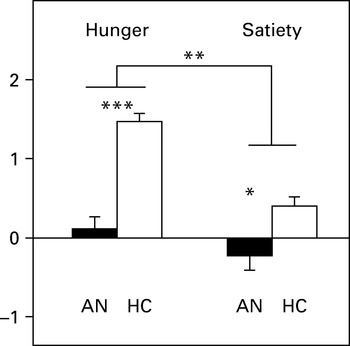
Significant main effects of group [F(1, 38)=4.33, p=0.04, η2 p=0.102], and hunger state [F(1, 38)=11.05, p=0.002, η2 p=0.225] emerged. These effects were qualified by an interaction between group and state [F(1, 38)=5.75, p=0.02, η2 p=0.131]. As illustrated in Fig. 1, the HC women reported higher hedonic reactions to food pictures than AN patients during both the hunger (p<0.001, d=0.51) and the satiety (p=0.03, d=0.21) sessions. Furthermore, only the HC subjects rated the food pictures as more pleasant in the hunger than in the sated session (p<0.0001). The prime effect failed to reach significance. Note that the lower subjective rating of pleasure in AN women than in HC during the pre-prandial condition was not solely due to hunger differences because the group effect remained significant after including hunger scores as a covariate in the ANCOVA [F(1, 37)=4.15, p=0.048, η2 p=0.10].
Mean ratings of self-reported pleasure (liking) and standard errors bars in response to food pictures during the hunger and satiety sessions in anorexic (AN) and healthy control (HC) participants (* p<0.05, ** p<0.01, *** p<0.001).

Wanting rating
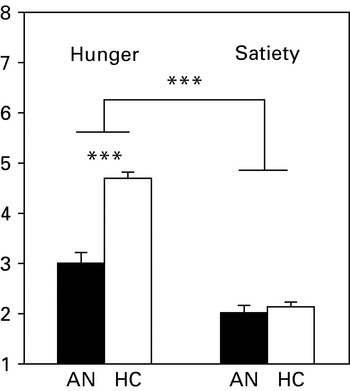
There were also significant effects of group [F(1, 38)=25.96, p<0.00001, η2 p=0.406] and hunger state [F(1, 38)=4.69, p=0.037, η2 p=0.11], and a significant group×state interaction [F(1, 38)=8.47, p=0.006, η2 p=0.182]. As shown in Fig. 2, this interaction reflects the fact that the HC responded to the food pictures with higher wanting scores than did the AN patients during the hunger state only (p<0.001, d=0.53). However, both the AN and the HC women reported higher wanting scores during the hungry than during the satiety sessions (p<0.001). No priming effect was detected.
Mean ratings of self-reported desire (wanting) and standard errors bars in response to food pictures during the hunger and satiety sessions in anorexic (AN) and healthy control (HC) participants (*** p<0.001).

Facial EMG
Corrugator muscle activity
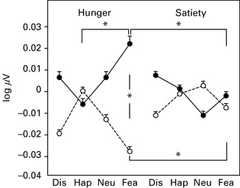
The ANCOVA did not reveal significant main effects for group, hunger state or priming. However, significant group×priming [F(3, 102)=6.00, p=0.0008, η2 p=0.15] and group×state×priming [F(3, 102)=2.91, p=0.038, η2 p=0.08] interactions were detected. Fig. 3 shows that these interactions reflect the fact that only subliminal fear expressions induced larger activity of the corrugator muscle region in AN than HC participants viewing food pictures during the hunger session (p=0.029, d=0.91). Furthermore, fear primes induced a larger increase in corrugator activity in AN patients during the hunger than during the satiety session (p=0.02, d=0.54), whereas in the HC the effect of fear primes on corrugator activity to food pictures was lower in the hunger than in the satiety sessions (p=0.04, d=0.42). A larger corrugator activity was also found in response to fear as compared to happy primes in AN participants during the hunger states (p=0.048, d=0.57). Note that the effect sizes were medium to large, suggesting that corrugator activity differences between the AN and HC participants can be accepted with confidence.
Mean electromyography (EMG) amplitudes and standard errors bars of corrugator muscle reactivity to food pictures primed by the subliminal presentation of emotional facial expressions (Dis, disgust; Hap, happy; Neu, neutral; Fea, fear) during the hunger and satiety sessions in anorexic (AN; –––) and healthy control (HC; – – –) participants (* p<0.05).

Zygomatic muscle activity
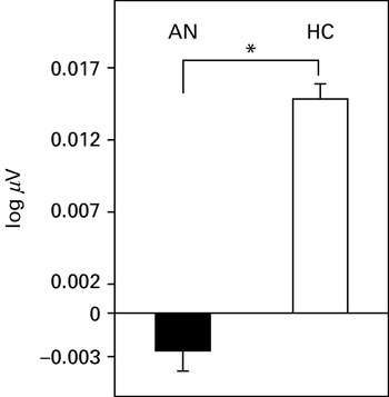
A significant main effect of group was detected [F(1, 34)=4.51, p=0.04, η2 p=0.12], revealing that, while viewing food pictures, the controls demonstrated a higher activity of zygomatic muscle region than the AN patients (d=0.78) (Fig. 4). There was a trend for a larger zygomatic activity during the hunger than during the satiety sessions [F(1, 34)=3.42, p=0.07, η2 p=0.09], more particularly for the HC group as compared to the AN group [F(1, 34)=2.94, p=0.09, η2 p=0.08, d=0.48]. Note, however, that the group×state interaction reached significance when depression was not entered as a covariate [F(1, 35)=8.15, p=0.007, η2 p=0.19]. This latter finding suggests that depression modulates positive responsiveness to food pictures in AN patients. There was no main priming effect, and no significant interaction effects between priming and the other factors. Of interest, the group effect on zygomatic activity persisted after entering the hunger score as a covariate [F(1, 33)=4.85, p=0.034, η2 p=0.128], suggesting that differences in motivational state between both groups of participants are not sufficient to explain the lower zygomatic activity to food pictures in AN patients.
Mean electromyography (EMG) amplitudes and standard errors bars of zygomatic muscle reactivity to food pictures in anorexic (AN) and healthy control (HC) participants (* p<0.05).

Autonomic reactivity
SCR changes
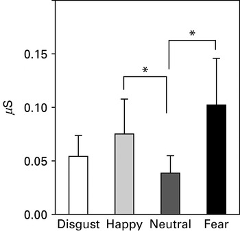
The ANCOVA yielded only a main priming effect [F(3, 111)=3.15, p=0.03, η2 p=0.08], revealing that, while the participants viewed food pictures, subliminal fear and happy primes elicited a higher increase in SCR than did the neutral primes (all p<0.05) (Fig. 5).
Mean amplitudes of skin conductance response (SCR) and standard errors bars to food pictures primed by the subliminal presentation of emotional facial expressions in all the participants (* p<0.05).

HR changes
HR was not affected either by motivational state, group or priming factors, or by their interactions. Both AN and HC groups evinced an overall decrease in HR in hunger (AN: −0.40±0.56, HC: −0.57±0.43) and satiety states (AN: −0.17±0.60, HC: −0.65±47), suggesting an orienting response to food pictures in all participants.
Videotaped facial behavior
Smiling
The ANCOVA revealed a main effect for group [F(1, 34)=4.31, p=0.045, η2 p=0.15] and state [F(1, 34)=9.03, p=0.005, η2 p=0.21]. HC subjects smiled significantly more than AN patients while viewing food pictures (0.61±0.11 v. 0.18±0.15, d=0.78) and the frequency of smiles was higher during hunger (0.58±0.22) than during satiety (0.21±0.16) sessions. There was no significant difference in the frequency of smiles as a function of primes. Controls also displayed more smiles than AN participants during the pre-prandial condition when the hunger level was entered as a covariate [F(1, 34)=7.51, p=0.009, η2 p=0.18], suggesting that differences in the motivational state did not account for the higher positive facial behavior to food pictures in the HC than in the AN group.
Negative facial expressions
When the participants were presented with food pictures, no main effects of state, group and priming or interaction effects of priming and the other factors were noted with this variable.
Discussion
This research aimed to explore the value of a positive incentive theoretical framework to further clarify psychological processes underlying salient symptoms in anorexia, such as the decline of appetite and food restriction. More specifically, we investigated hedonic reactivity to food pictures in AN, and the potential influence of unconscious emotional processes on the low sensitivity to the positive reinforcement of food that characterizes this eating disorder. To reach this objective, the following issues were addressed.
Do measures of hedonic processing differentiate between AN and HC women?
Our findings provide clear evidence that both subjective (verbal ratings) and bodily (facial EMG activity, videotaped facial expressions) measures of affective processes induced by food pictures discriminate AN patients from HC subjects. On the one hand, AN women reported similar hedonic ratings to food pictures (next to neutrality) during hunger and satiety whereas the controls declared higher pleasure when hungry than when sated. Additionally, in both pre- and post-prandial states, the AN group rated the sight of food pictures as less pleasant than did the HC group. Thus, regardless of current interoceptive signals of hunger/satiation, the present findings confirm a decline in the positive incentive value of foods in AN women, in line with the findings in a recent neuroimaging study (Santel et al. Reference Santel, Baving, Krauel, Munte and Rotte2006). On the other hand, facial measures of positive affect also evidenced a differential pattern of responsiveness. Compared to HC participants, AN patients exhibited lower zygomatic muscle activity and expressed fewer smiles in response to food pictures. This convergence of both implicit and explicit measures of hedonic processing supports the robustness of our data, and excludes explanations based on social desirability or disturbances in the accurate perception of internal states in AN (Garfinkel, Reference Garfinkel1974). It may be noted that lower declared pleasure and positive facial behavior in AN do not simply reflect a difference in prandial state because all participants obtained higher hunger scores before than after lunch, and the differences between both groups persisted after controlling for hunger ratings. Thus, these findings give credit to the hypothesis of a diminished capacity to express and experience pleasure in AN persons while viewing foods, which could be causal in reduced intake (Davis & Woodside, Reference Davis and Woodside2002). This is in accordance with a prior study in AN patients that used reaction time as an indicator of implicit processes in an affective priming paradigm (Roefs et al. Reference Roefs, Stapert, Isabella, Wolters, Wojciechowski and Jansen2005). The AN group was slower in responding, suggesting less sensitivity to the palatability of foods than controls. However, our finding is not entirely consistent with those of another study using a startle eyeblink modulation paradigm to gauge appetitive and aversive responses in eating disordered patients (Friederich et al. Reference Friederich, Kumari, Uher, Riga, Schmidt, Campbell, Herzog and Treasure2006). In this study, AN persons showed an EMG startle response to food pictures that was not significantly different from healthy controls. This lack of difference, which was not in favour of a dysfunctional appetitive system in AN, may be due to the lack of subjective (e.g. pleasantness ratings) or facial (i.e. zygomatic activity) measures of positive affect, or to several possible biases (e.g. restricting and purging AN subtypes regrouped, food intake prior to the experiment not monitored, depression not used as a covariate) in the Friederich et al. study.
Do subliminal primes influence subjective and psychophysiological reactivity to food pictures in AN and HC participants?
The major finding of our study is the differential impact of subliminal fear primes on negative facial EMG reactions in AN and healthy women. Namely, the unconscious perception of fear expressions potentiated the corrugator muscle responses to food pictures during the hunger state in AN patients only. This suggests that a social display signaling a potential threat/danger, which operates outside phenomenal awareness and controlled processes, can influence an implicit measure of negative emotion in AN patients viewing palatable food pictures during a motivational state (hunger) that usually generates a positive affect in healthy controls. By contrast, the effect of subliminal fear primes on corrugator activity was lower in hungry than in sated HC participants. This result is in line with the positive incentive theory that predicts that an affective prime presented during a congruent motivational state facilitates responses to target stimuli. However, contrary to our prediction, no subliminal prime effect of the happy faces on zygomatic muscle activity was found in hungry HC participants. This did not reflect unresponsiveness to the happy primes because an increase in the SCR, an indicator of sympathetic arousal, was evidenced in all participants subliminally exposed to happy faces. Moreover, in all participants the impact of subliminal fear expressions on this autonomic measure was higher than the impact of the other primes. This suggests that valence (indexed by facial EMG) and arousal (indexed by SCR) dimensions of affective processes may be dissociated in AN patients and HC subjects, and partly reflects the influence of motivational state. Although we have no clear explanation for the lack of priming effect of happy faces on an implicit measure of affective valence, the following proposal may be suggested. A ceiling effect cannot be ruled out because the food stimuli induced both a high level of self-reported pleasure and positive facial behaviors in the controls during the pre-prandial condition. Thus, it cannot be excluded that an affective prime potentially interferes with the strength of affective valence of a target, accounting for the ongoing facial muscular activity. It is interesting to note that previous studies using the backward-masking technique in healthy participants demonstrated priming effects on zygomatic EMG activity when the happy faces were associated with either neutral targets (Rotteveel et al. Reference Rotteveel, de Groot, Geutskens and Phaf2001) or no target at all (Dimberg et al. Reference Dimberg, Thunberg and Elmehed2000). Further research is needed to clarify this issue.
An unexpected finding of the present study is the lack of priming effect for the subliminal facial expressions of disgust. Neither psychophysiological nor subjective evidence was found that the participants reacted strongly to the covert presentation of disgust faces when they were associated with food pictures. Thus, our findings suggest that fear primes are more powerful than disgust primes to influence arousal and valence dimensions of affective processes and to discriminate AN from controls. This could be explained by the fact that fear, as a salient feature in AN (DSM-IV; APA, 1994), is probably a potent emotion that influences food restriction.
It can be hypothesized that the fear priming effect evidenced here probably reflects the involvement of brain structures, such as the amygdala, that are crucial for mediating autonomic and behavioral responses to the threat value of stimuli (Ledoux, Reference Ledoux1996; Öhman et al. Reference Öhman, Carlsson, Lundqvist and Ingvar2007), with attention and awareness having little impact on its reactivity to fear expressions (Whalen et al. Reference Whalen, Rauch, Etcoff, McInerney, Lee and Jenike1998; Pegna et al. Reference Pegna, Khateb, Lazeyras and Seghier2005; Ohrmann et al. Reference Ohrmann, Rauch, Bauer, Kugel, Arolt, Heindel and Suslow2007). Exaggerated amygdala responses may also be induced by masked presentation of fearful faces in patients with post-traumatic stress disorder, suggesting that affective upheavals might lead to an amygdalian hypersensitivity to potentially harmful stimuli (Rauch et al. Reference Rauch, Whalen, Shin, McInerney, Macklin, Lasko, Orr and Pitman2000). A tentative interpretation related to AN symptoms would be that an irrational fear of food intake/weight gain, resulting from the amygdalian hyperactivation, would negatively influence the appraisal of food cues. It can be noted that the supraliminal presentation of a high caloric drink to AN patients has been shown to produce a ‘caloric fear’, with activation of limbic/paralimbic regions (i.e. amygdala, insula, anterior cingulate gyrus) (Ellison et al. Reference Ellison, Foong, Howard, Bullmore, Williams and Treasure1998). This leaves the possibility that structures such as the amygdala react automatically to food stimuli evaluated as threatening by AN people. Additional studies are required to test this hypothesis in AN individuals exposed to food cues primed by subliminal fear and disgust faces in using both functional magnetic resonance imaging (fMRI) and backward masking techniques.
Are subjective measures of liking and wanting dissociated in AN patients?
Our results revealed that subjective measures of desire (wanting) and pleasure (liking) of food stimuli were partially dissociated in AN patients as compared to HC subjects. That is, whereas controls reported higher wanting and liking scores for the visually presented foods when hungry than when sated, AN patients expressed no food pleasure (next to neutrality) during both states, although their desire or motivation to eat a food was higher in the hunger than in the sated condition. Although wanting and liking normally work together, they can be split apart by neurochemical manipulations or under certain circumstances (Berridge, Reference Berridge2004a). Available psychopharmacological evidence on animal models indicates that both of these food reward components are mediated by partially separated brain substrates relying on dopamine or opioid neurotransmission (Berridge, Reference Berridge, Kirkham and Cooper2007). Our finding of an apparent dissociation between liking and wanting in AN patients is consistent with the view that a lower capacity to experience pleasure than desire to eat foods might be more involved in AN. This issue should be addressed in future studies using implicit measures of desire/motivation to eat (e.g. reaction time to select foods). It further remains to demonstrate whether candidate genes that have been hypothesized to be involved in the susceptibility to AN are differentially related to both components of food reward. Polymorphisms in both opioid δ receptor (Brown et al. Reference Brown, Bujac, Mann, Campbell, Stubbins and Blundell2007) and D2 dopamine receptor genes (Bergen et al. Reference Bergen, Yeager, Welch, Haque, Ganjei, van den Bree, Mazzanti, Nardi, Fichter, Halmi, Kaplan, Strober, Treasure, Woodside, Bulik, Bacanu, Devlin, Berrettini, Goldman and Kaye2005) have recently been reported in AN patients. Given the functional role of the dopamine and opioid systems in desire and pleasure of foods, studies using genotyping, neuroimaging and psychophysiological measures are needed to clarify the issue of the differential involvement of these neurochemical systems in the components of food reward in AN.
In conclusion, our psychophysiological study using a backward masking paradigm emphasizes the heuristic value of a positive-incentive framework for a better understanding of some phenotypic features of AN. First, in contrast to healthy controls, AN patients did not display objective and subjective indices of pleasure during the hunger state. Second, the experience of pleasure to food stimuli seemed to be more affected than the desire to eat in AN. Third, we provide the first evidence that unconscious fear cues potentiated negative facial reactions to food stimuli in AN patients. This leaves the possibility that a non-controlled emotional mechanism might bias the appraisal of alimentary cues and contribute to induce food aversion in persons with AN.
Acknowledgments
We thank C. Serrano for installing the experimental room, S. Martin for managing the participants during the experiments, and N. Mutin for her highly effective logistic and administrative support. We thank the participants who made this research possible. This research was funded by a grant from Conseil Régional de Bourgogne (to T.J.) and by the Centre National de la Recherche Scientifique.
Declaration of Interest
None.