INTRODUCTION
The 50th anniversary of the International Neuropsychological Society provides a golden opportunity to take stock of research on the neuropsychology of traumatic brain injury (TBI). TBI is a major public health and socioeconomic burden worldwide (Humphrey et al., 2013; Roozenbeek, Mass, & Menon, Reference Roozenbeek, Maas and Menon2013), as well as a primary focus for many neuropsychologists in their research and clinical practice (Podell et al., Reference Podell, Gifford, Bougakov and Goldberg2014). Entire volumes have been devoted to the role of neuropsychology in the assessment and management of TBI (Sherer & Sander, Reference Sherer and Sander2014).
Neuropsychologists and neuropsychological testing have played a critical role in research documenting the outcomes of TBI. The literature on the consequences of TBI is extensive, and a detailed review is beyond the scope of this study. In general, however, research indicates that TBI, especially when severe, can produce deficits in a variety of domains: alertness and orientation; language and communication skills; nonverbal skills; attention, memory, and executive functions; corticosensory and motor skills; academic achievement and work performance; social functioning; and adaptive functioning and emotional/behavioral adjustment (Roebuck-Spencer & Sherer, Reference Roebuck-Spencer and Sherer2008; Yeates, Reference Yeates2010). Our goal in this study is look back at three major scientific advances in research on TBI that we believe have been critical in pushing neuropsychological research on TBI forward over the past several decades, and peer ahead to discern two important directions that seems destined to influence the field over the next 50 years.
LOOKING BACK: ADVANCES IN NEUROPSYCHOLOGICAL RESEARCH ON TBI
Advance #1: Modern Neuroimaging
Structural imaging
One of the signature advances in neuropsychological research on TBI in the past 50 years was the advent of modern neuroimaging methods. Early neuroimaging techniques revolutionized our ability to visualize brain lesions, and have continued to advance our understanding of the pathophysiology associated with TBI.
Computed tomography
Introduced in the 1970s, computed tomography (CT) was the first imaging modality to characterize the heterogeneity in pathophysiology of acute TBI and elucidate its chronic neuropathology in vivo. Noninvasive and accessible to adults and children, CT enabled investigators in the National Institutes of Health (NIH) Trauma Coma Data Bank (TCDB) to classify severe TBI patients by specific types of pathology and improve prediction of global outcome (Eisenberg et al., Reference Eisenberg, Gary, Aldrich, Saydjari, Turner, Foulkes and Young1990; Marshall et al., Reference Marshall, Marshall, Klauber, Van Berkum Clark, Eisenberg, Jane and Foulkes1992). The TCDB also implemented central reading of the CT scans to ensure satisfactory reliability and developed a coding form that was a forerunner to the National Institutes of Health Common Data Elements for imaging.
Analysis of acute CT showed that focal left hemisphere lesions were associated with longer time until the patient began to obey commands (Levin, Gary, & Eisenberg, Reference Levin, Gary and Eisenberg1989), that is, the highest-level motor score on the Glasgow Coma Scale (GCS) (Teasdale & Jennett, Reference Teasdale and Jennett1974). Validated, predictive models for outcome of moderate to severe TBI have since included CT classification, combined with the total GCS score (or Motor score), age, and pupillary response (Roozenbeek et al., Reference Roozenbeek, Lingsma, Lecky, Lu, Weir, Butcher and Steyerberg2012). For mild TBI, acute CT showing contusions and other intracranial pathology (“complicated mild TBI”) was found to predict worse neurobehavioral outcome than in patients with normal scans (Williams, Levin, & Eisenberg, Reference Williams, Levin and Eisenberg1990). In chronic TBI, the ventricle-to-brain ratio provided an index of brain atrophy that was related to cognitive and behavioral sequelae (Turkheimer et al., Reference Turkheimer, Cullum, Hubler, Paver, Yeo and Bigler1984).
Magnetic resonance imaging
The introduction of magnetic resonance imaging (MRI) in the 1980s greatly expanded investigation of brain–behavior relations in TBI because it offered improved spatial resolution without exposure to ionizing radiation and was better suited to longitudinal studies. Heterogeneity in TBI pathology characterized by MRI was reported even in patients with the same GCS total score of 8 (Saatman et al., Reference Saatman, Duhaime, Bullock, Maas, Valadka and Manley2008). MRI also better identified pathology associated with TBI, facilitating investigation of neural mechanisms mediating neurobehavioral phenotypes. For example, basal ganglia pathology was identified in children who had mutism early after TBI (Levin et al., Reference Levin, Madison, Bailey, Meyers, Eisenberg and Guinto1983). Morphometric MRI enabled investigators to identify pathology in structures such as the corpus callosum, which atrophied in adults and lagged in development in children following moderate to severe TBI (Ewing-Cobbs et al., Reference Ewing-Cobbs, Prasad, Swank, Kramer, Cox, Fletcher and Hasan2008; Levin et al., Reference Levin, Benavidez, Verger-Maestre, Perachio, Song, Mendelsohn and Fletcher2000).
Powerful software for imaging analysis of MRI became widely accessible during the past 20 years, enabling investigators to more efficiently measure brain region volumes and parcellate tissue into gray matter, white matter, and cerebrospinal fluid compartments. Researchers have reliably measured brain region volumes in children and adults with TBI, finding reductions in volumes of specific regions that are related to neurobehavioral outcomes of moderate to severe TBI. Improved efficiency of pipelines for analyzing imaging data has also facilitated measurement of cortical thickness; investigators have found cortical thinning during early and chronic phases of recovery in a wide spectrum of acute TBI severity, including mild TBI (Govindarajan et al., Reference Govindarajan, Narayana, Hasan, Wilde, Levin, Hunter and McCarthy2016). Neuropsychologists have also reported that reduced cortical thickness is related to the cognitive and behavioral effects of TBI, again including mild TBI (Mayer et al., Reference Mayer, Ling, Dodd, Gasparovic, Klimaj and Meier2015).
Diffusion tensor imaging
More recently, diffusion tensor imaging (DTI) has provided a method for assessing the organization and robustness of white matter tracts and other regions by noninvasively measuring the directional coherence of water diffusion in tissue. With myelination continuing through the third decade, DTI is sensitive to brain development and to the effects of TBI in children on subsequent white matter maturation (Ewings-Cobbs et al., 2008). Animal models and clinical studies have supported the sensitivity of DTI to diffuse axonal injury, and suggested that TBI is a disorder of disrupted brain connectivity (Hayes, Bigler, & Verfaellie, Reference Hayes, Bigler and Verfaellie2016). DTI has been especially informative in mild TBI, as compared with the limited findings reported for clinical MRI (Wilde et al., Reference Wilde, Bouix, Tate, Lin, Newsome, Taylor and York2015). Major tracts that frequently have abnormal mean diffusivity (MD), suggesting axonal injury or demyelination, or altered fractional anisotropy (FA), indicative of reduced neuronal integrity after TBI, include the genu of the corpus callosum, the cingulum bundle, and the uncinate fasciculus (Hulkower, Poliak, Rosenbaum, Zimmerman, & Lipton, Reference Hulkower, Poliak, Rosenbaum, Zimmerman and Lipton2013).
Functional imaging
Functional imaging techniques, including task-related functional MRI (fMRI) and resting state fMRI (rs-fMRI), as well as positron emission tomography (PET), developed after structural techniques, and have been used to study patterns of neural activity underlying cognitive deficits and other neurobehavioral sequelae of TBI.
Task-related fMRI
Task-related fMRI, which became available to investigators earlier than rs-fMRI, has often shown increased brain activation associated with various cognitive, sensorimotor, emotional, and social cognitive functions following TBI, including both mild TBI (McAllister et al., Reference McAllister, Saykin, Flashman, Sparling, Johnson, Guerin and Yanofsky1999), and moderate to severe injuries (Scheibel et al., Reference Scheibel, Newsome, Troyanskaya, Steinberg, Goldstein, Mao and Levin2009). In contrast to task-related fMRI in uninjured persons, increments in activation may also be a nonlinear function of task difficulty following recent mTBI (McAllister et al., Reference McAllister, Saykin, Flashman, Sparling, Johnson, Guerin and Yanofsky1999). In tasks that assess functions such as working memory or cognitive control, brain injured participants may activate posterior cortical and subcortical regions that are not typically as active during performance by healthy subjects, who activate primarily lateral prefrontal cortex and inferior parietal cortex (Scheibel et al Reference Scheibel, Newsome, Troyanskaya, Steinberg, Goldstein, Mao and Levin2009). This aberrant pattern of activation after TBI has been interpreted as a compensatory mechanism, implicating reorganization of function (Newsome et al., Reference Newsome, Scheibel, Hanten, Chu, Steinberg, Hunter and Levin2010).
Resting state fMRI
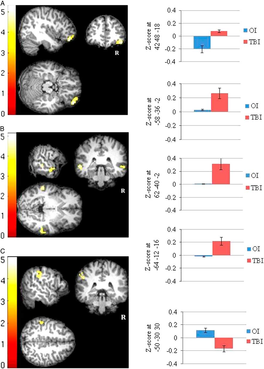
When a person rests and is not engaged in a task, brain regions in the default mode network (DMN) are activated. The DMN consists of regions (e.g., posterior cingulate cortex, medial prefrontal cortex, lateral parietal cortex, temporal parietal junction) that exhibit synchronous activation at rest, that is, are functionally connected (Fox et al., Reference Fox, Snyder, Vincent, Corbetta, Van Essen and Raichle2005). In contrast, these regions normally deactivate during performance of an external task. DMN dysfunction, such as failure to deactivate when an unexpected external event demands cognitive control, may result from (1) altered within-network functional connectivity (FC), as in reduced connectivity between posterior cingulate cortex with ventromedial prefrontal cortex (Sharp, Scott, & Leech, Reference Sharp, Scott and Leech2014) or (2) alteration of between-network FC, such as increased connectivity between one or more DMN regions and ventrolateral prefrontal cortex, a task-related region (Figure 1; Newsome et al., Reference Newsome, Scheibel, Hanten, Chu, Steinberg, Hunter and Levin2016).
Thirty days after sports related concussion (SRC), high school athletes differed in FC of the DMN from a demographically similar group of athletes who had sustained an orthopedic injury (OI). (A) Using a posterior cingulate cortex seed, FC to ventrolateral prefrontal cortex was greater in the SRC than OI group; (B) With a right lateral parietal cortex seed, FC with right and left middle temporal cortex was also increased in the SRC group relative to OI athletes. (C) In contrast, a right lateral parietal cortex seed showed greater FC with the right supramarginal gyrus in the OI than SRC group, possibly related to residual pain or other sensation following orthopedic injury. From Newsome et al., Reference Newsome, Li, Lin, Wilde, Ott, Biekman and Levin2016. Reproduced with permission by M. Newsome and Frontiers of Neurology.

Emerging evidence indicates that failure of task-related regions to deactivate during rest or of DMN regions to deactivate during task performance are related to residual cognitive dysfunction (Sharp et al., Reference Sharp, Scott and Leech2014). TBI may also produce aberrant connectivity between the DMN and the salience network (i.e., anterior insula and dorsal anterior cingulate cortex), a network that underpins selective attention to relevant stimuli and switching between attention to internally generated mental activity and external events, However, the long range implications of aberrant FC following TBI remain to be elucidated. Longitudinal imaging is necessary to elucidate the long-term effects on connectivity, evaluate whether DMN status is a useful prognostic biomarker, and determine if measurement of FC is useful for evaluating interventions in TBI patients.
Positron emission tomography
Epidemiological and neuropathological studies of TBI have produced strong evidence that a single moderate to severe TBI is a risk factor for Alzheimer’s disease (AD) and other dementias (Washington, Vilapol, & Burns, Reference Washington, Vilapol and Burns2016). Moreover, mild TBI in persons older than 65 years has also been linked to dementia (Gardner et al., Reference Gardner, Burke, Nettiksimmons, Kaup, Barnes and Yaffe2014). These reports have given impetus to research with PET, which can be used to measure the uptake of radioligands that selectively bind to beta-amyloid, providing an indirect index of amyloid plaques associated with dementia (Gatson et al., Reference Gatson, Stebbins, Mathews, Harris, Madden, Batjer and Minei2016). To prospectively evaluate in vivo p-tau deposits in former athletes, ongoing PET studies are using radioligands that selectively bind to p-tau (Dickstein et al., Reference Dickstein, Pullman, Fernandez, Short, Kostakoglu, Knesaurek and Gandy2016). PET research also is underway to evaluate the chronic inflammation that is thought to result from TBI and contribute to neurodegenerative processes.
Network dysfunction and multimodality brain imaging
Although early imaging research on TBI focused on specific lesions or regional volumetric changes, current imaging research supports the view that studying network dysfunction can elucidate the neural mechanisms of acute neurobehavioral disturbance, such as posttraumatic amnesia, as well as chronic impairments, including attention and memory deficits (Sharp et al., Reference Sharp, Scott and Leech2014). Neural mechanisms of social cognitive and behavioral sequelae can also be understood by analysis of the relevant brain networks. Local and global connectivity and other metrics of network efficiency may prove to be useful as outcome measures for clinical trials in TBI (Bullmore & Sporns, Reference Bullmore and Sporns2012).
Usage of multimodality imaging also is a current trend in TBI research, facilitated by reduced acquisition times and motivated by discovering how different neural mechanisms interact in injury and repair processes and contribute to neurodegenerative conditions. For example, the interrelationship of structural to functional connectivity provides converging data that enhances the validity of each technique in isolation (Bullmore & Sporns, Reference Bullmore and Sporns2009). Multimodality imaging could potentially characterize the underpinnings of response to interventions, including pharmacologic and non-pharmacologic treatments.
Advance #2: Non-injury Influences on TBI Outcomes
At the same time that neuroimaging began to allow for much more precise characterization of the pathophysiology of TBI, researchers were becoming increasingly cognizant that injury severity accounted for only a small proportion of the variance in outcomes after TBI. A second critical advance in the field, therefore, was the recognition of the importance of non-injury related influences on recovery. For instance, research on childhood TBI was heavily influenced by the seminal studies of Rutter and colleagues showing that psychosocial adversity moderated the behavioral outcomes of childhood TBI (Brown, Chadwick, Shaffer, Rutter, & Traub, Reference Brown, Chadwick, Shaffer, Rutter and Traub1981).
Pre-injury factors
A variety of pre-injury factors have been identified over the years as important in predicting the outcomes of TBI. For instance, demographic factors such as age at injury are known to affect outcomes, with poorer outcomes demonstrated for both the youngest and oldest segments of the population (Anderson, Catroppa, Morse, Haritou, & Rosenfeld, Reference Anderson, Catroppa, Morse, Haritou and Rosenfeld2000; Hukkelhoven et al., Reference Hukkelhoven, Steyerberg, Rampen, Farace, Habbema and Maas2003). Pre-injury cognitive ability also has been shown to moderate the effect of TBI, with higher cognitive ability fostering better outcomes (Fay et al., Reference Fay, Yeates, Taylor, Bangert, Dietrich, Nuss and Wright2010). Premorbid behavioral or psychiatric problems affect the outcomes of TBI as well. For instance, among adults with TBI, a prior history of depression predicts higher post-TBI rates of major depressive disorder (Alway, Gould, Johnston, McKenzie, & Ponsford, Reference Alway, Gould, Johnston, McKenzie and Ponsford2016; Bombardier et al., Reference Bombardier, Fann, Temkin, Esselman, Barber and Dikmen2010).
Similarly, premorbid attention problems heighten the likelihood of an increase in post-injury attention problems among children with moderate to severe TBI (Yeates et al., Reference Yeates, Armstrong, Janusz, Taylor, Wade, Stancin and Drotar2005). Research indicating that pre-injury factors such as IQ or behavioral adjustment moderate the outcomes of TBI fueled theories of cognitive and brain reserve capacity, which suggest that vulnerability to neurological insults varies as a function of pre-injury functioning and brain integrity (Dennis, Yeates, Taylor, & Fletcher, Reference Dennis, Yeates, Taylor and Fletcher2007; Stern, Reference Stern2000).
More recently, interest has turned to the potential role of genetics and epigenetics in modifying recovery from TBI (Kurowski, Martin, & Wade, Reference Kurowski, Martin and Wade2012; McAllister, Reference McAllister2015). A variety of genes have been studied to date, with apolipoprotein E receiving the most attention. Associations with recovery have been inconsistent, but much additional research is needed, with sample sizes large enough to explore not only main effects, but also gene X environment interactions (Treble-Barna et al., Reference Treble-Barna, Zang, Zhang, Martin, Yeates, Taylor and Kurowski2016).
Comorbid conditions
TBI often occurs alongside other conditions that can hamper recovery. For instance, post-traumatic stress disorder (PTSD) can co-occur with TBI, although its estimated incidence varies widely (Bryant & Harvey, Reference Bryant and Harvey1999), and PTSD is associated with poorer outcomes after TBI (Alway, McKay, Gould, Johnston, & Ponsford, Reference Alway, McKay, Gould, Johnston and Ponsford2016). The relationship between PTSD and TBI severity is uncertain, although the prevalence of PTSD is often higher in patients with mild TBI than moderate to severe TBI (Hoge et al., Reference Hoge, McGurk, Thomas, Cox, Engel and Castro2008; Zatzick et al., Reference Zatzick, Rivara, Jurkovich, Hoge, Wang, Fan and Mackenzie2010). Sleep disturbance, cognitive problems, or emotional distress associated with PTSD may negatively influence recovery from TBI (Lew et al., Reference Lew, Otis, Tun, Kerns, Clark and Cifu2009).
TBI is also frequently associated with comorbid pain, as a result of physical trauma, and possibly because of changes in brain functioning that affect sensory and motor functioning and, perhaps, perception of pain stimuli (Sherman, Goldberg, & Bell, Reference Sherman, Goldberg and Bell2006). A systematic review found a 75% prevalence of chronic pain among adults with mild TBI and 32% among those with moderate to severe TBI (Nampiaparampil, Reference Nampiaparampil2008). Among adolescents with TBI of all severities, 24% reported persistent pain 3 years after a TBI (Tham et al., Reference Tham, Palermo, Wang, Jaffe, Temkin, Durbin and Rivara2013). Chronic pain is often associated with other problems, including functional disability, psychological distress, and vocational issues (Lew et al., Reference Lew, Otis, Tun, Kerns, Clark and Cifu2009). In addition, pain and PTSD are often associated, as chronic pain can provoke PTSD-related thoughts and PTSD symptoms such as hyperarousal may increase pain intensity (Lew et al., Reference Lew, Otis, Tun, Kerns, Clark and Cifu2009).
Compensation-seeking behavior or litigation is another common comorbidity with TBI that is associated with poorer outcomes. TBI survivors who are actively engaged in litigation generally report more and longer-lasting symptoms, which in turn are associated with longer delays in return to work and higher levels of psychological stress (Paniak et al., Reference Paniak, Reynolds, Toller-Lobe, Melnyk, Nagy and Schmidt2002).
Contextual factors
Contextual influences are another potential source of non-injury related variance in outcomes. For example, a substantial literature demonstrates that the home environment is an important predictor of the outcomes of childhood TBI. Measures of socioeconomic status are consistent predictors, as are more proximal measures of family status and the home environment (Taylor et al., Reference Taylor, Yeates, Wade, Drotar, Klein and Stancin1999; Yeates et al., Reference Yeates, Taylor, Drotar, Wade, Klein and Stancin1997). Notably, the home environment moderates the impact of childhood TBI, by buffering or exacerbating consequences (Taylor et al., Reference Taylor, Yeates, Wade, Drotar, Klein and Stancin1999, 2002; Yeates et al., Reference Yeates, Taylor, Drotar, Wade, Klein and Stancin1997). Specifically, the effects of severe TBI relative to orthopedic injuries are more pronounced for children from dysfunctional families than for children from more functional families.
The relative importance of injury severity and family environment varies according to the specific outcome, however, with injury severity playing a larger role for cognitive outcomes and the family environment assuming more importance for behavioral and functional outcomes (Yeates, Taylor, Walz, Stancin, & Wade, Reference Yeates, Taylor, Walz, Stancin and Wade2010). Among adults, better family functioning also has been associated with improved outcomes after TBI, including reductions in overall disability, improvements in adaptive functioning, and increased employability; in contrast, family stress and unhealthy family communication can hamper recovery (Sander et al., Reference Sander, Caroselli, High, Becker, Neese and Scheibel2002).
Medical and non-medical intervention
The recognition that non-injury factors play a significant role in moderating the outcomes of TBI raised hopes that active interventions, ranging from acute medical care to education and workplace accommodations, could promote more rapid and complete recovery after TBI. However, the empirical evidence for most interventions remains limited. Guidelines exist for the acute medical management of TBI, but by and large are not based on rigorous evaluation through randomized controlled trials (RCT) (Jankowitz & Adelson, Reference Jankowitz and Adelson2006), and many RCTs that have been attempted have failed (Adelson, Reference Adelson2010). As the next section shows, cognitive rehabilitation has been a focus of substantial research, although empirical evidence of its effectiveness remains limited, especially for children (Ylvisaker, Adelson, et al., Reference Ylvisaker, Turkstra and Coelho2005). The inclusion of family members in the rehabilitation of both children and adults with TBI has received some support (Braga, Da Paz Júnior, & Ylvisaker, Reference Braga, Da Paz Júnior and Ylvisaker2005; Kreutzer et al., Reference Kreutzer, Stejskal, Ketchum, Marwitz, Taylor and Menzel2009). Family interventions may include educational, skill-building, and psychological support components. Much less is known regarding the effectiveness of educational or workplace interventions for persons with TBI (Mateer & Sira, Reference Mateer and Sira2006; Ylvisaker et al., Reference Ylvisaker, Todis, Glang, Urbanczyk, Franklin, DePompei and Tyler2001).
Advance #3: Non-physical Problems and Cognitive Rehabilitation
The heterogeneous nature of TBI results in considerable variability in the nature and severity of resultant impairments. Nevertheless, due to the high frequency of injury to fronto-temporal brain regions, as well as diffuse axonal injury, certain sequelae are particularly common. Over 50 years of research has shown that TBI often results in cognitive deficits in memory, attention, processing speed, word finding, and planning and problem-solving; behavioral difficulties such as lack of initiative, irritability, and poor temper control; somatic symptoms such as headaches, dizziness, fatigue, sleep disturbance, and poor balance and coordination; and psychological distress, including anxiety and depression (Dikmen, Machamer, Fann, & Temkin, Reference Dikmen, Machamer, Fann and Temkin2010). Although these difficulties usually resolve to some degree, they persist in many cases even decades after injury (Himanen et al., Reference Himanen, Portin, Isoniemi, Helenius, Kurkj and Tenovuo2006; Hoofien, Gilboa, Vakil, & Donovick, Reference Hoofien, Gilboa, Vakil and Donovick2001; Ponsford, Downing, et al., Reference Ponsford, Downing, Olver, Ponsford, Acher, Carty and Spitz2014), and result in long-term difficulties with independence in daily activities and participation in work or study, leisure activities, and social and personal relationships (Ponsford, Downing, et al., Reference Ponsford, Downing, Olver, Ponsford, Acher, Carty and Spitz2014; Sherer et al., Reference Sherer, Sander, Nick, High, Malec and Rosenthal2002).
In the 1980s, the growing realization that cognitive, emotional, and behavioral deficits had a greater impact on long-term functional outcomes than physical impairments led to the birth of the field of cognitive rehabilitation. The field has experienced tremendous growth since then. Early systematic reviews and guidelines initially identified only a handful of studies with rigorous methodology (Cicerone et al., Reference Cicerone, Dahlberg, Kalmar, Langenbahn, Malec, Bergquist and Morse2000), but the number has grown steadily (Cicerone et al., Reference Cicerone, Dahlberg, Malec, Langenbahn, Felicetti, Kneipp and Catanese2005, Reference Cicerone, Langenbahn, Braden, Malec, Kalmar, Fraas and Ashman2011). Even so, few specific techniques are yet underpinned by strong evidence. Cognitive rehabilitation interventions can be broadly classified as either restorative, aiming to restore impaired functions, or compensatory, aiming to develop strategies or use residual strengths to minimize the functional impact of impairments.
Fatigue and sleep disturbance
Fatigue and sleep disturbance are debilitating and persistent problems for individuals with TBI of all severities (Ponsford et al., Reference Ponsford, Ziino, Parcell, Shekleton, Roper, Redman and Rajaratnam2012). Until recently, these problems have not been the focus of treatment. Blue light therapy has been shown to reduce fatigue and daytime sleepiness in one pilot RCT (Sinclair, Ponsford, Taffe, Lockley, & Rajaratnam, Reference Sinclair, Ponsford, Taffe, Lockley and Rajaratnam2014). Cognitive behavioral therapy has also shown some promise for addressing both sleep disturbance and fatigue (Nguyen et al., Reference Nguyen, McKay, Wong, Spitz, Mansfield, Williams and Ponsford2017).
Attentional problems
Both children and adults with TBI display a range of attention problems (Ponsford, Bayley, et al., Reference Ponsford, Downing, Olver, Ponsford, Acher, Carty and Spitz2014; Yeates et al., Reference Yeates, Armstrong, Janusz, Taylor, Wade, Stancin and Drotar2005). Substantial research has been devoted to evaluating methods of rehabilitating attention. Many studies in adults, and more recently in children, have focused on retraining attention using computer-mediated tasks. These reveal gains on the trained tasks, with generalization to similar cognitive measures but not to everyday attentional behavior (Laatsch et al., Reference Laatsch, Harrington, Hotz, Maracantuomo, Mozzoni, Walsh and Hersey2007; Ponsford, Bayley, et al., Reference Ponsford, Downing, Olver, Ponsford, Acher, Carty and Spitz2014). Stronger evidence suggests that metacognitive strategies can be applied to everyday tasks to compensate for everyday attention problems in adults, with evidence of potential for training in multi-tasking (Ponsford, Bayley, et al., Reference Ponsford, Downing, Olver, Ponsford, Acher, Carty and Spitz2014). Variability in nature and severity of cognitive impairments, self-awareness, and capacity to implement compensatory strategies are likely important determinants of success of any rehabilitative intervention for attention, although the influence of these factors has not been systematically studied. No studies have evaluated changes to the individual’s environment to alleviate attention problems, although such interventions are frequently recommended for both adults and children (Savage, DePompei, Tyler, & Lash, Reference Savage, DePompei, Tyler and Lash2005).
Memory problems
The use of restorative techniques such as computer-based training to enhance impaired memory processes after TBI has received little support (Velikonja et al., Reference Velikonja, Tate, Ponsford, McIntyre, Janzen and Bayley2014). However, increasing evidence supports the use of compensatory strategies for memory problems. Studies have shown that internal memory strategies, such as mnemonics, visual imagery, and self-instructional methods, can enhance performance on neuropsychological tests (Velikonja et al., Reference Velikonja, Tate, Ponsford, McIntyre, Janzen and Bayley2014). Self-instructional methods that also enhance the individual’s understanding of manifestations of their memory problems have been successfully applied to prospective memory tasks in adults (Shum, Fleming, Gill, Gullo, & Strong, Reference Shum, Fleming, Gill, Gullo and Strong2011; Velikonja et al., Reference Velikonja, Tate, Ponsford, McIntyre, Janzen and Bayley2014) and children (Lawson & Rice, Reference Lawson and Rice1989).
Individuals with less severe cognitive deficits have shown more benefit from such memory strategies than those with more severe deficits, presumably reflecting the need for executive skills, including self-awareness, to implement strategies. Stronger evidence indicates that environmental supports, including diaries, notebooks, paging devices, mobile/smart phones, and Google calendar, are helpful to adults with memory impairments (Sohlberg et al., Reference Sohlberg, Kennedy, Avery, Coelho, Turkstra, Ylvisaker and Yorkston2007). Use of picture prompts or the Neuropage has been effective in children (Lawson & Rice, Reference Lawson and Rice1989; Wilson, Emslie, Quirk, & Evans, Reference Wilson, Emslie, Quirk and Evans2001). An important determinant of successful use of such aids is the ability to manage the sophistication of the device, as influenced by age, executive skills, prior experience, access to supportive technology, and having a family member available to support and monitor use of the device (Velikonja et al., Reference Velikonja, Tate, Ponsford, McIntyre, Janzen and Bayley2014). Errorless learning, spaced retrieval, and vanishing cues techniques also have all been shown to be effective methods of learning in individuals with TBI (Ehlhardt et al., Reference Ehlhardt, Sohlberg, Kennedy, Coelho, Ylvisaker, Turkstra and Yorkston2008).
Disorders of communication and social cognition
Many communication disturbances associated with TBI involve changes in pragmatic skills that impair social competence for both children and adults with TBI (Togher et al., Reference Togher, Wiseman-Hakes, Douglas, Stergiou-Kita, Ponsford, Teasell and Turkstra2014). The recent development of theories of social communication after TBI has led to new approaches to assessment and treatment (Coelho, Ylvisaker, & Turkstra, Reference Coelho, Ylvisaker and Turkstra2005). Social communication training for adults with TBI, predominantly in group formats, has been shown to be effective for individuals 6–24 months post-injury (Togher et al., Reference Togher, Wiseman-Hakes, Douglas, Stergiou-Kita, Ponsford, Teasell and Turkstra2014). Training that incorporates everyday communication partners has been more successful, encouraging practice of skills beyond the period of the training (Ylvisaker, Turkstra, & Coehlo, Reference Ylvisaker, Turkstra and Coelho2005). RCTs focusing on emotion recognition and social inference have shown benefits (McDonald et al., Reference McDonald, Togher, Tate, Randall, English and Gowland2013; Radice-Neumann, Zupan, Tomita, & Willer, Reference Radice-Neumann, Zupan, Tomita and Willer2009). Recognition is growing that interventions should be provided and evaluated in the natural environments of the person’s everyday life, including with communication partners (Togher et al., Reference Togher, Wiseman-Hakes, Douglas, Stergiou-Kita, Ponsford, Teasell and Turkstra2014).
Executive dysfunction
Interventions for executive impairments have addressed self-awareness, planning, problem solving, self-monitoring, abstract reasoning, and cognitive flexibility. Metacognitive strategy training methods have some support, including goal-management training (Levine et al., Reference Levine, Schweizer, O’Connor, Turner, Gillingham, Stuss and Robertson2011), multifaceted strategy training (Spikman, Boelen, Lamberts, Brouwer, & Fasotti, Reference Spikman, Boelen, Lamberts, Brouwer and Fasotti2010), and training in problem orientation and problem solving in adults (Tate et al., Reference Tate, Kennedy, Ponsford, Douglas, Velikonja, Bayley and Stergiou-Kita2014). As with other cognitive interventions, the likelihood of generalization is limited for individuals with severe executive and memory deficits, and is maximized by application in everyday contexts (Dawson et al., Reference Dawson, Gaya, Hunt, Levine, Lemsky and Polatajko2009; Grant, Ponsford, & Bennett, Reference Grant, Ponsford and Bennett2012). Several RCTs have successfully promoted reasoning abilities, including gist reasoning and categorization (Tate et al., Reference Tate, Kennedy, Ponsford, Douglas, Velikonja, Bayley and Stergiou-Kita2014). Self-awareness deficits have been treated with involvement in contextualized activities, training to anticipate obstacles, feedback, self-monitoring, and self-evaluation techniques (Fleming & Ownsworth, Reference Fleming and Ownsworth2006). Direct, within-task feedback also has been supported by RCTs (Tate et al., Reference Tate, Kennedy, Ponsford, Douglas, Velikonja, Bayley and Stergiou-Kita2014).
Behavioral problems
Interventions for behavioral problems after TBI have received limited attention (Cattelani Zettin, & Zoccolotti, Reference Cattelani, Zettin and Zoccolotti2010; Ylvisaker et al., Reference Ylvisaker, Turkstra, Coehlo, Yorkston, Kennedy, Sohlberg and Avery2007). In the 1980s and 1990s, programs using structured contingency approaches to behavior modification were favored (Eames & Wood, Reference Eames and Wood1985). In more recent years, advocates have promoted contextual approaches with Positive Behavior Support interventions, designed to teach or shape adaptive and cooperative social behaviors as well as to promote physical and functional independence (Ylvisaker, Jocobs, & Feeney, Reference Ylvisaker, Jacobs and Feeney2003). However, evidence for these interventions remains limited. Cognitive behavioral approaches to anger control were successful in one small RCT (Medd & Tate, Reference Medd and Tate2000). In children with TBI, the focus has been on training social problem-solving, often in conjunction with parents, whose stress levels and parenting styles are strongly predictive of children’s’ behavior problems. Successful Interventions for behavioral problems have focused on families, delivered in person or online (Wade et al., Reference Wade, Walz, Carey, McMullen, Cass, Mark and Yeates2011).
Anxiety and depression
The negative impact of psychological distress on functional outcome after TBI is increasingly recognized. Anxiety and depressive disorders occur in a significant proportion of adults with TBI (Alway, Gould, et al., Reference Alway, Gould, Johnston, McKenzie and Ponsford2016). In children, attention deficit hyperactivity disorder and depression predominate (Bloom et al., Reference Bloom, Levin, Ewing-Cobbs, Saunders, Song, Fletcher and Kowatch2001). Although results have been mixed, recent evidence from RCTs suggests that cognitive behavioral therapy adapted to circumvent the effects of memory and executive impairments may reduce anxiety and depression symptoms after TBI (Ponsford et al., Reference Ponsford, Lee, McKay, Wong, Haines, Alway and O’Donnell2016). Adapted cognitive behavioral interventions for social anxiety are also being trialed in children.
PEERING AHEAD: THE FUTURE OF TBI RESEARCH
Although we do not claim to have a crystal ball, we agree that two important trends can be discerned in the field that are likely to shape neuropsychological research on TBI in the coming decades. One is the growing emphasis on “big data,” as reflected in the development of large, multi-site research consortia and adoption of common data elements. The other trend is the increasing focus on knowledge translation, or how to translate research on TBI into clinical practice, especially in the domain of cognitive rehabilitation.
Big Data and Big Science: The Age of Consortiums and Common Data Elements
Early research on TBI often suffered from small, unrepresentative samples, disagreements about definitions of TBI severity and associated inclusion and exclusion criteria, and inconsistencies in predictor and outcome measurement. In recent years, the field has begun to overcome those problems by conducting large, multi-site studies with very large samples, in the context of both prospective observational research and RCTs, using common data elements that are collected consistently across all studies. Examples of individual studies that represent this movement include the TRACK-TBI study (Yue et al., Reference Yue, Vassar, Lingsma, Cooper, Okonkwo, Valadka and Manley2013), the CARE consortium (Broglio, 2015), the 5P study (Zemek, Osmond, & Barrowman, Reference Zemek, Osmond and Barrowman2013) and the ADAPT trial (Larsen et al., Reference Larsen, Schober, Fabio, Wisniewski, Grant, Shafi and Bell2016). The selection and use of common data elements have been promoted by government funding agencies, such as NIH and Canadian Institutes of Health Research (e.g., Hicks et al., Reference Hicks, Giacino, Harrison-Felix, Manley, Valadka and Wilde2013; McCauley et al., Reference McCauley, Wilde, Anderson, Bedell, Beers, Campbell and Yeates2012), as well as by large research consortia, such as the International Initiative for Traumatic Brain Injury Research (InTBIR) (Tosetti et al., Reference Tosetti, Hicks, Theriault, Phillips, Koroshetz and Draghia-Akli2013).
Recent multicenter studies of TBI also have implemented uniform imaging protocols and quality assurance procedures that have enhanced the feasibility of aggregating imaging data across different MRI platforms (Yuh et al., Reference Yuh, Mukherjee, Lingsma, Yue, Ferguson, Gordon and Manley2013). Central analysis of the data has also mitigated site differences in the review of pathology. Common data elements for reporting pathology based on MRI has enhanced harmonization of data across different centers (Duhaime et al., Reference Ewing-Cobbs, Prasad, Swank, Kramer, Cox, Fletcher and Hasan2010). TRACK-TBI investigators have reported that punctate lesions seen with a gradient echo sequence are indicative of hemorrhagic axonal injury and, along with the presence of contusions, improve prediction of three month outcome of mild TBI even after taking into account CT results and other clinical data (Yuh et al., Reference Yuh, Mukherjee, Lingsma, Yue, Ferguson, Gordon and Manley2013). This finding suggests that imaging biomarkers may enrich studies of mild TBI patients (and other TBI groups). This strategy could facilitate clinical trials by selecting those patients who are at high risk for persistent problems and reducing heterogeneity in the samples.
Translating Research Into Clinical Practice
A second important trend in TBI research involves attempts to translate research into clinical practice. Although substantial gains have been made in understanding the impairments that impede adults and children who sustain TBI, and developing and evaluating interventions to overcome them, a significant gulf remains between research evidence and clinical practice. Almost all cognitive rehabilitation studies have been completed in experimental settings using artificial tasks, and have evaluated treatment efficacy using neuropsychological tests bearing little resemblance to the demands of everyday life.
Some recent studies suggest that a greater focus on making interventions relevant to the individual goals and needs of the adult or child with TBI and embedding them in everyday contexts will maximize the durability of gains (Togher et al., Reference Togher, Wiseman-Hakes, Douglas, Stergiou-Kita, Ponsford, Teasell and Turkstra2014). More studies in real world contexts are badly needed, especially studies that also examine what factors moderate response to intervention, including non-injury factors such as age, education, IQ, psychiatric status, family and social support, nature and severity of cognitive deficits, motivation, and environmental demands and supports.
Successful treatments need to be manualized, and methods of training clinicians and barriers to implementation evaluated. Over the last two decades, some clinical practice guidelines (CPG) have been developed in the domain of cognitive rehabilitation, guided by best available evidence (e.g., Cicerone et al, Reference Cicerone, Langenbahn, Braden, Malec, Kalmar, Fraas and Ashman2011; Bayley et al., Reference Bayley, Tate, Douglas, Turkstra, Ponsford, Stergiou-Kita and Bragge2014). Some evidence suggests that such guidelines can improve decision making and ultimately, clinical outcomes (Keris, Lavendelis, & Macane, 2007). However, a recent study auditing the use of the International Cognitive Rehabilitation working group guidelines by clinicians (Bayley et al., Reference Bayley, Tate, Douglas, Turkstra, Ponsford, Stergiou-Kita and Bragge2014) suggested a significant gap between the availability of CPGs and their use in clinical practice, even in specialized brain injury rehabilitation centers. An important next step is to evaluate the use of CPGs and associated clinical pathways using rigorous methods drawn from implementation science (Grimshaw, Eccles, Lavis, Hill, & Squires, Reference Grimshaw, Eccles, Lavis, Hill and Squires2012), with the goal of increasing evidence-based practice and improving patient outcomes.
CONCLUSION
The past 50 years have been a period of exciting progress in neuropsychological research on TBI. The advent of modern neuroimaging tools, the recognition of the importance of non-injury factors in determining recovery from TBI, and the explosion of research on cognitive rehabilitation, have all helped to push the field forward significantly. We now have a better understanding of the pathophysiology of TBI and how recovery from the injury is shaped by pre-injury, comorbid, and contextual factors, as well as a growing body of evidence suggesting that active interventions, including cognitive rehabilitation, may help to promote better outcomes. Future large-scale observational studies and RCTs using common data elements should capitalize on these past advances to provide critical evidence regarding the outcomes of TBI and its treatment; and formal knowledge translation efforts built on rigorous implementation science methods should help to disseminate and implement the knowledge gained from research into health care and health policy, to the eventual betterment of the quality of life of persons with TBI.
ACKNOWLEDGMENTS
K.O.Y. is supported by the Ronald and Irene Ward Chair in Pediatric Brain Injury from the Alberta Children’s Hospital Foundation, as well as by a Canadian Institutes of Health Research Foundation Grant. H.S.L. is supported by grants from the NIH (R21NS086714-01), DoD (PT13078), and VA (B1320-I). J.P. is supported by grants from NHMRC, Institute for Safety Compensation Recovery Research and the Transport Accident Commission. The authors have no conflicts of interest to report.