Introduction
Most zoological gardens are dedicated to the preservation of endangered animals through captive breeding and reintroduction programs (Maesano et al. Reference Maesano, Capasso, Ianniello, Cringoli and Rinaldi2014; Mitchell and Tully Reference Mitchell and Tully2009). The preservation of these animals in captivity within zoological gardens can exacerbate the issue of parasitic infections (Muoria et al. Reference Muoria, Muruthi, Rubenstein, Oguge and Munene2005). Indeed, captive-bred animals are more susceptible to pathogens compared to their wild counterparts (Kolodziej-Sobocinska et al. Reference Kolodziej-Sobocinska, Demiaszkiewicz, Pyziel and Kowalczyk2018). This is often attributed to animals experiencing higher concentration of parasites due to the confined living conditions and the increased environmental contamination associated with maintaining animals in enclosed areas (Citino Reference Citino, Fowler and Miller2003; Gracenea et al. Reference Gracenea, Gómez, Torres, Carné and Fernández-Morán2002). In all instances, transmission mainly takes place via the faecal-oral route, either through direct contact with infected hosts (or their faecal material) or indirectly by consuming contaminated water or food (Köster et al. Reference Köster, Martínez-Nevado, González, Abelló-Poveda, Fernández-Bellon, de la Riva-Fraga, Marquet, Guéry, Knauf-Witzens, Weigold, Dashti, Bailo, Imaña, Muadica, González-Barrio, Ponce-Gordo, Calero-Bernal and Carmena2022). Moreover, gastrointestinal parasitic infections represent a significant issue, leading to diseases and even mortality (Thawait et al. Reference Thawait, Maiti and Dixit2014). However, to ensure optimal health and welfare in animal zoo species, effective captive management is crucial. This management should encompass preventive healthcare, as well as optimal husbandry and nutrition practices (Mellor et al. Reference Mellor, Hunt and Gusset2015).
Despite the implementation of various control and prevention measures in these facilities, the identification of parasites in captive species within zoos will facilitate the development of effective husbandry practices aimed at maintaining the health status of managed species (Carrera-Játiva et al. Reference Carrera-Játiva, Morgan, Barrows and Wronski2018). Therefore, numerous mammals within zoos play an essential role as hosts and reservoirs for various zoonotic parasites (Fagiolini et al. Reference Fagiolini, Lia, Laricchiuta, Cavicchio, Mannella, Cafarchia, Otranto, Finotello and Perrucci2010; Mirzapour et al. Reference Mirzapour, Kiani, Mobedi, Spotin, Seyyed Tabaei and Rahimi2018; Purcell and Philipp Reference Purcell, Philipp and Sonia2005). Among various parasites, Trichuris spp. infections are widespread among captive animals globally (Aviruppola et al. Reference Aviruppola, Rajapakse and Rajakaruna2016; Barbosa et al. Reference Barbosa, Pinheiro, Dos Santos, de Lima, Dib, Echarte, Augusto, Bastos, Antunes Uchôa, Bastos, Santos, Fonseca and Amendoeira2020).
The genus Trichuris comprises intestinal nematode parasites that infect mammals, including humans. Human trichuriasis is caused by the Trichuris trichiura species, which belongs to the group of causative agents of geohelminthiasis or soil-transmitted helminths (STHs) (WHO 2023). It is estimated that approximately 800 million people of the world’s population is parasitized by T. trichiura (CDC 2017). Evidence suggests that T. trichiura is a complex of cryptic species capable of infecting both humans and non-human primates (NHP) living in the wild and in captivity (Hawash et al. Reference Hawash, Andersen, Gasser, Stensvold and Nejsum2015; Ravasi et al. Reference Ravasi, O’Riain, Davids and Illing2012; Rivero et al. Reference Rivero, García-Sánchez, Zurita, Cutillas and Callejón2020, Reference Rivero, Cutillas and Callejón2021a). Moreover, they are frequently documented as the most prevalent species affecting primates residing in zoological gardens in China (Li et al. Reference Li, Zhao, Li, Wang, Niu, Deng, Gu, Peng, Wang and Yang2015) and have also been found in other zoos in Italy (Cavallero et al. Reference Cavallero, De Liberato, Friedrich, Di Cave, Masella, D’Amelio and Berrilli2015, Reference Cavallero, Nejsum, Cutillas, Callejón, Dolezalová, Modry and D’Amelio2019), and in a UK safari park (Juhasz et al. Reference Juhasz, Spiers, Tinsley, Chapman, Shaw, Head, Cunningham, Archer, Jones, Haines, Davies Walsh, Johnson, Quayle, Jones, LaCourse, Cracknell and Stothard2023). These infections can be highly persistent due to egg’s high environmental resistance and the long lifespan of adults. Consequently, eradicating them from a colony of captive NHPs requires specific treatment strategies and significant economic resources (Reichard et al. Reference Reichard, Wolf, Carey, Garrett and Briscoe2007).
Parasitic diseases that can be transmitted from zoo animals to humans, known as zoonotic diseases, are a major area of concern (Pérez Cordón et al. Reference Pérez Cordón, Hitos Prados, Romero, Sánchez Moreno, Pontes, Osuna and Rosales2008). There are studies on zoonotic transmission between NHP and their zookeepers in European zoos, where both NHP and zookeepers were found to be infected with Trichuris spp. (Levecke et al. Reference Levecke, Dorny, Vercammen, Visser, Van Esbroeck, Vercruysse and Verweij2015). Furthermore, several studies have investigated the prevalence of Trichuris infection in NHPs in zoos, revealing high values, making it one of the most commonly found helminths (Juhasz et al. Reference Juhasz, Spiers, Tinsley, Chapman, Shaw, Head, Cunningham, Archer, Jones, Haines, Davies Walsh, Johnson, Quayle, Jones, LaCourse, Cracknell and Stothard2023; Kamel and Abdel-Latef Reference Kamel and Abdel-Latef2021; Levecke et al. Reference Levecke, Dorny, Vercammen, Visser, Van Esbroeck, Vercruysse and Verweij2015). Hence, microscopic examination for morphological identification remains the reference method for diagnosis parasites in faecal samples (de Waal Reference de Waal2012). However, to achieve a more comprehensive understanding of the Trichuris species identified in these samples, the implementation of alternative diagnostic methods to achieve species-level identification is essential. This is crucial in determining whether these Trichuris species belong to T. trichiura – parasites specific to humans. However, recently, it was also believed to be specific to several NHPs. It is noteworthy that other authors have suggested that multiple Trichuris species could be present among host species, and there is a possibility that Trichuris in NHPs might have zoonotic potential (Cavallero et al. Reference Cavallero, Nejsum, Cutillas, Callejón, Dolezalová, Modry and D’Amelio2019). Recent studies, employing morpho-biometric and molecular criteria, have led to the identification of new Trichuris species in primates. For instance, Trichuris rhinopiptheroxella was discovered in the golden snub-nosed monkey (Rhinopithecus roxellana) (Wang et al.,2019), Trichuris colobae in Colobus guereza kikuyuensis (Cutillas et al. Reference Cutillas, De Rojas, Zurita, Oliveros and Callejón2014), and Trichuris ursinus in Papio ursinus (Callejón et al. Reference Callejón, Halajian and Cutillas2017). These findings affirm that T. trichiura is not the exclusive whipworm species found in primates.
Based on this background, the main aim of the current study was to determinate the prevalence of Trichuris spp. in several NHPs within multiple zoological gardens in Spain, utilizing coprological analysis of feacal samples. Additionally, we conducted a statistical evaluation to assess the significance of this parasitism and to achieve a precise species-level identification of the Trichuris species, including the molecular characterization of the species found in the samples of NHPs analyzed. This information is crucial for implementing necessary control measures to enhance the health of the animals and ensure the safety of zoo visitors and caretakers.
Materials and methods
Ethical considerations
This study does not require approval from an ethics committee. All primates were handled and housed in a zoo in strict accordance with best practices for animal care.
Sampling
A total of 315 fecal samples were collected from 47 NHP species within 28 genera at 13 zoological gardens in Spain during the period from November 2017 to July 2023. The zoological gardens involved in the study were the Zoo Barcelona (Barcelona), Bioparc Fuengirola (Málaga), Bioparc Valencia (Valencia), La Reserva del Castillo de las Guardas (Seville), Parque de la Naturaleza de Cabárceno (Cantabria), Zoo de Castellar (Cadiz), Zoo de Córdoba (Córdoba), Zoobotánico de Jerez (Cadiz), Marcelle Natureza (Lugo), Selwo Aventura (Málaga), Río Safari Elche (Alicante), Mundo Park (Seville), and Zoológico El Bosque (Principado de Asturias). These zoos were named using the letters A to M in the designation ‘Zoo’ (Supplementary Table S1).
Anthelmintic medications have been periodically administered to all primates in the zoological gardens as part of a routine healthcare regimen. Fecal samples were placed into a cooler with ice packs immediately after collection and fetched to the Department of Microbiology and Parasitology, to the Parasitology Laboratory of the Faculty of Farmacy, University of Seville, the same day. Stools were refrigerated (2–6ºC) until analysis (within 2 days).
Coprological examinations or parasitological analysis
First, stools were visually exanimated for macroscopic parasitic structures and for the presence of blood, mucus, adults, etc. All samples underwent two types of analysis. The first was based on a direct examination of the sample according to Pourrut et al. (Reference Pourrut, Diffo, Somo, Bilong Bilong, Delaporte, LeBreton and Gonzalez2011). The second method involved a formalin-ether concentration method following a standardized protocol (Becker et al. Reference Becker, Lohourignon, Speich, Rinaldi, Knopp, N’goran, Cringoli and Utzinger2011).
The Trichuris specimens were collected from fecal samples after administering treatment, then washed with physiological saline solution (0.9% w/v), and subsequently stored at a temperature of -20ºC until they were needed for additional analysis. Moreover, the parasitological analysis revealed the presence of Trichuris eggs. Afterward, to conduct further molecular analyses, the eggs were collected and concentrated using a Sheather’s sugar solution (Gupta and Singla Reference Gupta, Singla, Gupta, Garg, Nehra and Lather2013) and subsequently embryonated at 32ºC for 3 to 4 weeks with the addition of a 0.2% w/v potassium dichromate solution to create humidity in the medium and prevent the growth of fungi and bacteria (Horii and Usui Reference Horii and Usui1985).
Morphological identification and molecular analysis
The adult whipworms were morphologically identified according to previous studies (Callejón et al. Reference Callejón, Halajian and Cutillas2017; Cutillas et al. Reference Cutillas, Callejón, de Rojas, Tewes, Ubeda, Ariza and Guevara2009, Reference Cutillas, De Rojas, Zurita, Oliveros and Callejón2014; Rivero et al. Reference Rivero, García-Sánchez, Zurita, Cutillas and Callejón2020, Reference Rivero, Cutillas and Callejón2021a). Genomic DNA was extracted from adults and batches of 45–200 isolated eggs using DNeasy Blood and Tissue Kit (Quiagen) following to the manufacturer’s protocol. The quality of extractions was evaluated through 0.8% agarose gel electrophoresis with SYBR Safe DNA gel stain. The polymerase chain reaction (PCR) was employed, using a thermal cycler (Eppendorf AG; Hamburg, Germany) to amplify all molecular markers sequenced, including rrnL mitochondrial DNA (mtDNA) (~386–395 base pairs (bp)), as well as ITS2 ribosomal DNA (rDNA) (~436–611 bp). Negative controls (no template DNA) were included in all runs. PCR was conducted in a total volume of 50 μL containing 25 μL GoTaq G2 Green Master Mix (Promega, Madison, WI, USA), 5 μL of each primer pair (10μM), 5 μL of template DNA, and nuclease-free water to 50 μL. For the amplification of the rrnL partial gene, the cycling conditions used were 95ºC for 15 min (initial denaturation), then 95ºC for 30 s (denaturation), 55ºC for 30 s (annealing), and 72ºC for 1 min (extension) for 35 cycles, and a final extension at 72ºC for 10 min, using the following primers: TTrrnLF (5’-TAAATGGCCGTCGTAACGTGACTGT-3’) and TTrrnLR (5’-AAAGAGAATCCATTCTATCTCGCAACG-3’) according to Liu et al. (Reference Liu, Gasser, Su, Nejsum, Peng, Lin, Li, Xu and Zhu2012). And for the amplification of ITS2 rDNA, the cycling conditions used were 94ºC for 3 min (initial denaturation), then 94ºC for 1 min (denaturation), 50ºC for 1 min (annealing), and 72ºC for 1 min (extension) for 35 cycles, and a final extension at 72ºC for 10 min, using the following primers: 5.8SF (5’-TGAAGAACGACGTGACACTC-3’) (Robles et al. Reference Robles, Cutillas, Panei and Callejón2014) and ITS2R (5’-TTAGTTTCTTTTCCTCCGCT -3’) (Ellis and Horvitz Reference Ellis and Horvitz1986). The PCR products were visualized on agarose gels stained with SYBR Safe (2% w/v Tris-borate-EDTA, TBE). Bands were eluted and purified from the agarose gel using QWizard SV Gel and PCR Clean-Up System Kit (Promega, Madison, WI, USA). After purification and concentration, sequencing was performed in both directions used for PCR by Stab Vida (Lisbon, Portugal). Then, for the identification of the analyzed species, the sequences obtained were compared with those previously deposited in GenBank using Standard Nucleotide BLAST (BLASTn) in the National Center for Biotechnology Information (NCBI) (https://blast.ncbi.nlm.nih.gov/Blast.cgi).
Statistical analysis
The results obtain from the zoological gardens were subject to statistical analysis. First, percent prevalence was determined according to Carrera-Játiva et al. (Reference Carrera-Játiva, Morgan, Barrows and Wronski2018), calculating the number of positive samples times 100 divided by the number of total samples. Statistical analysis was performed in the case of Trichuris-infested animals for which a data set of a suitable sample size was available (Norman and Streiner, Reference Norman and Streiner2008). In particular, a Chi-square test – or Fisher’s exact test where appropriate – was used to assess whether the different zoological gardens had an overall comparable percentage of, on the one hand, infected species and positive samples and, on the other hand, a comparable frequency of infection among the orders Cercopithecidae and Hominidae, with p < 0.05 regarded as statistically significant. All statistical analyses of the present study were performed with R commander (Rcmdr), the graphical user interface for R software, version 4.3-1. Due to the impossibility to determine the number of parasites per infected host, as faecal samples predominantly contain eggs, the calculation of parasite intensity was determined by dividing the positive total faecal samples by the positive number of infected hosts among the zoological gardens.
Results
Prevalence analysis
In the present study, a total of 315 samples collected from 47 different primate species in 13 Spanish zoological gardens were examined for the presence of Trichuris parasite infection (Figure 1). The overall prevalence of parasitic infection by Trichuris species was 19.05% (60/315) (see in Table 1).
Egg detected in a faecal sample of Papio papio from Parque de la Naturaleza de Cabárceno.

Infections identified in faecal samples and the prevalence obtained among captive NHPs in zoological gardens from Spain

Out of the 13 zoological gardens included in the study, Trichuris infection was found in NHP hosts in 7 of them (53.85%). However, there was remarkable differences in the prevalence between the analyzed zoos. ‘Zoo I’ showed the highest parasitic infection rate, reaching 50%, followed by ‘Zoo F’ at 39.39% and ‘Zoo J’ at 34.15%. Conversely, the lowest prevalence was recorded in the ‘Zoo G’, with only 1.89%. Additionally, in 6 out of the 13 zoological gardens analyzed, there was no infection by Trichuris spp. (46.15%) (see in Table 1).
‘Zoo J’ and ‘Zoo I’ were found to have the highest levels of parasite intensity among the zoological gardens analyzed (Table 1). Therefore, out of all the analyzed faecal samples, only three Trichuris adults were found in two faecal samples collected from Papio papio at Parque de la Naturaleza de Cabárceno.
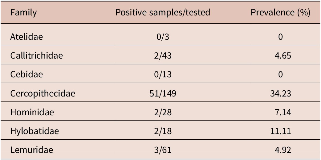
Statistical analysis revealed that the Trichuris infections in the different zoos were significantly different (χ2 = 19.08, p = 0.004). However, there were no significant differences observed in the patterns of parasitic infection between the species from the zoos (p = 0.32). When examining the data based on the primate families, significant differences were recorded in the frequency of Trichuris infections among the order Cercopithecidae (χ2 = 12.91, p = 0.04). On the contrary, no significant differences were found among the order Hominidae (p = 1) (see in Table 2).
Summary of positive faecal samples among the tested captive NHPs and the prevalence, categorized by their respective families, from various zoological gardens analyzed in Spain

Molecular analysis
Not all batches of eggs completed the process of embryonation; as a result, we were unable to obtain DNA from those specific samples. The details of the Trichuris samples, encompassing both adult whipworms and eggs, are detailed in Table 3.
Summary of Trichuris positive NHP hosts analyzed, type of samples (adult or eggs), Trichuris species obtained

*: DNA extraction was unsuccessful.
The identification of Trichuris species were carried out through the utilization of BLASTn in NCBI. The results demonstrated a high degree of similarity, approaching 100% in all sequences analyzed (Table 3). This analysis involved both mtDNA using the rrnL marker and rDNA using the ITS2 marker (Supplementary Table S2).
Discussion
Spanish zoological gardens are a major tourist attraction, hosting millions of visitors every year. They employ specialized veterinarians who provide regular preventive and curative treatments for the animals. However, despite these efforts, many animals still exhibit parasitic infections. Furthermore, there is a lack of comprehensive data regarding the prevalence of parasitic zoonoses within these zoological gardens. The existence of zoonotic parasites represents a potential hazard to the health of NHPs, thereby raising the possibility of zoonotic transmission occurring between visitors and NHPs.
In this study, we initially carried out epidemiological research to assess the prevalence of Trichuris infections in NHPs residing in captivity within zoological gardens in Spain. Subsequently, we proceeded with a species-level identification of the Trichuris species present in the analyzed NHP hosts.
Zoo animals are susceptible to a wide range of diseases, with helminth infestations being a major concern, including the genus Trichuris, that infect captive mammals worldwide (Barbosa et al. Reference Barbosa, Pinheiro, Dos Santos, de Lima, Dib, Echarte, Augusto, Bastos, Antunes Uchôa, Bastos, Santos, Fonseca and Amendoeira2020; Karim et al. Reference Karim, Li, Rume, Sumon, Selim, Hoda and Zhang2021; Khatun et al. Reference Khatun, Begum, Mamun, Mondal and Azam2014; Levecke et al. Reference Levecke, Dorny, Geurden, Vercammen and Vercruysse2007; Li et al. Reference Li, Zhao, Li, Wang, Niu, Deng, Gu, Peng, Wang and Yang2015; Mir et al. Reference Mir, Dua, Singla, Sharma and Singh2016; Naz et al. Reference Naz, Arooj, Ali and Farooq2021). Hence, in the different hosts, the presence of these parasites may lead to morbidity and mortality (Nath et al. Reference Nath, Islam and Chakraborty2012) and may also serve as reservoirs of parasites. Additionally, some of these infections can be zoonotic, meaning they can be transmitted to humans (Bogale et al. Reference Bogale, Chanie, Melaku, Fentahun and Berhanu2014) and raise concerns for public health (Levecke et al. Reference Levecke, Dorny, Geurden, Vercammen and Vercruysse2007). Parasites can be introduced into zoological gardens through various pathways, including contaminated food, intermediate and paratenic hosts, acquisition of new parasites by the animals, or through infected staff or visitors (Panayotova-Pencheva Reference Panayotova-Pencheva2013).
Zoo mammals play a crucial role as hosts and reservoirs for many zoonotic parasites (Mirzapour et al. Reference Mirzapour, Kiani, Mobedi, Spotin, Seyyed Tabaei and Rahimi2018). A study of mammals from Beni-Suef Zoo in Egypt revealed that the prevalence of intestinal parasitic infection among various zoo mammals was 11.69% positive with helminths (Kamel and Abdel-Latef Reference Kamel and Abdel-Latef2021). However, this result is not in concordance with those reported in Malaysia, where a higher prevalence of helminths (34.5%) was detected (Lim et al. Reference Lim, Ngui, Shukri, Rohela and Mat Naim2008). Nonetheless, when the investigation is among NHPs, T. trichiura infections is one the most common intestinal parasites, in addition to Giardia intestinalis and Entamoeba histolytica / dispar (Arafa et al. Reference Arafa, Mahrous, Aboelhadid and Abdel-Ghany2013; Arizono et al. Reference Arizono, Yamada, Tegoshi and Onishi2012; Gillespie et al. Reference Gillespie, Greiner and Chapman2005; Kamel and Abdel-Latef Reference Kamel and Abdel-Latef2021). Prevalence of trichuriasis in NHPs in a UK safari park was 48% (Juhasz et al. Reference Juhasz, Spiers, Tinsley, Chapman, Shaw, Head, Cunningham, Archer, Jones, Haines, Davies Walsh, Johnson, Quayle, Jones, LaCourse, Cracknell and Stothard2023) and in Beni-Suef Zoo in Egypt was 31.3% (Kamel and Abdel-Latef Reference Kamel and Abdel-Latef2021), while in a population of African green monkey on St. Kitts (West Indies), it was 91% (Yao et al. Reference Yao, Walkush, Shim, Cruz and Ketzis2018). In accordance with the mentioned above, in a study in five zoos in Belgium and the Netherlands, the prevalence of Trichuris infection was 17.9% (Levecke et al. Reference Levecke, Dorny, Vercammen, Visser, Van Esbroeck, Vercruysse and Verweij2015). The prevalence we have obtained in this study falls within the range previously reported (19.05%), aligning closely with the prevalence reported in European zoos. These variations may be attributed to climatic, seasonal, and geographical differences, as well as variations in husbandry practices among the different regions where the studied animals are located (Kamel and Abdel-Latef Reference Kamel and Abdel-Latef2021). Several factors influence the proliferation of parasites in mammals housed within zoological gardens, such as the intensive husbandry practices of wild animals in zoos that may contribute to higher animal densities within enclosures and closer proximity to other animals, thereby increasing the potential for accelerated transmission of parasites (Moudgil and Singla Reference Moudgil and Singla2013). However, it is essential to consider that prolonged and regular deworming treatments are linked to the development of drug resistance (Kaplan and Vidyashankar Reference Kaplan and Vidyashankar2012; Pyziel et al. Reference Pyziel, Björck, Wiklund, Skarin, Demiaszkiewicz and Höglund2018).
Conversely, it was crucial to achieve species-level identification of the Trichuris species parasitizing the analyzed hosts due to the aforementioned significance and the potential transmission of parasites between hosts and humans. Thus, the Trichuris species analyzed in this study were not all the same; three different species emerged across the analyzed hosts. This importance also lies in whether humans can be parasitized by Trichuris species that can develop in NHPs. Previously, Trichuris species were identified based on the host being parasitized. For instance, the species parasitizing both humans and primates was identified as T. trichiura, while that in pigs was Trichuris suis (Cutillas et al. Reference Cutillas, Callejón, de Rojas, Tewes, Ubeda, Ariza and Guevara2009). However, for many years, it was believed that the species T. trichiura and T. suis were the same species, given their morphological similarities. Over the past decade, novel diagnostic techniques have been introduced, departing from conventional morphological identification methods and embracing contemporary molecular techniques. This transition has revealed the existence of new Trichuris spp. as well as a complex of Trichuris species parasitizing both humans and NHPs, such as T. colobae, T. ursinus, or T. rhinopiptheroxella (Callejón et al. Reference Callejón, Halajian and Cutillas2017; Cutillas et al. Reference Cutillas, De Rojas, Zurita, Oliveros and Callejón2014; Wang et al. Reference Wang, Zhang, Song, Zhu, Chen, Ren, Liu and Zhao2019).
For identification of taxa, we employed various molecular markers. The utilization of molecular markers proves especially advantageous in differentiating taxa, particularly when morphological distinctions are insufficient for closely related species. This is particularly relevant when most variations in morphological features are linked to apparent adaptation or when species separation is relatively recent (Cavallero et al. Reference Cavallero, De Liberato, Friedrich, Di Cave, Masella, D’Amelio and Berrilli2015). The application of nuclear rDNA coding and non-coding regions has been widespread in molecular systematics and phylogenetic analyses of parasitic nematodes. These regions display greater variability between species than within species, facilitating the characterization of new species, the revelation of cryptic species, and the identification of potential hybrids among closely related species (Callejón et al. Reference Callejón, Halajian and Cutillas2017; Cavallero et al. Reference Cavallero, Nejsum, Cutillas, Callejón, Dolezalová, Modry and D’Amelio2019). Nonetheless, recent studies have observed that nuclear markers of rDNA are useful for differentiation among species but less so for closely related species, as noted by Rivero et al. (Reference Rivero, García-Sánchez, Zurita, Cutillas and Callejón2020) in a population of Trichuris from M. sylvanus. In this study, they suggested the presence of two cryptic species; however, the nucleotide similarity was high in nuclear rDNA markers, while there was a greater difference in mtDNA markers, such as cox1 and cytb partial genes. The findings corroborate those of Cavallero et al. (Reference Cavallero, Nejsum, Cutillas, Callejón, Dolezalová, Modry and D’Amelio2019), where the phylogenetic results from nuclear and mtDNA markers seemed incongruent. However, these authors propose that combining these markers could aid in elucidating relationships among closely related species within the Trichuris genus (Cavallero et al. Reference Cavallero, Nejsum, Cutillas, Callejón, Dolezalová, Modry and D’Amelio2019; Rivero et al. Reference Rivero, García-Sánchez, Zurita, Cutillas and Callejón2020, Reference Rivero, Callejón and Cutillas2021b). This is in agreement with Chan et al. (Reference Chan, Chaisiri, Morand, Saralamba and Thaenkham2020), who assessed the utility of mtDNA and rDNA genes for the study of parasitic nematode systematics, concluding that different genetic lineages should be delimited exclusively using mtDNA gene analyses. This is attributed to the slower substitutions rate and higher nucleotide saturation observed in ITS regions compared to mtDNA genes. Furthermore, they assessed the effectiveness of mtDNA markers and highlighted that, among the 18S and 28S rRNA genes, as well as the 12S (rrnS), 16S (rrnL) rRNA and cox1 mtDNA genes, the latter five demonstrated greater resolution for phylogenetic studies, suggesting that these genes have the potential to be employed as markers (Chan et al., Reference Chan, Chaisiri, Morand, Saralamba and Thaenkham2020).
Typically, genetic distance values below 2–6% are considered indicative of cryptic species, while values exceeding 10% suggest the presence of distinct morphospecies (Cavallero et al. Reference Cavallero, Nejsum, Cutillas, Callejón, Dolezalová, Modry and D’Amelio2019). However, in recent studies conducted on Trichuris species parasitizing closely related NPH host species, the percentages of genetic diversity were approximately ranged from 20–27% (Cavallero et al. Reference Cavallero, Nejsum, Cutillas, Callejón, Dolezalová, Modry and D’Amelio2019; Hawash et al. Reference Hawash, Andersen, Gasser, Stensvold and Nejsum2015; Rivero et al. Reference Rivero, García-Sánchez, Zurita, Cutillas and Callejón2020, Reference Rivero, Cutillas and Callejón2021a; Wang et al. Reference Wang, Zhang, Song, Zhu, Chen, Ren, Liu and Zhao2019). These results, previously published, are comparable to genetic diversity observed among different Trichuris species, such as T. trichiura and T. suis, and Trichuris muris and Trichuris arvicolae (Cavallero et al. Reference Cavallero, Nejsum, Cutillas, Callejón, Dolezalová, Modry and D’Amelio2019; Rivero et al. Reference Rivero, Cutillas and Callejón2021a). Consequently, the presence of a complex Trichuris species in the zoological gardens analyzed, potentially capable of parasitizing humans. Specifically, there is a coexistence of T. trichiura and T. colobae species, along with a cryptic species related to T. trichiura, which originates from M. sylvanus (Rivero et al. Reference Rivero, García-Sánchez, Zurita, Cutillas and Callejón2020). Furthermore, through molecular characterization, we can dismiss the hypothesis that primates were infected through an alternative route, such as an infestation with murine or porcine whipworms facilitated by rats or contaminated food. This is substantiated by the likelihood that they were infected by their specific wild whipworms prior to captivity.
Anthelminthic drugs represent a common strategy for treating and preventing parasitic diseases in zoos (Moreno Mañas et al. Reference Moreno Mañas, Gonzálvez, Ruiz de Ybáñez Carnero, Gilbert, Ortiz, Espeso, Benzal, Ibáñez and Valera Hernández2019) and are often administered to animals to limit the excretion of eggs in feces, ensuring that wildlife in captive breeding programs are treated to maintain proper health conditions (Lahat et al. Reference Lahat, Ortiz, Tizzani, Ibáñez, Valera, Moreno, Espeso and Ruiz de Ybáñez2021). Nevertheless, in captivity, their efficacy is limited to a brief duration, as animals often experience reinfection shortly after therapy cessation. Factors like high animal density and challenges in removing dung from enclosures contribute to soil and food contamination, fostering conditions conducive to reinfections (Goossens et al. Reference Goossens, Dorny, Boomker, Vercammen and Vercruysse2005). Consequently, captive animals typically host a diverse parasite community, with anthelmintic treatments serving primarily for control rather than complete elimination of parasitic infections (Lahat et al. Reference Lahat, Ortiz, Tizzani, Ibáñez, Valera, Moreno, Espeso and Ruiz de Ybáñez2021). While effective in terms of health management, this practice may, on the other hand, hinder the development of effective immunity against parasites in captive animals (Lahat et al. Reference Lahat, Ortiz, Tizzani, Ibáñez, Valera, Moreno, Espeso and Ruiz de Ybáñez2021). The extensive use of anthelmintics has resulted in the development of resistance in nematodes of veterinary significance, particularly those responsible for gastrointestinal parasitism (Kaplan and Vidyashankar Reference Kaplan and Vidyashankar2012; Matthews Reference Matthews2014; Sargison Reference Sargison2012; Sutherland and Leathwick Reference Sutherland and Leathwick2011). Furthermore, the documented issue of anthelmintic drug resistance, including multidrug resistance (MDR) encompassing benzimidazoles, imidothiazoles–tetrahydropyrimidines, and macrocyclic lactones, poses a significant challenge for nematode parasites control across various regions worldwide (Sangster et al. Reference Sangster, Cowling and Woodgate2018). The transmission of drug-resistant strains may be heightened through interactions among livestock and susceptible wildlife species (Gałązka et al. Reference Gałązka, Klich, Filip-Hutsch, Olech, Anusz and Pyziel2023). Indeed, various authors have reported a notable resistance of the Trichuris genus to administered anthelmintics (Bennett et al. Reference Bennett, Anderson, Barker, Michael and Bundy2002; Diawara et al. Reference Diawara, Drake, Suswillo, Kihara, Bundy, Scott, Halpenny, Stothard and Prichard2009, Reference Diawara, Halpenny, Churcher, Mwandawiro, Kihara, Kaplan, Streit, Idaghdour, Scott, Basáñez and Prichard2013). There is scant information available on the effectiveness of deworming with anthelmintics in captive NHPs in zoological gardens. Furthermore, supplementary factors encompass protocols for disease prevention and the nature of the administered treatment (Lim et al. Reference Lim, Ngui, Shukri, Rohela and Mat Naim2008). Hence, in instances when humans and NHPs cohabit the same ecosystems, sporadic outbreaks of zoonosis diseases may transpire (Lee et al. Reference Lee, Kang, Kim, Lee, Ahn, Kwon, Park and Kim2010).
The findings of this study indicate that Trichuris infections are prevalent among zoo primates, and these animals may potentially serve as reservoirs capable of transmitting this parasite to humans. Moreover, the research confirmed the existence of various Trichuris species, other than T. trichiura, infecting NHPs living in captivity. The results underscore the imperative requirement for an immediate parasite control program within the zoos. Additionally, there is a pressing need to continue investigating from the perspective of minimizing the risk of spreading drug-resistant parasite strains within the facilities where the hosts reside.
Supplementary material
The supplementary material for this article can be found at http://doi.org/10.1017/S0022149X24000774.
Acknowledgements
The authors acknowledge the significant support received from the collaborating zoological gardens, including the Zoo Barcelona, Bioparc Fuengirola, Bioparc Valencia, La Reserva del Castillo de las Guardas, Parque de la Naturaleza de Cabárceno, Zoo de Castellar, Zoo de Córdoba, Zoobotánico de Jerez, Marcelle Natureza, Selwo Aventura, Río Safari Elche, Mundo Park, Zoológico El Bosque. The authors also thank the caretakers and veterinarians.
Author contribution
Conceptualization: JR and RC; Methodology: JR and RC; Validation: JR and RC; Formal Analysis: JR and AMGS; Investigation: JR; Data Curation: JR and AMGS; Writing – Original Draft Preparation: JR; Writing – Review & Editing: JR, RC, and AMGS; Supervision: RC.
Financial support
This research received no specific grant from any funding agency, commercial or not-for-profit sectors.
Competing interest
The authors declare none.