INTRODUCTION
The Gulf of California is recognized as one of the most diverse hotspots of marine biodiversity (Roberts et al., Reference Roberts, McClean, Veron, Hawkins, Allen, McAllister, Mittermeier, Schueler, Spalding, Wells, Vyme and Werner2002). The major contributions to the actinofauna of the Gulf of California were those made by Carlgren (Reference Carlgren1940a, Reference Carlgren1951) in which he described 11 new species and reported seven known species for the region. In the last two decades, several contributions have increased the number of known taxa for the region (López-González et al., Reference López-González, Rodríguez, Gili and Segonzac2003; Daly, Reference Daly2004; Sanamyan & Sanamyan, Reference Sanamyan and Sanamyan2007; Eash-Loucks & Fautin, Reference Eash-Loucks and Fautin2012; Hendrickx et al., Reference Hendrickx, Hinojosa-Corona and Ayón-Parente2016; Barragán et al., in press). Currently, the Gulf of California has 24 species of sea anemones distributed within 10 families (Barragán et al., in press).
During recent expeditions to the southern Gulf of California, we found one specimen of an undescribed species of sea anemone belonging to the family Haloclavidae Verrill, Reference Verrill1899. Members of Haloclavidae are characterized by having a single strong siphonoglyph, relatively low number of tentacles and mesenteries, and lacking basilar muscles; in addition, some members of Haloclavidae might have conchula, cinclides in the column and/or in the aboral end, capitate tentacles, fosse and rarely, an endodermal marginal sphincter muscle (revised in Gusmão, Reference Gusmão2016). Currently Haloclavidae encompasses 11 genera and 34 species worldwide distributed from intertidal to deep waters (Gusmão, Reference Gusmão2016; Izumi et al., Reference Izumi, Yanagi and Fujita2016, Reference Izumi, Fujita and Yanagi2017).
In this study, we describe a new species of sea anemone, Tenactis riosmenai gen. et sp. nov. based on a single specimen from the intertidal in the southern Gulf of California. We erect Tenactis gen. nov. to place this specimen; Tenactis gen. nov. is characterized by having mesenteries decamerously arranged, endodermal marginal sphincter, a single pair of directives attached to a single and strong siphonoglyph, conchula, basilar muscles, column with pseudoacrorhagi and verrucae, cinclides in the proximal column and pedal disc, and p-mastigophores A2 and p-mastigophores B1 and B2a in filaments.
MATERIALS AND METHODS
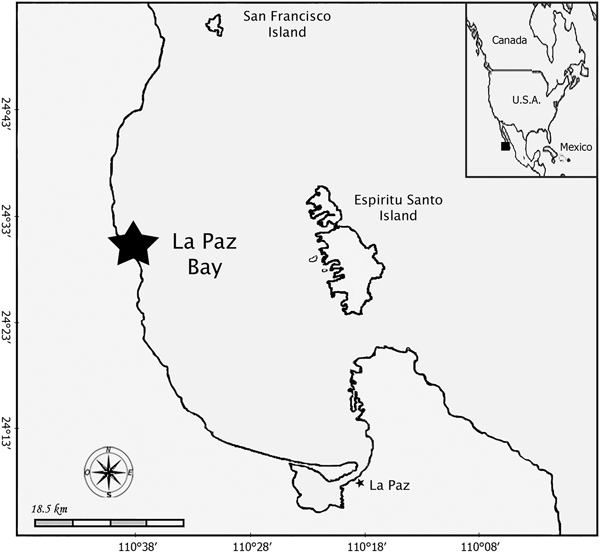
The studied material was collected in the intertidal rocky shore in Punta Coyote Sur, south of Gulf of California (Mexico) during the summer of 2013 (Figure 1). The only collected specimen (despite several field trips to the area to locate more specimens) was fixed in 10% seawater formalin. The preserved specimen was examined whole, in dissection and as serial sections. Fragments of the specimen were dehydrated in graded ethanol series and then embedded in paraffin. Histological sections 10 µm thick were stained with Heidenhain's Azan Triple Stain (Presnell & Schreibman, Reference Presnell and Schreibman1997).
Map with locality (asterisk) where Tenactis riosmenai gen. et sp. nov. has been collected.

Measurements of cnidae were made from preserved material; small pieces of tissue were smeared on slides and examined using differential interference microscopy (DIC) at 1000 × magnification. We scanned through the slides and haphazardly measured 40 undischarged capsules of each type (when possible) to generate a range: frequencies given are subjective impressions based on all the cnidae seen on the slides. Presence of cnidae types was confirmed in histological slides of the specimen. We agree with and use here the cnidae terminology detailed and used by Sanamyan et al. (Reference Sanamyan, Sanamyan and Tabachnick2012) but we amend their incorrect citation to Carlgren (Reference Carlgren1949) as the work modifying Weill's (Reference Weill1934) classification (see correct citation below); we also add some considerations to the use of cnidae terminology and its evolutionary implications as follow: cnidae nomenclature follows the classification of Weill (Reference Weill1934) modified by Carlgren (Reference Carlgren1940b), but Schmidt's (Reference Schmidt1969, Reference Schmidt1972) classification without the modification of den Hartog (Reference den Hartog1995) is used to differentiate ‘mastigophore’ morphologies. Using this compromise, we succeed in differentiating ‘basitrich’ and ‘b-mastigophore’ morphologies using Weill (Reference Weill1934) modified by Carlgren (Reference Carlgren1940b) while better capturing the underlying variation seen in ‘rhabdoids’ described by Schmidt (Reference Schmidt1969, Reference Schmidt1972), which is imperative to actiniarian systematics. In addition, we concur with Sanamyan et al. (Reference Sanamyan, Sanamyan and Tabachnick2012) and use the term ‘mastigophore’ instead of ‘rhabdoid’ to preserve stability and consistency (instead of the terms ‘penicillus’ and ‘spirula’ of Stephenson (Reference Stephenson1928) which have priority but have not been frequently used) but we acknowledge that the category ‘p-mastigophore’ does not exist in opposition to ‘amastigophores’ as defined by Weill (Reference Weill1934) because the latter (similarly to p-mastigophores) have a distal tubule but this tubule is small and breaks off in the discharged capsule (Cutress, Reference Cutress1955; Schmidt, Reference Schmidt1969; Östman, Reference Östman2000). We emphasize that we use a combination of classifications to better capture the underlying variation in cnidae morphology but do not imply any evolutionary relationship among these types. Finally, because of the complexity of several of the terminology issues stated above we consider that any nematocyst comparison among taxa going forward should be based on images (drawings or photographs) in order to be considered reliable. The studied material has been deposited in the American Museum of Natural History (AMNH) in New York City.
RESULTS
SYSTEMATICS
Order ACTINIARIA Hertwig, Reference Hertwig1882 Suborder ENTHEMONAE Rodríguez & Daly, 2014 in Rodríguez et al., Reference Rodríguez, Barbeitos, Brugler, Crowley, Grajales, Gusmão, Häussermann, Reft and Daly2014 Family HALOCLAVIDAE Verrill, Reference Verrill1899
DIAGNOSIS (MODIFIED FROM GUSMÃO, 2016; MODIFICATIONS IN BOLD, ADDITION IN ITALICS)
Body elongate, with or without basilar musculature; aboral end pedal disc or physa-like, physa sometimes broadly adherent. Column rarely divisible into physa, scapus and scapulus; may have parapet and deep fosse; with or without pseudoacrorhagi with basitrichs; smooth or with hollow or solid papillae, minute adherent rugae or verrucae, with or without cinclides. No marginal sphincter muscle or rarely an endodermal weak or strong developed diffuse one. Tentacles short, sometimes capitate, usually simple, never numerous, inner shorter or of same length as outer ones. Single, ventral, very strong siphonoglyph, occasionally more or less completely separated from rest of actinopharynx and drawn out at its oral end into a more or less folded conchula. Perfect pairs of mesenteries vary in number, usually all macrocnemes. One or two pairs of directives. Retractors usually strong. Parietobasilar musculature usually distinct. Cnidom: basitrichs, b-mastigophores, p-mastigophores A and p -mastigophores A2, p-mastigophores B1 and B2a, and spirocysts.
REMARKS
The diagnosis has been modified to include morphological characteristics of Tenactis gen. nov. within the family but also to capture the recent newly described diversity within Haloclavidae (see Izumi et al., Reference Izumi, Yanagi and Fujita2016, Reference Izumi, Fujita and Yanagi2017) and to reflect the followed terminology of the cnidae (see rationale in Materials and Methods section). Distinct verrucae (adhesive cup-shape evaginations in the column), a single pair of directives attached to the single siphonoglyph, pseudoacrorhagi bearing only basitrichs, pedal disc with basilar muscles, strong endodermal sphincter and p-mastigophores B1 in the filaments are the novel features for the family observed in Tenactis gen. nov. Similarly, we included having p-mastigophores A2 and p-mastigophores B2a in the diagnosis because they are present in Antennapeachia Izumi, Yanagi & Fujita, Reference Izumi, Yanagi and Fujita2016 and in Tenactis gen. nov (see Izumi et al., Reference Izumi, Yanagi and Fujita2016, Reference Izumi, Fujita and Yanagi2017). We have also included ‘minute adherent rugae’ in the diagnosis because this is the most accurate way to define the minute adhesive areas found in the column of Stephanthus Rodríguez & López-González, Reference Rodríguez and López-González2003 and Antennapeachia. We have eliminated the term ‘sucker’ because after the redescription of Metapeachia Carlgren, Reference Carlgren1943 by Gusmão (Reference Gusmão2016) it does not reflect any character currently present within the family. Similarly, we eliminated microbasic amastigophores from the diagnosis (in addition to reflect the terminology used here) because nematocysts corresponding to this type have not been found within the family so far: the presence of microbasic amastigophores described in the filaments of Metapeachia by Yap et al. (Reference Yap, Fautin, Ramos and Tan2014) could not be confirmed in the type material or other species of the genus (see Gusmão, Reference Gusmão2016) and those named as microbasic amastigophores in the column and filaments of Antennapeachia setouchi Izumi, Yanagi & Fujita, Reference Izumi, Yanagi and Fujita2016 correspond to p-mastigophores A (see Figure 5F and J, respectively in Izumi et al., Reference Izumi, Yanagi and Fujita2016); we believe that the cnidae named as microbasic amastigophores in the tentacles, actinopharynx and filaments of Antennapeachia jambio Izumi, Yanagi & Fujita, Reference Izumi, Fujita and Yanagi2017 (see figure 4E, G and M in Izumi et al., Reference Izumi, Fujita and Yanagi2017) correspond to immature developmental stages of p-mastigophores A in which the capsule and basal tubule are not completely compacted yet (see Östman et al., Reference Östman, Kultima, Roat and Rundblom2010), which do not correspond to the category microbasic amastigophores under Weill's (Reference Weill1934) system (Reft, Reference Reft2012).
Genus Tenactis gen. nov.
DIAGNOSIS
Haloclavidae with aboral end well-developed as pedal disc, with basilar musculature and cinclides. Column cylindrical, not divisible into regions, with longitudinal rows of verrucae and pseudoacrorhagi with basitrichs; cinclides in proximal column. Tentacles to 20, without acrospheres. Mesenteries not differentiated into macro- and microcnemes; decamerously arranged. All pairs of mesenteries perfect and fertile. One single pair of directives attached to a single, strong and well-developed siphonoglyph, with oral end drawn out into a poorly developed conchula not forming distinct tube separated from actinopharynx. Same number of mesenteries proximally and distally. Endodermal marginal sphincter muscle diffuse. Retractors restricted. Parietobasilar musculature strong, well developed. Cnidom: basitrichs, b-mastigophores, p-mastigophores A and p-mastigophores A2, p-mastigophores B1 and B2a, and spirocysts.
TYPE SPECIES
Tenactis riosmenai sp. nov., by original designation.
ETYMOLOGY
From the English word ‘ten’ referring to the number of mesenteries of the type species. It is combined with the Greek ‘–actis’ meaning ray, a common suffix in sea anemone generic names. Gender feminine.
Tenactis riosmenai sp. nov. (Figures 2–4, Table 1)
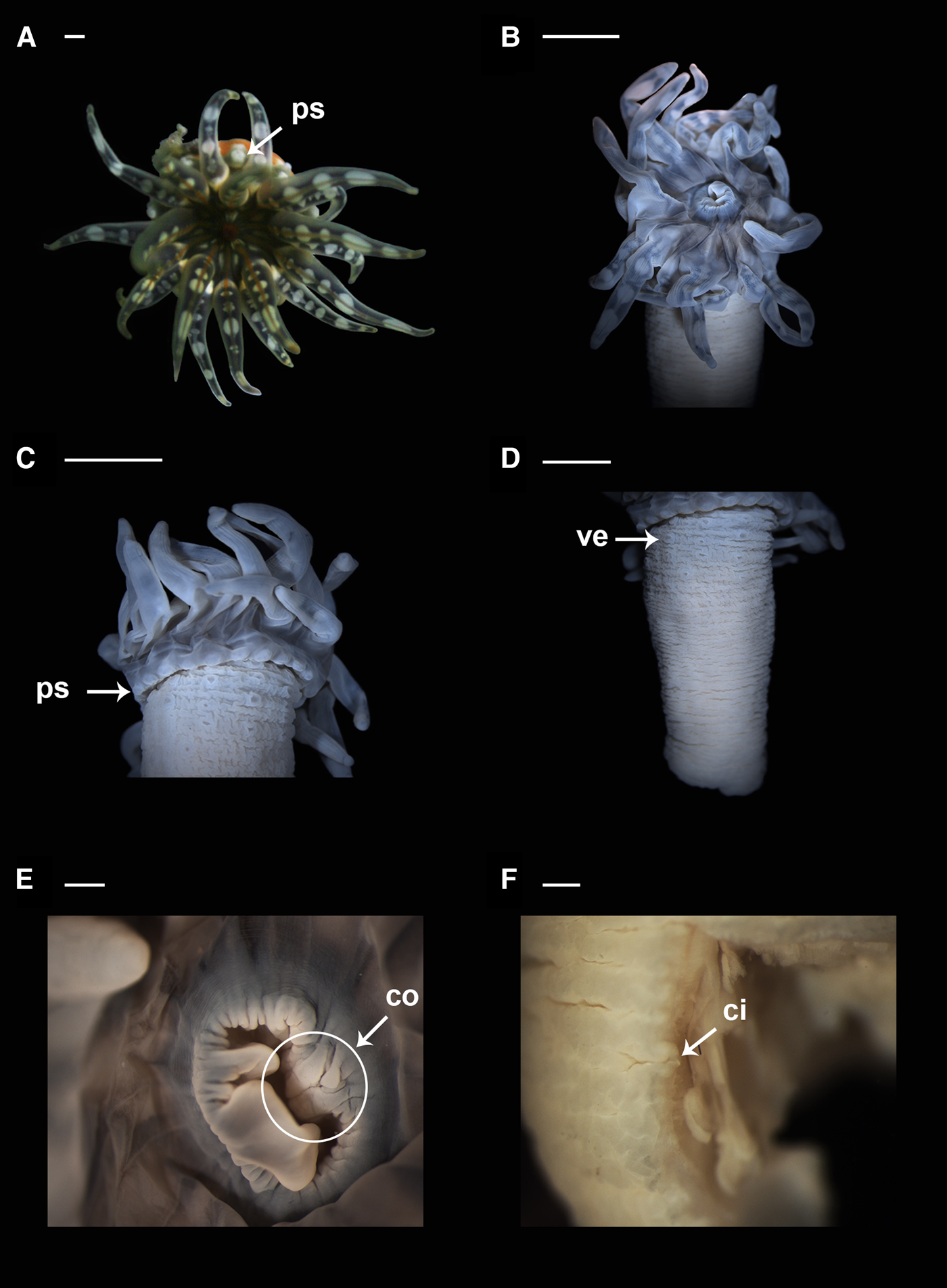
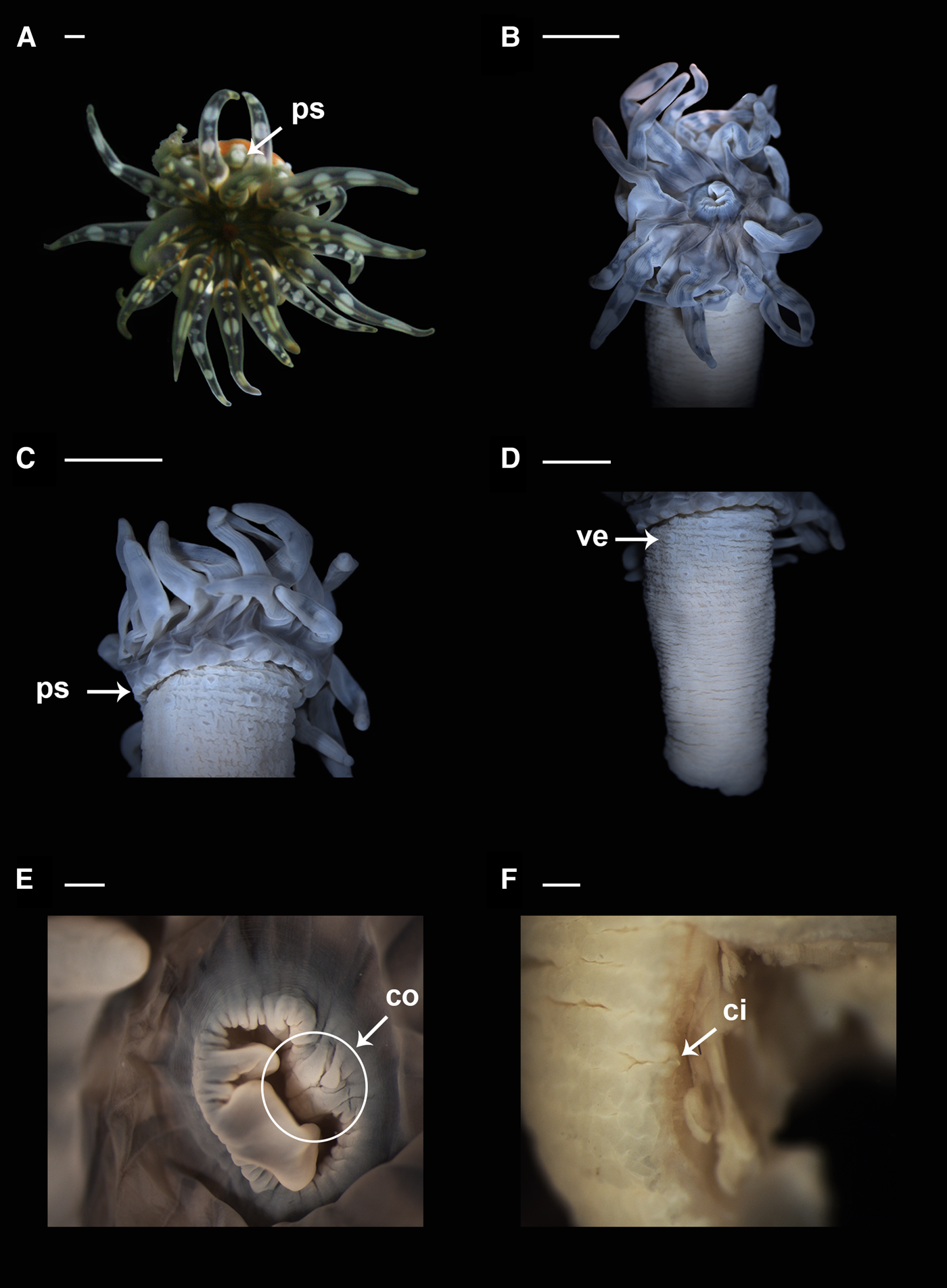
Tenactis riosmenai gen. et sp. nov., external anatomy: (A) oral view of the living specimen; (B) oral view of the preserved specimen; (C) detail of the pseudoacrorhagi and distal column; (D) detail of the column and verrucae; (E) detail of the poorly developed conchula (circle); (F) detail of one cinclid in the pedal disc. ci, cinclid; co, conchula; ps, pseudoacrorhagi; ve, verruca. Scale bars: A–F, 0.5 mm.
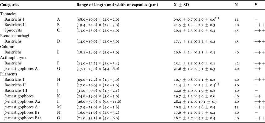
Size ranges of the cnidae of Tenactis riosmenai gen. et sp. nov. Letters correspond with images in Figure 4.

N, total number of capsules measured; F, Frequency; +++ = very common; ++ = common; + = rather common; − = sporadic. Different categories of same type of nematocysts indicated with roman numbers; (*) mean and standard deviation values marked with an asterisk are based on fewer than 40 capsules.
TYPE MATERIAL
Holotype: adult male (spermatic cysts developed), partially dissected and sectioned in several slides. (Coordinates: 24°31.44′N 110°44.2′W; water depth: 0–5 m); (AMNH_IZC 250219); coll. Y. Barragán, 25 July 2013.
DESCRIPTION
External anatomy
Well-developed pedal disc, 7 mm diameter in preserved specimen, with cinclides (Figures 2F & 3H). Column cylindrical, to 26 mm height and 9 mm diameter in preserved specimen. Column with 20 longitudinal rows of verrucae from middle column to most distal column; a ring of 20 endo- and exo-coelic pseudoacrorhagi with basitrichs (see Figures 2C, D & 3B). Deep fosse. Fully expanded oral disc to 8 mm diameter in preserved specimen. Poorly developed conchula, with three slightly thickened lobes; two lateral lobes of similar size, middle one smaller (Figure 2E). Tentacles 20, arranged in two cycles; simple, smooth, tapering toward tips, all of similar length. Mesenterial insertions visible in oral disc (Figure 2A).
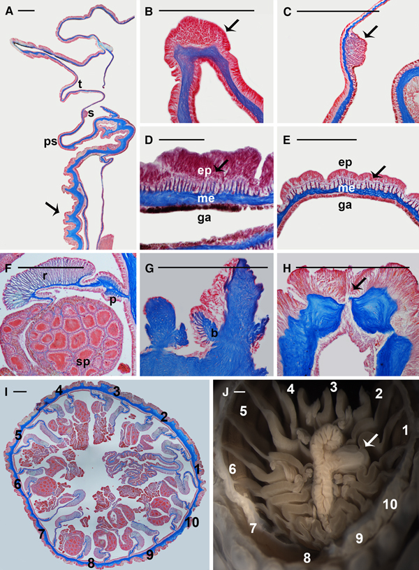
Tenactis riosmenai gen. et sp. nov., internal anatomy: (A) longitudinal section of distal part of the column, notice the verrucae (arrow); (B) detail of a pseudoacrorhagi, notice the spherical structure at the tip of the projection (arrow); (C) detail of the endodermal marginal sphincter muscle (arrow); (D) and (E) cross section of the tentacle and the oral disc (respectively) showing the ectodermal longitudinal musculature (arrow); (F) detail of retractor and parietobasilar muscles, and gametogenic tissue; (G) detail of the proximal end showing the basilar muscles; (H) detail of the pedal disc showing cinclides (arrow); (I) histological cross section of the column at the level of the actinopharynx showing the 10 pairs of perfect mesenteries and the single pair of directives (arrow); (J) cross section of the column at the level of the actinopharynx showing the 10 pairs of perfect mesenteries and the single siphonoglyph (arrow). b, basilar muscles; ep, epidermis; ga, gastrodermis; me, mesoglea; p, parietobasilar muscles; ps, pseudoacrorhagi; r, retractor, t, tentacle; s, sphincter; sp, spermatic cysts. Scale bars: A–C, 0.5 mm; D, 0.1 mm; E–I, 0.5 mm.

Internal anatomy
Same number of mesenteries distally and proximally. Ten pairs of mesenteries arranged in one cycle, all perfect and fertile. One single pair of directives attached to one single, well-developed, strong siphonoglyph (Figures 3I, J). Siphonoglyph deep but not enlarged or separated from actinopharynx. Gonochoric, developed spermatic cysts 0.09–0.20 mm in diameter in specimen collected in August (Figure 3F). Retractors restricted, strong (Figure 3F). Parietobasilar muscles well developed, with long and thick free mesogleal lamella (Figure 3F). Basilar muscles well developed, with short but thick mesogleal processes (Figure 3G). Endodermal marginal sphincter muscle diffuse and strong (Figures 3A, C). Longitudinal muscles of tentacles and radial muscles of oral disc ectodermal (Figures 3D, E, respectively).
Cnidom
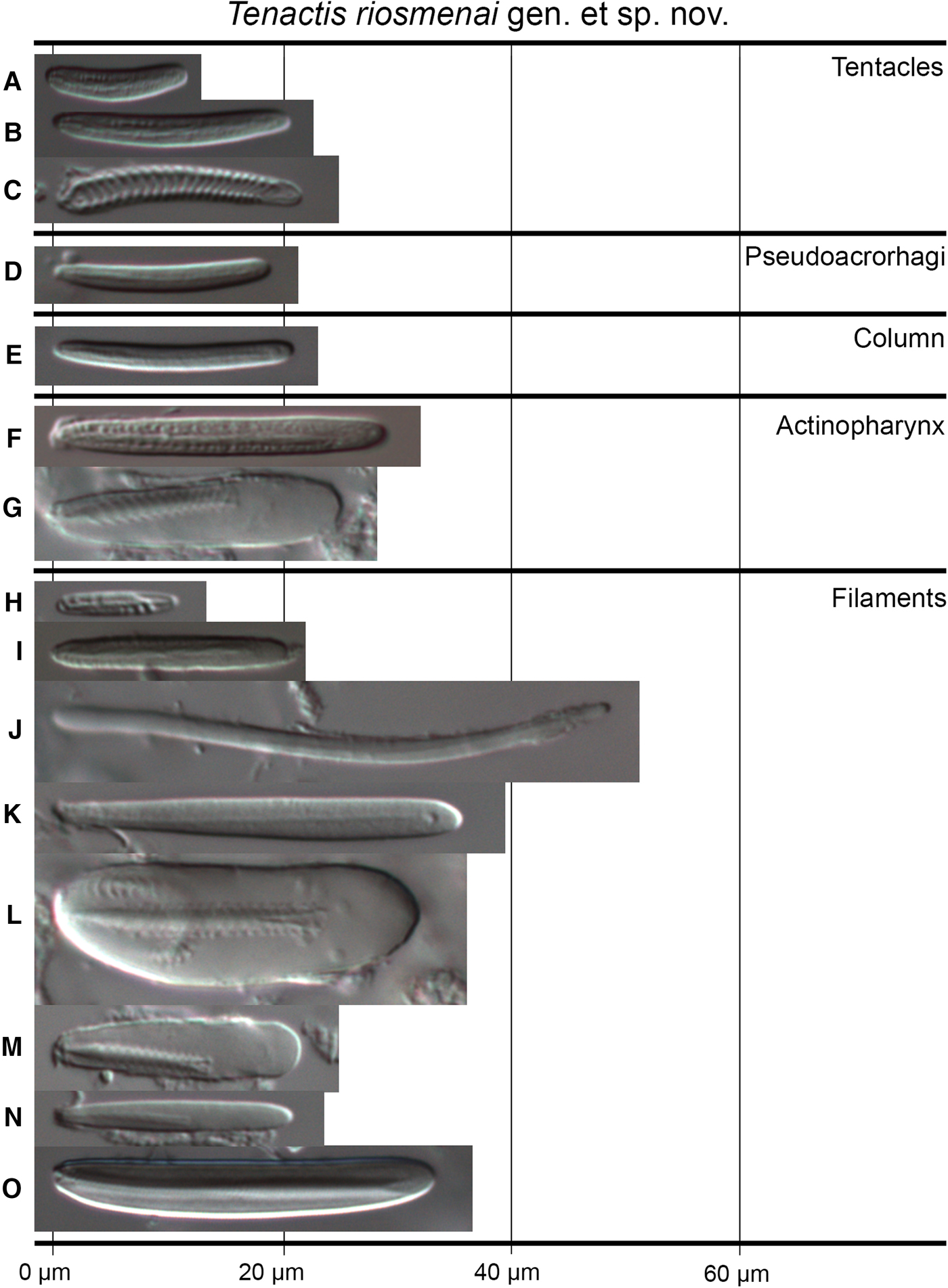
Basitrichs, b-mastigophores, p-mastigophores A and p-mastigophores A2, p-mastigophores B1 and B2a, and spirocysts. See Figure 4 and Table 1 for size and distribution.
Tenactis riosmenai gen. et sp. nov., cnidae: basitrichs (A, B, D, E, F, H, I, J); b-mastigophores (K); p-mastigophores A2 (L); p-mastigophores A (G, M); p-mastigophores B1 (N); p-mastigophores B2a (O); spirocysts (C).

Color
Living specimen with pedal disc and column orange. Verrucae and pseudoacrorhagi white (Figure 2A). Oral disc grey with orange radial lines from the mouth to the middle of oral side of tentacles of first cycle; spaces of oral disc between orange lines with white lines. Mouth orange. Tentacles grey, with several white spots along oral side of the tentacle; tentacles of second cycle with a light grey line in middle of oral side; these lines are orange in first cycle tentacles (Figure 2A). Preserved material with uniform beige column; pseudoacrorhagi white, oral disc and tentacles light grey fading to beige; tentacles maintain chromatic pattern with spots and middle radial lines but grey in colour (Figure 2B–D).
ETYMOLOGY
The species is named after the recently deceased professor Rafael Riosmena Rodríguez (Universidad Autónoma de Baja California Sur (UABCS), Mexico) for his unconditional support and assistance to YB and others, facilitating the fieldwork in which the studied specimen was collected.
GEOGRAPHIC AND BATHYMETRIC DISTRIBUTION
Tenactis riosmenai gen. et sp. nov. has been collected in the intertidal rocky shore (2 m) in La Paz Bay, southern Gulf of California, Mexico (see Figure 1).
DISCUSSION
Differential diagnosis of Tenactis gen. nov. within Actiniaria
Tenactis gen. nov. is clearly distinct because it has a single cycle of mesenteries decamerously arranged, one single pair of directives attached to a single siphonoglyph, conchula, diffuse endodermal marginal sphincter muscle, basilar muscles, p-mastigophores A2 and p-mastigophores B1 and B2a in the filaments, verrucae and cinclides.
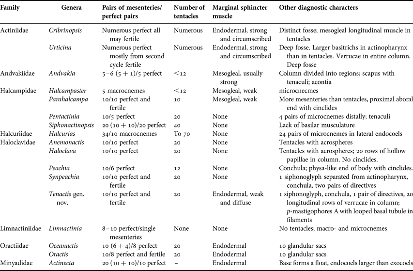
Although the most widespread mesenterial arrangement within actiniarians is the hexamerous one, penta-, hepta-, octa- and decameral representatives also exist within the order (Stephenson, Reference Stephenson1928; Carlgren, Reference Carlgren1949). However, most of the deviations to the hexamerous mesenterial pattern could be regarded as irregular variations in the hexameral method of mesenterial cycle development, affected by synchronous appearance of the first, second or later cycles (Grebelny, Reference Grebelny1982). Decamerous mesenterial arrangements in actiniarians are not very common; only 16 genera within eight families are reported to have decamerous mesenteries within the order (see Table 2).
Genera within Actiniaria with mesenteries decamerously arranged.

Within Actiniidae Rafisnesque, Reference Rafisnesque1815 two genera resemble Tenactis gen. nov. in the decamerous mesenterial arrangement: Cribrinopsis (Carlgren, Reference Carlgren1921) and Urticina (Ehrenberg, Reference Ehrenberg1834). These two genera are heterogeneous (Sanamyan & Sanamyan, Reference Sanamyan and Sanamyan2006), have a well-developed pedal disc with basilar muscles, a strong and circumscribed endodermal marginal sphincter muscle, fosse and numerous perfect mesenteries usually decamerously (or irregularly) arranged. Cribrinopsis may or may not present verrucae and pseudoacrorhagi (=marginal projections with no holotrichs) (Daly, Reference Daly2003) in the column, and all mesenteries may be fertile. Urticina bears verrucae in the entire column (although some species might lack them) and the mesenteries are fertile mostly starting from the second cycle (Sanamyan et al., Reference Sanamyan, Sanamyan and McDaniel2013); in addition, species of Urticina are characterized by having larger basitrichs in the actinopharynx than in the tentacles (Sanamyan & Sanamyan, Reference Sanamyan and Sanamyan2006). Although most of these features are present in Tenactis gen. nov. (e.g. endodermal marginal sphincter and basilar muscles, verrucae, larger basitrichs in the actinopharynx than in the column) having a single pair of directives attached to a single, strong siphonoglyph, conchula, cinclides, and p-mastigophores A2 and p-mastigophores B1 and B2a in filaments distinguishes Tenactis gen. nov. from the decamerously arranged actiniid species.
Other actinioidean genera with decamerous mesenteries are those within Oractiidae Riemann-Zürneck, Reference Riemann-Zürneck2000 (original spelling of the name must be maintained per Article 29.4 of the International Code of Zoological Nomenclature 1999 (ICZN, 1999); see Table 2). Although members of this family share with Tenactis gen. nov. having 20 tentacles and an endodermal marginal sphincter muscle, the presence of 10 distinct invaginations of the oral disc (or ‘glandular sacs’) is unique to this family (see Riemann-Zürneck, Reference Riemann-Zürneck2000; Sanamyan, Reference Sanamyan2003). Members of Limnactiniidae Carlgren, Reference Carlgren1921 are differentiated from Tenactis gen. nov. because they lack tentacles and endodermal marginal sphincter muscle, and have 8–10 perfect but single mesenteries (not pairs). Members of Minyadidae Milne Edwards, Reference Milne Edwards1857 can be easily differentiated from Tenactis gen. nov. because their base forms a float and their arrangement of mesenteries is distinct with endocoels being larger than exocoels (Carlgren, Reference Carlgren1949).
Another genus resembling Tenactis gen. nov. in a decamerous mesenterial arrangement is Halcurias (McMurrich, Reference McMurrich1893). This genus as well as Tenactis gen. nov. has a mesenterial arrangement in pairs of 10 and a single siphonoglyph. However, Halcurias – as a member of the suborder Anenthemonae Rodríguez & Daly, 2014 – has an uncommon arrangement of mesenteries within Actiniaria (Rodríguez et al., Reference Rodríguez, Barbeitos, Brugler, Crowley, Grajales, Gusmão, Häussermann, Reft and Daly2014): it has a first cycle of 10 pairs of macrocnemes, but the consecutive cycles of mesenteries (microcnemes) arise in the lateral endocoels (instead of arising in the exocoels) with the retractors oriented as in the directives. In addition, Halcurias has more mesenteries distally than proximally, no marginal sphincter muscle or a very weak mesogleal one, and no basilar muscles (reviewed in Rodríguez et al., Reference Rodríguez, Mendoza and Häussermann2013).
Despite several genera within Halcampidae Andres, Reference Andres1883 and Andvakiidae Danielssen, Reference Danielssen1890 sensu Rodríguez et al. (Reference Rodríguez, Barbeitos, Daly, Gusmão and Häussermann2012) have decamerous pairs of mesenteries (e.g. Pentactinia Carlgren, Reference Carlgren1900, Parahalcampa Carlgren, Reference Carlgren1921, Andvakia Danielssen, Reference Danielssen1890, see Table 2), members in these families usually have a mesogleal marginal sphincter muscle or none, and an elongate body differentiated into regions, often provided with some cuticulate structure (e.g. tenaculi); in addition, having acontia distinguishes members of Andvakia from Tenactis gen. nov. Within Halcampidae, Parahalcampa resembles Tenactis gen. nov. the most by having exactly 10 pairs of perfect mesenteries, low number of tentacles, cinclides in the proximal aboral end, and a single siphonoglyph; however, Parahalcampa has only 10 tentacles, a weak siphonoglyph and a weak mesogleal sphincter muscle whereas in Tenactis gen. nov. tentacles are twice the number of pairs of mesenteries (i.e. 20), has a very strong single siphonoglyph, and an endodermal marginal sphincter muscle.
Tenactis gen. nov. resembles the most to members of Haloclavidae; several haloclavid genera have 10 pairs of mesenteries, one single siphonoglyph, conchula and cinclides.
Differential diagnosis of Tenactis gen. nov within Haloclavidae
Tenactis gen. nov. is particularly distinguishable by having 10 pairs of mesenteries, a single pair of directives attached to the siphonoglyph, conchula, and p-mastigophores A2 and p-mastigophores B1 and B2a in the filaments.
Within the currently 11 valid haloclavid genera (see Table 3 in Gusmão, Reference Gusmão2016; Izumi et al., Reference Izumi, Yanagi and Fujita2016), only four present 10 pairs of mesenteries (i.e. Anemonactis Jourdan, Reference Jourdan1880; Haloclava Verrill, Reference Verrill1899; Peachia Gosse, Reference Gosse1855; and Synpeachia Yap, Fautin, Ramos & Tan, Reference Yap, Fautin, Ramos and Tan2014). Anemonactis, Haloclava and Peachia have two cycles of mesenteries (6 + 4) whereas Synpeachia has only one cycle. Distinguishing both cycles of mesenteries is easy in Peachia due to the presence of imperfect mesenteries forming the second cycle. However, in Anemonactis and Haloclava this distinction is not that clear because all 10 pairs of mesenteries are perfect. Verrill (Reference Verrill1899) in the original description of Haloclava separated the mesenteries into two cycles due to its length (i.e. first cycle larger than the second one); later on, Carlgren (Reference Carlgren1921) provided more detail about this issue, mentioning that the first cycle is attached for the entire length of the actinopharynx whereas the second cycle is only attached for about half the length of the actinopharynx. There is no information about the differences within the cycles of mesenteries in Anemonactis nor in Synpeachia (Andres, Reference Andres1881; Carlgren, Reference Carlgren1921; Stephenson, Reference Stephenson1935; Fautin, Reference Fautin, Scott and Blake1998; Yap et al., Reference Yap, Fautin, Ramos and Tan2014), and we did not revise the type material of these species. In Tenactis gen. nov. all mesenteries are attached for the entire length of the actinopharynx; although some pairs of mesenteries in T. riosmenai gen. et sp. nov. seem to be less developed than others (i.e. pairs of mesenteries #4, 6, 8 and 10 vs pairs #1, 5, 7 and 9, see Figure 3I) and could be attributable to a second cycle of mesenteries, not all the pairs of mesenteries follow this pattern (i.e. pairs of mesenteries #2 and 3, see Figure 3I). This, together with the fact that we only have one specimen and character variation is difficult to assess in this case, made us be rather conservative and not distinguish into two cycles of mesenteries in Tenactis gen. nov. In addition, because it is often hard to determine whether the perfect mesenteries are attached for the entire length of the actinopharynx in contracted or poorly preserved specimens and this information is often omitted in the literature, we do not consider it as a reliable way to differentiate the cycles of mesenteries. In this sense, we consider Anemonactis, Haloclava, Synpeachia and Tenactis gen. nov. (but Peachia) as having only one cycle of 10 pairs of mesenteries.
Although Anemonactis, Haloclava and Synpeachia resemble Tenactis gen. nov. in having one cycle of 10 pairs of perfect and fertile mesenteries and 20 tentacles, Anemonactis and Haloclava can be distinguished from Tenactis gen. nov. because they have acrospheres in the tentacles and lack a conchula whereas Tenactis gen. nov. lacks acrospheres but has conchula. The morphology of the conchula is variable, being more or less developed in different genera and species (reviewed in Gusmão, Reference Gusmão2016). The conchula in Tenactis gen. nov. is poorly developed and not as distinct in comparison to those found in Metapeachia, Peachia and Synpeachia. However, it is relatively similar to other less distinctly developed conchulae described for some specimens of Peachia hastata Gosse, Reference Gosse1855 (Stephenson, Reference Stephenson1928), Anemonactis clavus (Quoy & Gaimard, Reference Quoy and Gaimard1833) (Stephenson, Reference Stephenson1935) or Antennapeachia. Thus, we have considered the thickened folds in the distal part of the siphonoglyph in Tenactis gen. nov. as a poorly developed conchula. Synpeachia resembles Tenactis gen. nov. the most because they both have a conchula but lack acrospheres; however, in Synpeachia the conchula is well developed with five lobes and the single siphonoglyph is completely separated from the actinopharynx (forming a tube) whereas in Tenactis gen. nov. the conchula is poorly developed and the siphonoglyph is connected to the actinopharynx.
The most common internal pattern within Actiniaria is having two pairs of directives with both pairs either attached to one siphonoglyph each, or if a single siphonoglyph is present, only one pair of directives is attached to the sole siphonoglyph (Stephenson, Reference Stephenson1928; Carlgren, Reference Carlgren1949). Only several species within a few families (e.g. Actiniidae, Halcampidae, Andvakiidae Danielssen, Reference Danielssen1890) deviate from this pattern (see Carlgren, Reference Carlgren1949). Most of these instances (e.g. Anemonia Risso, Reference Risso1826; Amphianthus Hertwig, Reference Hertwig1882) are related to the species reproducing asexually or regeneration processes after injuries and as a result they have irregular arrangements of mesenteries, siphonoglyphs and directives (Stephenson, Reference Stephenson1928). Having a single siphonoglyph is relatively common within Actiniaria (e.g. members of Edwardsiidae Andres, Reference Andres1881; Halcuriidae Carlgren, Reference Carlgren1918; Halcampidae sensu Rodríguez et al. (Reference Rodríguez, Barbeitos, Daly, Gusmão and Häussermann2012)). However, having only one pair of directives is a rare condition within Actiniaria; so far, only four genera (without asexual reproduction documented) had been described as having a single pair of directives (see Haddon & Shackleton, Reference Haddon and Shackleton1893; Haddon & Duerden, Reference Haddon and Duerden1896; Carlgren, Reference Carlgren1938, Reference Carlgren1949; England, Reference England1987, Reference England and Morton1992). Haddon & Shackleton (Reference Haddon and Shackleton1893) reported one single pair of directives for specimens from Australia of Isactinia citrina (Haddon & Shackleton, Reference Haddon and Shackleton1893). Later on, England (Reference England1987) redescribed the species and noted that the specimens he studied (from Singapore), which belong undoubtedly to the same species as that from Haddon & Shackleton (Reference Haddon and Shackleton1893), had two directive pairs. In their description of Actinothoë carlgreni Haddon & Duerden, Reference Haddon and Duerden1896 (based on two specimens), the authors reported the species as having only one pair of directives attached to one siphonoglyph but without further detail (Haddon & Duerden, Reference Haddon and Duerden1896). Similarly, Carlgren (Reference Carlgren1938, Reference Carlgren1949) reported Litophellia Carlgren, Reference Carlgren1938 as having a single pair of directives but without further discussion. The monotypic genus Gymnophellia England, Reference England and Morton1992 is also reported as having a single pair of directives attached to the single siphonoglyph present; similarly, to the other genera there is no discussion about this fact (England, Reference England and Morton1992). Another genus, Scytophorus Hertwig, Reference Hertwig1882 also appears to have a single pair of directives; however, once examined closely, this appearance can be explained as an artifact of species in this genus having 14 perfect mesenteries arranged as 6 pairs + 1 couple of single perfect mesenteries, one in each side of the dorsal siphonoglyph with retractors facing towards the siphonoglyph (see Carlgren, Reference Carlgren1949; Grebelny, Reference Grebelny1982). In Tenactis riosmenai gen. et sp. nov. the presence of a single pair of directives is not an artifact of the arrangement of mesenteries as in Scytophorus nor due to asexual reproduction or regeneration: the single collected specimen has developed gametogenic tissue (spermatic cysts), does not present evidence of asexual reproduction, and it was collected without damage. Thus, having only one pair of directives is a diagnostic feature for Tenactis gen. nov. within Haloclavidae.
The cnidom (types of cnidae of a taxon) is considered useful and has been used to distinguish groups at different taxonomic levels within Actiniaria (e.g. Carlgren, Reference Carlgren1949; Schmidt, Reference Schmidt1974; Riemann-Zürneck, Reference Riemann-Zürneck1978, Reference Riemann-Zürneck1991). However, the evolutionary significance of having different types of cnidae is not yet well understood, especially because of conflicts among different cnidae terminologies but also because of the lack of homology and phylogenetic studies of the cnidae (Reft, Reference Reft2012). Nevertheless, recent work has shown that cnidae carry some phylogenetic information (e.g. Lauretta et al., Reference Lauretta, Häussermann, Brugler and Rodríguez2013; Grajales & Rodríguez, Reference Grajales and Rodríguez2014, Reference Grajales and Rodríguez2016). For example, Rodríguez et al. (Reference Rodríguez, Barbeitos, Brugler, Crowley, Grajales, Gusmão, Häussermann, Reft and Daly2014) found that having p-mastigophores B distinguished the superfamily Metridioidea (although some metridioidean genera also have p-mastigophores A) from the other superfamilies (Actinernoidea Stephenson, Reference Stephenson1922; Actinioidea Rafinesque, 1815; Actinostoloidea Carlgren, Reference Carlgren, Römer, Schaudinn, Brauer and Arndt1932; and Edwardsioidea Stephenson, Reference Stephenson1922) which usually lack this type of nematocyst. However, there are some exceptions to this observation: p-mastigophores B1 had been documented in the filaments of a few members within the superfamily Actinioidea (e.g. genera Anthopleura Duchassaing de Fonbressin & Michelotti, Reference Duchassaing de Fonbressin and Michelotti1860; Aulactinia Agassiz in Verrill, Reference Verrill1864; and Urticina, see Schmidt, Reference Schmidt1972; Sanamyan et al., Reference Sanamyan, Sanamyan and McDaniel2013). Tenactis gen. nov. has a distinct cnidom: we found p-mastigophores A2 and p-mastigophores B1 and B2a in the filaments. We discarded artifacts due to contamination because we confirmed the presence of these nematocysts in histological slides of the tissue. This is the first report of a species of Haloclavidae having p-mastigophores B1 in the filaments, the first report of an actinioidean having p-mastigophores B2a in the filaments and the fourth genera within Actinioidea having p-mastigophores B1 in the filaments. Although we cannot discard that this fact might be an artifact due to the problem of the different cnidae terminologies used along the history of the Actiniaria and the need for image-based re-evaluation of the cnidom of known species (e.g. Izumi et al., Reference Izumi, Fujita and Yanagi2017), we consider these findings remarkable; they also point out the fact that the diversity of the group is not yet well understood, in particular at the cnidae level. Another remarkable feature of the cnidom of Tenactis gen. nov. is the presence of p-mastigophores A with a looped basal tubule (p-mastigophores A2) in the filaments. This nematocyst is considered a subcategory of p-mastigophores A (A2) and it was mentioned and depicted (but not described) by Schmidt (Reference Schmidt1972, Reference Schmidt1974). Nematocysts with a looped basal tubule in the undischarged capsule are not common within the order, appearing only in seven genera within six actiniarian families (see Belém et al., Reference Belém, Herrera and Schlenz1996; Rodríguez et al., Reference Rodríguez, López-González and Daly2009; Izumi et al., Reference Izumi, Yanagi and Fujita2016). These types of mastigophores (rhabdoids sensu Schmidt (Reference Schmidt1969)) are classified into three categories according to Schmidt (Reference Schmidt1969, Reference Schmidt1974): (i) p-mastigophores C (those found within the metridioidean family Aliciidae Duerden, Reference Duerden1895, but this category also includes nematocysts without a looped basal tubule, e.g. nematocysts in Bunodeopsis Andres, Reference Andres1881 and Boloceroides Carlgren, Reference Carlgren1899); (ii) p-mastigophores B2b (those in Antipodactinidae Rodríguez, López-González & Daly, Reference Rodríguez, López-González and Daly2009; and Diadumenidae Stephenson, Reference Stephenson1920) and (iii) p-mastigophores A (those found in Actiniidae, Haloclavidae and Stichodactylidae Andres, Reference Andres1883). However, Schmidt's (Reference Schmidt1969) terminology for p-rhabdoibs (used here) does not consider the length of the shaft as a relevant character for classification (he prioritized the ultrastructure of the armature to distinguish types), because very similar capsules with similar spine armature get assigned in very different categories if the consideration between terms macro- and microbasic is used (Schmidt, Reference Schmidt1969, Reference Schmidt1974; Reft, Reference Reft2012). The p-mastigophores A with a looped basal tubule found in Tenactis gen. nov. resemble those of the column of members of Isoaulactinia Belém, Herrera & Schlenz, Reference Belém, Herrera and Schlenz1996 (Actiniidae) and those in the filaments of Antennapeachia setouchi (Izumi et al., Reference Izumi, Yanagi and Fujita2016), corresponding in our terminology to p-mastigophores A2. Because in most p-mastigophores A the basal tubule does not loop (see Schmidt, Reference Schmidt1969, Reference Schmidt1972, Reference Schmidt1974), we considered that is important to make the distinction even if Schmidt never described this subcategory in detail. Regrettably, we did not find any discharged capsule to provide further details on the ultrastructure of this type of mastigophore and make further conclusions. This is the second record of this type of nematocyst within Haloclavidae.
Tenactis gen. nov. presents a unique combination of characters within members of the family Haloclavidae. We consider that the presence of an endodermal marginal sphincter muscle, a single pair of directives, and p-mastigophores A2 and p-mastigophores B1 and B2a in filaments warrant erecting a new genus for Tenactis riosmenai gen. et sp. nov.
ACKNOWLEDGEMENTS
Special thanks to all members of the laboratory ‘Programa de Investigación para la Conservación de la Fauna Arrecifal’ (PFA) of the Universidad Autónoma de Baja California Sur, (UABCS), specially to Z. Hoffman, E. Alcalá, R. Arcos, L. Cruz, F. Calderón, D. Corona, D. Gamero, A. Tamayo, E. Medina, I. Osuna, G. González and J. Hardy for their valuable assistance during fieldwork when the specimen was collected. We also thank L. Gusmão (AMNH) and A. Reft (US National Museum of Natural History) for their valuable comments and discussions. Finally, we thank the two anonymous reviewers who helped to improve this manuscript.
FINANCIAL SUPPORT
Yamaly Barragán was supported by a master degree fellowship from the Consejo Nacional de Ciencia y Tecnología of Mexico 414444 (CONACYT). This work was conducted as part of the ‘Programa de Investigación para la Conservación de la Fauna Arrecifal’ (PFA) of the UABCS. This project was partially supported by NSF DEB-1456196 grant to E. Rodríguez, CONABIO JF190/2013 [‘Inventory of sessile marine life in the Pacific Islands of Baja California Sur’], The Gulf of California Marine Program (GCMP): UABCS-Scripps Institution of Oceanography-Centro para la Biodiversidad Marina y la Conservación [‘Ecological Monitoring Project – ProMonitor-ProMares’] and DataMares-ProMares from 1998 to date (http://datamares.ucsd.edu/eng/).