Bovine mastitis is an inflammatory response of bovine mammary glands due to a variety of factors with a high incidence and widespread occurrence (Bradley, Reference Bradley2002; Fugui et al., Reference Fugui, Xueyan, Zhengui, Zhiyong and Yun2018). It is one of the most common and serious diseases in dairy farms, causing significant financial losses to farming in many countries and regions around the world (Stuhr and Aulrich, Reference Stuhr and Aulrich2010; Hu et al., Reference Hu, Yin, Lin, Yan and Wang2015). There exist many factors that lead to mastitis, which are mainly divided into biological and abiotic factors, with microbial infection dominant (Contreras and Rodriguez, Reference Contreras and Rodriguez2011).
Staphylococcus aureus (S. aureus) is a Gram-positive bacterium and one of the main microorganisms that cause mastitis (Foster, Reference Foster2005). It exists in the daily environment and can colonize the mammary gland, skin, and respiratory mucosa (Martin and Bhanu, Reference Martin and Bhanu2012). It also has strong immunosuppressive characteristics, facultative intracellular infection and considerable drug resistance. It can cause persistent infection of cow mastitis, which is difficult to cure and has a high recurrence rate (Barkema et al., Reference Barkema, Schukken and Zadoks2006). Previous research has indicated that S. aureus can invade MAC-T cells and induce autophagosome formation, transformation of LC3I/II, and degradation of p62/SQSTM1. Furthermore, intracellular S. aureus can utilize autophagy to survive in bovine mammary epithelial cells (Geng et al., Reference Geng, Liu, Lu, Xu, Wang, Wang, Liu, Liu and Han2020).
Autophagy is essentially an emergency mechanism for cells in a high-stress environment, aiming to maintain the homeostasis of the internal environment of cells. Unlike apoptosis, autophagy does not necessarily lead to cell death (apoptosis is a process in which cells are bound to die under the control of the caspase pathway). Autophagy mainly includes the initiation of autophagosomes, the extension of autophagosome membranes, the maturation and transport of autophagosomes, the fusion of lysosomes and the degradation of autophagy lysosomes, each of which is strictly regulated by a series of autophagy-related genes (ATG: Cuervo, Reference Cuervo2004; Takuo et al., Reference Takuo, Jahangir, Alam and Noda2018; Yujuan et al., Reference Yujuan, Qinqin, Guihua, Xingpo, Yingli, Yihong and Sidang2018). According to the function of autophagy in the elimination of intracellular bacteria, it can be divided into antibacterial autophagy, non-bacterial autophagy and pro-bacterial autophagy (Mostowy, Reference Mostowy2012). Autophagy is an important weapon for cells to resist intracellular pathogens. Formation of a double-layered autophagosome around the pathogen is followed by combined action with lysosomes to degrade tha autophagosome, helping to prevent the pathogen from spreading in the host cell (Shahnazari and Brumell, Reference Shahnazari and Brumell2011). Evidence has shown that bacteria have produced a variety of mechanisms against autophagy during evolution. Some virulence factors can interact with host autophagy molecules, thereby interfering with autophagy (Shahnazari et al., Reference Shahnazari, Namolovan, Mogridge, Kim and Brumell2011) or even using host autophagy to build survival capability and promote self-growth (Niu et al., Reference Niu, Yamaguchi and Rikihisa2008). Others prevent fusion of autophagosomes and lysosomes (Chargui et al., Reference Chargui, Cesaro, Mimouna, Fareh, Brest, Naquet, Darfeuille-Michaud, Hébuterne, Mograbi and Vouret-Craviari2012), or can disguise the autophagosome as a host protein to avoid recognition of autophagy molecules (Dortet et al., Reference Dortet, Mostowy and Cossart2012).
The gene for MAPK14, p38α, is a part of the p38 MAPK family (Li et al., Reference Li, Li, Li, Lin, Hu, Meng, Wu, He and Feng2018). At present, it has been found that p38 MAPK has 5 isomers, p38α, p38β1, p38β2, p38γ, and p38δ, with about 60% amino acid homology, whose distribution is tissue-specific (Schnöder et al., Reference Schnöder, Hao, Qin, Liu, Tomic, Liu, Fassbender and Liu2016). p38α, p38β1, p38β2 exist widely in various tissues, p38γ only exists in skeletal muscle cells and p38δ mainly exists in glandular tissues (Fu et al., Reference Fu, Liu and Li2017). The MAPK pathway is widely used by eukaryotic cells to interpret extracellular stimuli (such as ionizing radiation), in particular cellular stress signals. Once activated, p38α can phosphorylate diverse substrates, which subsequently regulate a variety of cellular physiological functions (Burotto et al., Reference Burotto, Chiou, Lee and Kohn2014; Liu et al., Reference Liu, Wang, Liu, Tang, Fan, Zhao, Jiao, Wang, Sun and Lin2017). There is evidence that p38α can activate chaperone-mediated autophagy by phosphorylating the lysosomal membrane protein LAMP2A (Li et al., Reference Li, Zhu, Dou, She, Tao, Xu, Yang and Mao2017). However, another study pointed out that p38α can inhibit both basal and starvation-induced macroautophagy, either through binding to p38IP, or by phosphorylating ATG5 (Webber and Tooze, Reference Webber and Tooze2009). In addition, there is growing evidence that suggests that p38α is involved in regulating autophagy in a variety of ways. Therefore, this study investigated the intrinsic relationship of p38α in S. aureus-induced autophagy in bovine mammary epithelial cells.
Materials and methods
Antibodies and reagents
Antibodies used in this research: anti-GAPDH, anti-p38α, anti-LC3B and anti-SQSTM1/p62 were obtained from Abcam (Cambridge, MA, USA), anti-phospho-p38α (Thr180/Tyr182; DF39) were purchased from Cell Signaling Technology (Danvers, MA, USA), anti-ULK1 and phospho-ULK1(ser757) were procured from Affinity Biosciences, peroxidase-conjugated AffiniPure goat anti-mouse IgG was from ZSGB-BIO, Beijing, China and goat anti-rabbit IgG was from CWBIO, Beijing, China.
The reagents used in this study: 4′, 6-diamidino-2-phenylindole dihydrochloride DAPI and Triton × 100 were obtained from Solarbio (Beijing, China), gentamycin sulfate was from Biotopped, Beijing, China, anisomycin from Beyotime, China, SB203580 from Sigma-Aldric, USA and BCA protein assay kit from Thermo Fisher Scientific Pierce, Waltham, MA, USA).
Bacterial strains and culture conditions
S. aureus strain ATCC25923 and another three mastitis S. aureus isolates were used in this research, the preparation method comprised the following steps: first, the bacteria were inoculated on manitol salt agar and cultured at 37°C; 24 h later, a single colony was randomly selected and placed in LB broth and then placed on a shaker at 37°C, 200 rpm. After 12 h of culture, the OD600nm value of the bacteria was determined. The cultured S. aureus was kept at −80°C before use.
Cell culture
The bovine mammary epithelial cell line MAC-T was placed in a cell culture dish and cultured in Dulbecco's modified Eagle's medium (DMEM) supplemented with 10% (v/v) FBS, and placed in a cell incubator (37°C, 5% CO2). Cells cultured to monolayer fusion were used for the following experiments:
Intracellular infection model
The intracellular infection model was established as illustrated in preceding research (Neumann et al., Reference Neumann, Bruns, Rohde, Prajsnar and Schmitz2016). In brief, MAC-T was seeded on a cell culture plate, and when the cells were cultured to a monolayer confluent state, S. aureus was added with a multiplicity of infection (MOI) of 8:1 (Schnaith et al., Reference Schnaith, Kashkar, Leggio, Addicks, Kronke and Krut2007). After 2 h of co-incubation, the cells were washed 3 times with PBS before being added to fresh medium supplemented with gentamicin (50 μg/ml) and lysostaphin (10 μg/ml) for 12 min (Mohamed et al., Reference Mohamed, Sommer, Sethi, Domann and Alt2014). Extracellular fluid was gathered for bacterial culture experiments to verify that extracellular S. aureus had been completely removed (Neumann et al., Reference Neumann, Bruns, Rohde, Prajsnar and Schmitz2016). Then the cells were washed by PBS for 3 times to keep the extracellular fluid clean, and finally the cells were cultured with DMEM containing 10% FBS. Cells treated with only gentamicin and lysostaphin was set as treatment control, and cells with no treatment was set as blank control. SB203580 pretreatment for 1 h was used as the p38α inhibition group, and anisomycin pretreatment for 1 h was used as the p38α activation group.
Western blot analysis
To analyze protein expression, Western blot was implemented as described in Zhou et al., Reference Zhou, Ning, Shuai, Shuhong and Zhizhong2018. Cells were collected and lysed in radioimmunoprecipitation assay (RIPA) buffer containing 1 mm phenylmethanesulfonyl fluoride, and then were placed in the centrifuge at 4 °C for 30 min. After centrifugation at 12 000 rpm for 10 min, the supernatant was aspirated for experiment. BCA protein assay kit was used to measure the protein concentration. After heat denaturation for 5 min, 15 mg proteins were separated by 12% SDS-polyacrylamide gel electrophoresis, and moved to 0.22 μm PVDF membrane. The membrane was sealed at room temperature for 1 h with TBST (TBS of 0.1% Tween 20) containing 5% skimmed milk powder, then incubated with the primary antibodies of p38α (1: 1000), P-p38α (1: 1000), ULK1 (1: 500), p-ULK1ser757(1: 500), LC3-II (1: 1000) or P62 (1: 1000) overnight at 4°C. After being washed with TBS three times for 10 min each, the membrane continued to incubate with secondary antibody at 25 °C for 1.5 h, subsequently, ECL Western detection reagent was used to measure the target protein. Finally, the protein level was analyzed by image J.
Immunofluorescence
After treatment, the MAC-T cells were washed with PBS, then 4% paraformaldehyde was added to fix the cells. Thirty minutes later, cells were infiltrated with 5% Triton X-100 for 25 min and soaked in PBS containing 4% BSA for 1.5 h. After washing with PBS, cells were incubated successively with the primary antibodies LC3 (1: 300) at 4°C overnight and secondary antibody conjugated with Alexa Fluor 594 and CoraLite 488 at 25 °C for 1 h. After a further wash with PBS, the cells were stained with DAPI for 14 min. Finally, all slides were observed under the Leica TCS SPE confocal microscope.
Intracellular survival assay
After establishing the S. aureus intracellular infection model as described above, every two hours, the medium was replaced by that containing lysostaphin and gentamicin and placed in a cell incubator for 12 min to ensure that there was no viable S. aureus in the extracellular fluid. In order to study the influence of p38α on the intracellular survival of S. aureus, SB203580 and anisomycin were used to pre-treat for 1 h in subsequent experiments, and at 6 h and 12 h after S. aureus infection, aliquots were incubated with 0.25% (m/v) trypsin for 6 min. Cells were gathered by centrifugation, and 0.2% Triton X-100 was added to increase cell permeability and release intracellular bacteria. The bacterial solution was cultured in the medium for 24 h. Colony counting was used to determine the number of intracellular bacteria.
Image analysis
Image analysis was performed using Imaris x64 v9.5.0 (Bitplane, Switzerland). Autophagosomes per cell were quantified as previously described (Irving et al., Reference Irving, Mimuro, Kufer, Lo, Wheeler, Turner, Thomas, Malosse, Gantier, Casillas, Votta, Bertin, Boneca, Sasakawa, Philpott, Ferrero and Kaparakis-Liaskos2014) using Imaris Cell to detect cell boundaries and nuclei, followed by vesicle detection and thresholding based on autophagosome size (number of voxels), intensity (max intensity, intensity center) and the built-in ‘quality’ threshold. Colocalization analysis was performed as previously described (Irving et al., Reference Irving, Mimuro, Kufer, Lo, Wheeler, Turner, Thomas, Malosse, Gantier, Casillas, Votta, Bertin, Boneca, Sasakawa, Philpott, Ferrero and Kaparakis-Liaskos2014) using the built-in Imaris Coloc feature. Thresholding was performed using a reference image of the positive control and maintained for all subsequent analyses. Three biological replicates were examined, and for each replicate three separate fields of view were imaged per treatment, containing a minimum of 10 cells per field of view and a minimum of 50 cells per biological replicate.
Statistics analysis
All statistical analyses were performed using Statistical program version 19.0 (SPSS Inc. Chicago, IL, USA) and histograms were produced with GraphPad Prism 7.0 (GraphPad Software, CA, USA). The significance of differences among different groups were analyzed by one-way ANOVA. Data were expressed as mean ± standard deviation (sd), a level of 0.05 was accepted as significant (P < 0.05).
Results
Activation of p38α
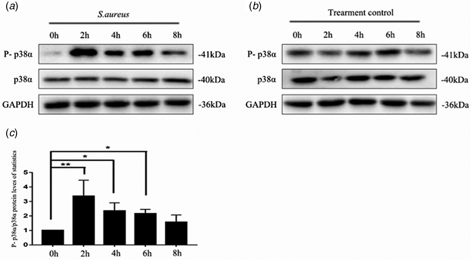
The previous study has shown that intracellular S. aureus induced autophagy in bovine mammary epithelial cell (Geng et al., Reference Geng, Liu, Lu, Xu, Wang, Wang, Liu, Liu and Han2020). To examine whether p38α was activated by S. aureus in MAC-T, we selected multiple time points in the process of S. aureus infection of MAC-T cells, and then analyzed the protein expression level of p38α. As shown in Fig. 1, it is worth noting that S. aureus induced a robust activation of p38α, indicated by its increased phosphorylation, reaching a peak at 2 h, after which the phosphorylation level gradually decreased with time.
Detection of p38α activation after S.aureus infection in MAC-T cells: (A) Western blot analysis of p38α and P-p38α of the cells in experimental group. (B) p38α and P-p38α of the cells in control group. (C) Quantitative analysis of p38α and P-p38α protein levels. 0, 2, 4, 6, 8 h after S. aureus infection with MAC-T cells, and the statistical value = (test group xh/0 h – treatment control xh/0 h) + 100% (x equals 0, 2, 4, 6, 8). Values are presented as mean ± sd, N = 3. (*, P < 0.05; **, P < 0.01).

Autophagic flux
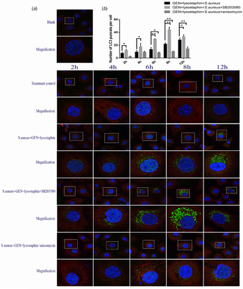
LC3 is an autophagosome-specific marker that is located on the autophagosome membrane and can be used for monitoring the autophagy process (Schaaf et al., Reference Schaaf, Keulers, Vooijs and Rouschop2016). LC3 is labeled by cellular immunofluorescence to detect positive focal points (Neumann et al., Reference Neumann, Bruns, Rohde, Prajsnar and Schmitz2016). As shown in Fig. 2A, after S. aureus infection of MAC-T cells, autophagosomes gradually increased with time, reaching a peak at 12 h. It can be seen from Fig. 2 that when the inhibitor SB203580 was added, it could effectively promote the generation of green fluorescent spots and reach the peak of the LC3 positive aggregation point at 8 h. In contrast, the addition of activator anisomycin could inhibit the formation of green fluorescent spots to a certain extent.
Effect of p38α activity on autophagy flow: (A) Autophagosomes under confocal microscopy. SB203580 and anisomycin were added to observe LC3 positive aggregation points under confocal microscope. (B) Quantitative analysis of autophagosomes. Quantification was conducted by counting 100 cells per condition. Statistics value = (Test group-Blank1) – (Treatment control – Blank2) + mean value of Blank, and average numbers of puncta per cell are presented as means ± sd, N = 3. (*, P < 0. 05; **, P < 0. 01).

Changes of LC3 and p62 proteins
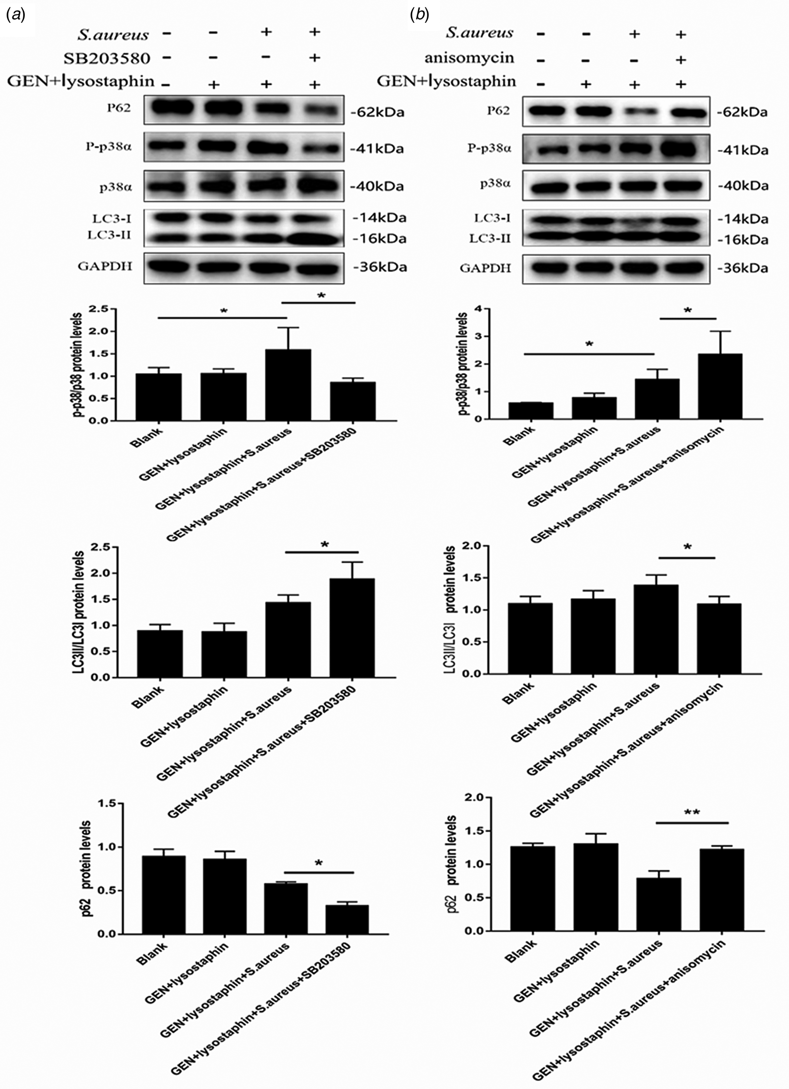
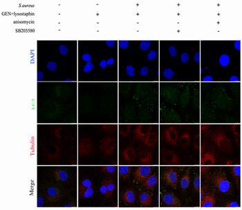
LC3-II is the lipid form and the autophagic marker of LC3 in mammals, whilst p62 is a ubiquitin-binding protein and a protein marker for autophagy, which is involved in the degradation of the autophagolysosomal system. To verify the function of p38α in S. aureus-induced inhibition of autophagy, we co-treated MAC-T cells with S. aureus and SB203580. The result indicated that, in connection with its effects on LC3-II and p62, S. aureus induced strong activation of p38α, which was indicated by its increased phosphorylation. These S. aureus-induced changes were significantly attenuated by SB203580 (Fig. 3A). However, increasing p38α activity by its small molecule activator anisomycin substantially diminished the level of endogenous LC3-II (Fig. 3B) (Zhu et al., Reference Zhu, Carneiro, Dostmann, Hewlett and Blakely2005). To reinforce these findings, we also used immunofluorescent labeling LC3 to detect LC3 positive aggregation points under confocal conditions (Fig. 4).
Effects of p38α activity on LC3 and p62 protein: (A) Effects of p38α inhibition on LC3 and p62 protein levels. SB203580 (30 μm) added to detect p38α/P-p38α, LC3-II/LC3-I, and p62 protein levels of MAC-T cells infected with S. aureus at 6th h. (B) Effects of p38α activation on LC3 and p62 protein levels. Anisomycin (10 μm) added to detect p38α/P-p38α, LC3-II/LC3-I, and p62 protein levels of MAC-T cells infected with S. aureus at 6th h. Values are presented as mean ± sd, N = 3. (*, P < 0.05; **, P < 0.01).

Effects of p38α activity on the formation of autophagosomes: After additions of SB203580 (30 μm) and Anisomycin (10 μm), LC3 positive aggregation points were detected by immunofluorescence.

Phosphorylation of ULK1ser757
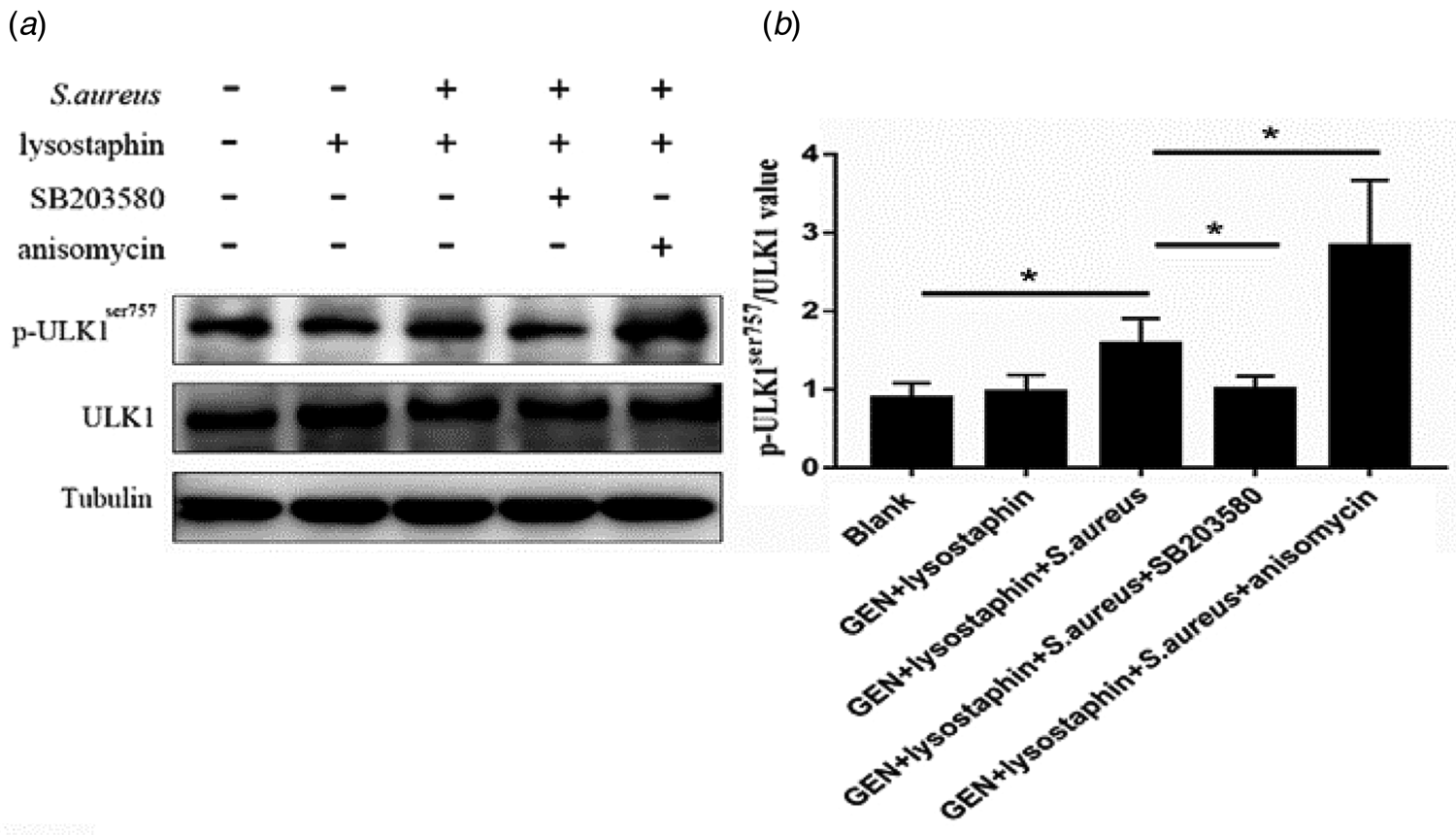
ULK1 is a homologous protein of ATG1 in mammals (Mizushima, Reference Mizushima2010). The activation of ULK1 determines the initiation of autophagy while the phosphorylation of ULK1ser757 prevents the process of cell autophagy (Kim and Guan, Reference Kim and Guan2011; Jie et al., Reference Jie, Zhao, Hu, Sun and Kong2018). So it is important to verify whether changes of p38α activity in S. aureus-infected MAC-T cells affected the phosphorylation of ULK1ser757 site and thus affected S. aureus-induced autophagy. The results showed that when S. aureus infected MAC-T cells for 6 h, the level of phosphorylation of ULK1ser757 was significantly higher than that of the blank control group. Then we used SB203580 and anisomycin to change p38α activity. As shown in Fig. 5, after adding SB203580 to inhibit p38α activity, the level of phosphorylation of the S. aureus-induced ULK1ser757 site was significantly reduced, and after the addition of anisomycin activating p38α activity, the level of phosphorylation at the ULK1ser757 site induced by S. aureus increased significantly (Fig. 5). It can be shown that p38α activation can inhibit MAC-T cell autophagy by altering the phosphorylation of ULK1ser757.
Effect of p38α activity on p-ULK1ser757 protein level: (A) Western blot analysis. SB203580 (30 μm) and anisomycin (10 μm) were used to change p38α activity, and ULK1 and p-ULK1ser757 were detected by Western Blot. (B) Quantitative analysis of p-ULK1ser757/ULK1. Values are presented as mean ± sd, N = 3. (*, P < 0.05; **, P < 0.01).

Survival of intracellular S. aureus
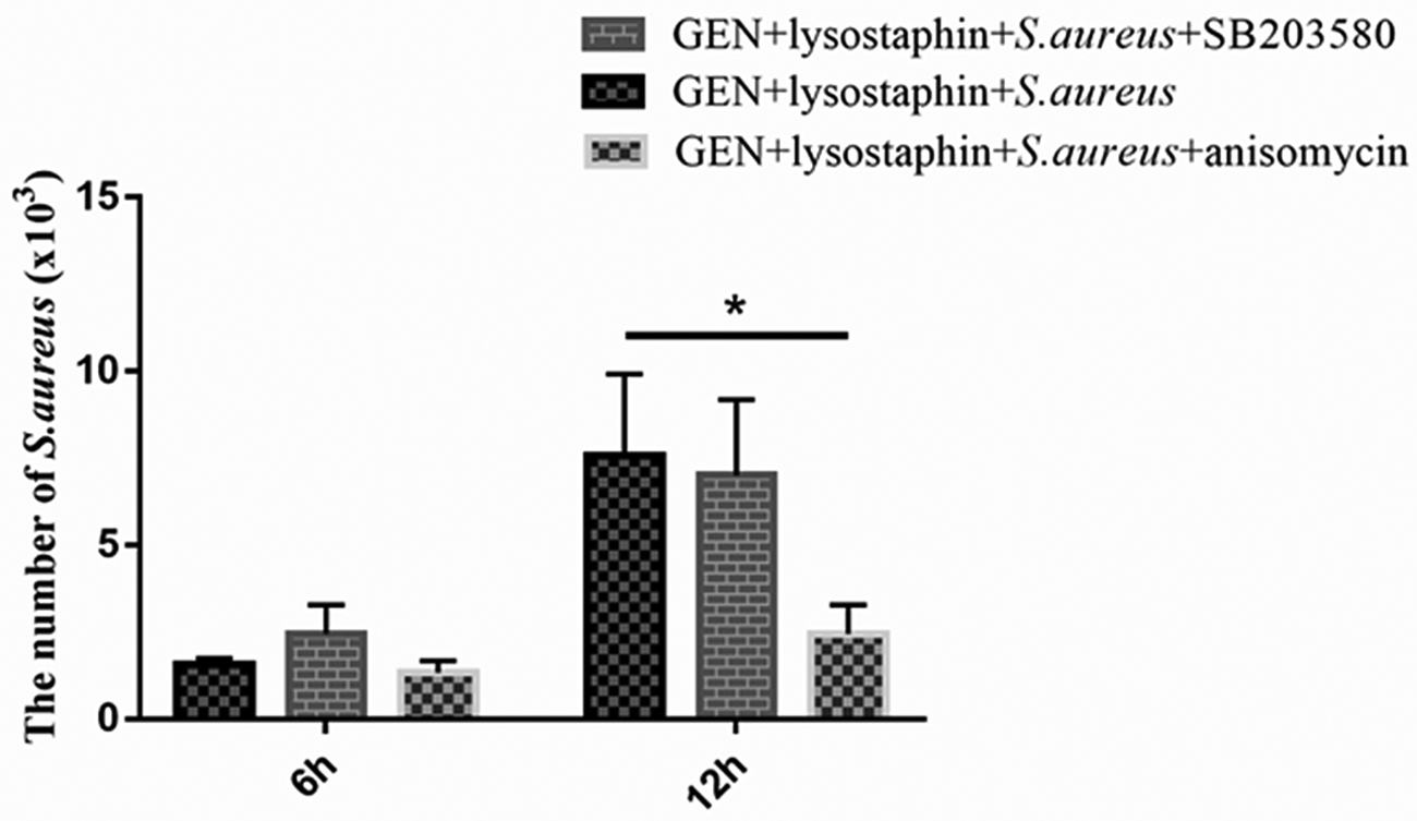
To study the impact of p38α activation on the survival of intracellular S. aureus, p38α inhibitor SB203580 (30 μm) and activator anisomycin (10 μm) were used. The intracellular bacteria were taken and counted respectively when MAC-T cells were infected by S. aureus for 6 h and 12 h. The results in Fig. 6 showed that, compared with the test group, intracellular S. aureus in the anisomycin group was significantly decreased at 12 h. This indicated that excessive activation of p38α may be disadvantageous for S. aureus survival in bovine mammary epithelial cells.
The number of intracellular S. aureus: MAC-T were infected with S. aureus at a MOI of 8:1. After pretreatment of SB203580 and anisomycin, the intracellular S. aureus load was evaluated at 6th h and 12th h after infection. Values are presented as mean ± sd, N = 3. (*, P < 0.05; **, P < 0.01).

Discussion
Autophagy is a conservative and complex process that plays a vital role in maintaining cell stability and survival, and is regulated by a variety of upstream signaling pathways, such as MAPK and AKT pathways (Ning and Karantza-Wadsworth, Reference Ning and Karantza-Wadsworth2009; Wang et al., Reference Wang, Miao, Xu, Meng, Cui, Wang, Zhu, Sha, Wei and Zhu2019). Our team has demonstrated that S. aureus can induce autophagy in bovine mammary epithelial cells (Geng et al., Reference Geng, Liu, Lu, Xu, Wang, Wang, Liu, Liu and Han2020). p38α is widely expressed in various cell types, and can be activated under related stimuli. The p38MAPK pathway plays a pivotal role in cell autophagy (Choi et al., Reference Choi, Jung and Oh2010). In rat hepatocyte cells, p38 was activated by osmotic stress to inhibit autophagy degradation of proteins; p38α was evidently phosphorylated and involved in the autophagy of S. aureus-infected mouse fibroblasts (Neumann et al., Reference Neumann, Bruns, Rohde, Prajsnar and Schmitz2016). Other studies have shown that p38α inhibits autophagy and encourages microglial inflammatory autophagy by phosphorylating ULK1 (He et al., Reference He, She, Zhang, Xu, Cheng, Yepes, Zhao and Mao2017). Therefore, we selected p38α and established whether it was activated when S. aureus infect mammary epithelial cells. As shown in the present study, p38α was significantly activated when S. aureus infected MAC-T cells, and reached a peak at 2 h, thus confirming that S. aureus infection of MAC-T cells activated the p38MAPK signaling pathway.
LC3 is a homologous protein of mammalian ATG8 (Zhai et al., Reference Zhai, Dong, Feng, Li and Hu2019). Generally, LC3I is localized in the cytoplasm but when converted to LC3II type by binding to phosphatidylethanolamineit relocates to the inner and outer surfaces of the autophagosome membrane (Yang et al., Reference Yang, Wilkie-Grantham, Yanagi, Shu, Matsuzawa and Reed2015; Ravikumar et al., Reference Ravikumar, Sarkar, Davies, Futter, Garcia-Arencibia, Green-Thompson, Jimenez-Sanchez, Korolchuk, Lichtenberg, Luo, Massey, Menzies, Moreau, Narayanan, Renna, Siddiqi, Underwood, Winslow and Rubinsztein2010). This transformation process is important for autophagy. The amount of LC3II is a key and important indicator of autophagosome formation. SQSTM1/p62 has been identified as a substrate and regulatory protein for mammalian autophagy (Pankiv et al., Reference Pankiv, Clausen, Lamark, Brech and Johansen2007). SQSTM1/p62 is continuously produced under normal conditions and is degraded by selective autophagy. In response to the accumulation of autophagy substrates, SQSTM1/p62 performs the function of connexin to deliver ubiquitinated proteins into autophagy vesicles for degradation in autolysosomes (Lamark et al., Reference Lamark, Svenningand and Johansen2017). Therefore, in our study, as shown in Fig. 3, application of the p38α inhibitor SB203580 increased the level of LC3 II/I and decreased the expression of p62 protein compared to the S. aureus group. Later, when we significantly activated p38α activity, LC3 II/I levels were significantly reduced and p62 protein levels were up-regulated. Immunofluorescence is an important method for detecting autophagy. By detecting the immunofluorescent staining of the critical autophagy protein LC3, we can assess the number of autophagosomes in the samples(Li et al., Reference Li, Li, Li, Lin, Hu, Meng, Wu, He and Feng2018). After inhibiting p38α activity, LC3 positive aggregation sites caused by S. aureus had a faster generation rate and increased number. Subsequently, the addition of anisomycin significantly activated p38α activity, which slowed down the formation of LC3 positive aggregation sites and reduced the number of LC3 positive aggregation sites. This indicated that activation of p38α has an inhibitory effect on autophagy caused by S. aureus infection of MAC-T cells.
ULK1 is a mammalian homologue of ATG1 and is the core protein of autophagy. Previous studies have found that during starvation, AMPK can phosphorylate Ser317 and Ser777 of ULK1, which directly activate autophagy. When nutrition is adequate, the ser757 of ULK1 was phosphorylated, inhibiting the activation of ULK1, thereby inhibiting the occurrence of autophagy (Kim and Guan, Reference Kim and Guan2011; Jie et al., Reference Jie, Zhao, Hu, Sun and Kong2018). When S. aureus infects cells, changes of ULK1-ATG13 signaling pathway demonstrated that S. aureus-induced autophagy and cell death could be reduced through inhibition of the ULK1 protein complex (Radhi et al., Reference Radhi, Davidson, Scott, Zeng, Jones, Tomkinson, Yu and Chan2019). At the same time, other studies have shown that activation of p38α can phosphorylate the ULK1ser757 site to inhibit the interaction between ULK1 and ATG13 and thus inhibit microglial inflammation and autophagy (He et al., Reference He, She, Zhang, Xu, Cheng, Yepes, Zhao and Mao2017). Therefore, we speculated that after S. aureus invades mammary epithelial cells, it may activate p38α to up-regulate the phosphorylation level of ULK1ser757, thereby altering the activity of ULK1 and inhibiting autophagy of bovine mammary epithelial cells. In the current study, we also found that intracellular S. aureus promoted the phosphorylation of ULK1ser757. After p38α activity was inhibited by SB203580, the phosphorylation level of ULK1ser757 decreased in contrast with the S. aureus group, whereas p38α was activated, ULK1ser757 phosphorylation was up-regulated. Therefore, we hypothesized that following S. aureus invasion of MAC-T cells, p38α was activated and then inhibited autophagy by altering the phosphorylation level of ULK1ser757.
As for the effect of p38α activity on the intracellular survival of S. aureus, the quantity of intracellular S. aureus decreased significantly after 12 h of infection when anisomycin was applied to strongly activate the activity of p38α. It indicated that excessive activation of p38α may be negative for the intracellular survival of S. aureus. In conclusion, in this study we provide a theoretical basis for S. aureus to inhibit autophagy of mammary epithelial cells so as to promote its survival in those cells. This is achieved through an inhibition of autophagy flow concomitant on activation of p38α.
Acknowledgments
The project was supported by the National Natural Science Foundation of China (31802259, 31872535), Shandong Key R&D Program (2019GNC106141), Shandong Natural Science Foundation of China (ZR2018MC027, ZR2016CQ25), China Postdoctoral Science Foundation (2018M632704, 2019T120601) and Funds of Shandong ‘Double Tops’ Program.