Introduction
Bovine tuberculosis (bTB), caused by Mycobacterium bovis (Mycobacterium tuberculosis complex), is an established zoonotic disease which affects cattle and wildlife worldwide (Buddle et al., Reference Buddle, Wedlock and Denis2006). Several wildlife species are naturally infected with M. bovis, but European wild boar (Sus scrofa) is a major reservoir host in Spain (Gortazar et al., Reference Gortazar, Vicente, Samper, Garrido, Fernández-De-Mera, Gavín, Juste, Martín, Acevedo, De La Puente and Höfle2005; Vicente et al., Reference Vicente, Höfle, Garrido, Fernandez-de-Mera, Juste, Barral and Gortazar2006). In this species, most tuberculous lesions are located in the mandibular lymph nodes (MLN) and oropharyngeal tonsils (OT) and disseminated lesions are occasionally observed in the liver, spleen, lungs and other organs (Gortazar et al., Reference Gortazar, Vicente and Gavier-Widén2003; Martín-Hernando et al., Reference Martín-Hernando, Höfle, Vicente, Ruiz-Fons, Vidal, Barral, Garrido, de la Fuente and Gortazar2007). The presence of common M. bovis genotypes in both cattle and wild boar suggests a threat of interspecies transmission of the pathogen (Gortazar et al., Reference Gortazar, Vicente, Samper, Garrido, Fernández-De-Mera, Gavín, Juste, Martín, Acevedo, De La Puente and Höfle2005).
In European wild boar as in other species, genetic factors have been associated with resistance to bTB (Acevedo-Whitehouse et al., Reference Acevedo-Whitehouse, Vicente, Gortazar, Höfle, Fernández de Mera and Amos2005; Naranjo et al., Reference Naranjo, Höfle, Vicente, Martín, Ruiz-Fons, Gortazar, Kocan and de la Fuente2006a, Reference Naranjo, Ayoubi, Vicente, Ruiz-Fons, Gortazar, Kocan and de la Fuenteb). However, limited information is available on the protective response of this and other wildlife reservoirs to mycobacterial infection. Recent studies in cultured cells and laboratory or domestic animals using functional genomic analysis have provided data toward understanding the mechanisms involved in host–pathogen interactions and host cell responses to mycobacteria (reviewed by Koul et al., Reference Koul, Herget, Klebl and Ullrich2004). However, little is known about the immune response and the genes differentially expressed in naturally infected animal populations (Naranjo et al., Reference Naranjo, Höfle, Vicente, Martín, Ruiz-Fons, Gortazar, Kocan and de la Fuente2006a, Reference Naranjo, Villar, Martín-Hernando, Vidal, Höfle, Gortazar, Kocan, Vázquez and de la Fuente2007; Thacker et al., Reference Thacker, Palmer and Waters2006). Analysis of genes differentially expressed in tissue biopsies collected from infected and uninfected individuals exposed to mycobacteria in endemic areas would contribute to the understanding of the cellular response to natural mycobacterial exposure (Naranjo et al., Reference Naranjo, Höfle, Vicente, Martín, Ruiz-Fons, Gortazar, Kocan and de la Fuente2006a; Orlova et al., Reference Orlova, Majorov, Lyadova, Eruslanov, M'lan, Greenwood, Schurr and Apt2006). These results provide information on genes that may correlate with resistance to tuberculosis and/or contribute to the development of protective responses in wildlife reservoirs of infection.
In this paper we review the application of genomic and proteomic analyses to the study of tissue-specific response and function in natural wild boar M. bovis infections and the results were analyzed considering previous reports of experimental infections in laboratory and domestic animals. For this study, we used the comparative analysis between MLN and OT, which may have different exposure to mycobacteria in wild boar, a natural reservoir of infection. These analyses may impact on the understanding of the pathobiology of M. bovis infection and the control of bTB.
Methodological approach
The wild boar data reviewed herein was obtained from the genomic and proteomic analyses in OT and MLN of M. bovis-infected and uninfected European wild boar (Naranjo et al., Reference Naranjo, Höfle, Vicente, Martín, Ruiz-Fons, Gortazar, Kocan and de la Fuente2006a, Reference Naranjo, Villar, Martín-Hernando, Vidal, Höfle, Gortazar, Kocan, Vázquez and de la Fuente2007). The analysis of differential gene expression and global protein patterns in OT and MLN of M. bovis-infected (N=4) and uninfected (N=3) European wild boar was done by suppression-subtractive hybridization (SSH) and two-dimensional gel electrophoresis followed by matrix-assisted laser desorption/ionization–time of flight mass spectrometry, respectively (Naranjo et al., Reference Naranjo, Höfle, Vicente, Martín, Ruiz-Fons, Gortazar, Kocan and de la Fuente2006a, Reference Naranjo, Villar, Martín-Hernando, Vidal, Höfle, Gortazar, Kocan, Vázquez and de la Fuente2007). Infected animals were diagnosed by pathology, culture and spoligotyping of mycobacteria (Naranjo et al., Reference Naranjo, Höfle, Vicente, Martín, Ruiz-Fons, Gortazar, Kocan and de la Fuente2006a). The extremely detailed necropsies performed, in parallel with the complete histopathology and the immediate culture of fresh lymphoid and non-lymphoid tissues warrant an optimal sensitivity to detect mycobacterial infections in mammals that almost rules out infection in the uninfected animals (Naranjo et al., Reference Naranjo, Höfle, Vicente, Martín, Ruiz-Fons, Gortazar, Kocan and de la Fuente2006a). Some of the differentially expressed genes and regulated proteins were validated by quantitative real-time RT–PCR, immunohistochemistry and protein level determinations in 15–90 samples from infected and uninfected individuals (Naranjo et al., Reference Naranjo, Höfle, Vicente, Martín, Ruiz-Fons, Gortazar, Kocan and de la Fuente2006a, Reference Naranjo, Villar, Martín-Hernando, Vidal, Höfle, Gortazar, Kocan, Vázquez and de la Fuente2007; Vidal et al., Reference Vidal, Naranjo, Mateo, Gortazar and de la Fuente2006). Protein ontology was determined using protein reference databases (http://www.hprd.org and http://www.proteinlounge.com). Results were compared between M. bovis-infected and uninfected European wild boar and between OT and MLN.
Comparison of the differential gene expression profiles in MLN and OT of M. bovis-infected and uninfected European wild boar
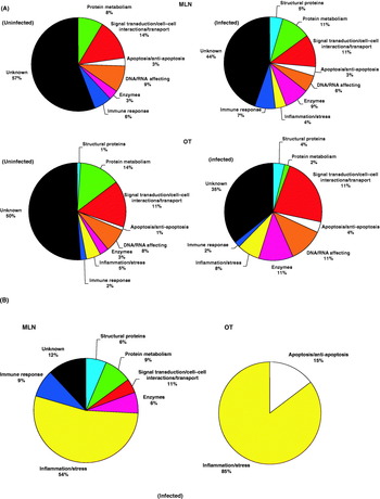
The genomic analysis resulted in 270 and 199 genes differentially regulated in MLN and OT, respectively. These genes affected cellular functions such as cell structure, protein and DNA/RNA metabolism, signal transduction, cell–cell interactions, transport, growth, inflammation/stress, immune response and enzymatic processes (Fig. 1A). However, most of the differentially expressed genes identified were of unknown function (Fig. 1A).
Protein ontology assignments of (A) unique and (B) redundant (not unique) genes differentially expressed in MLN and OT of uninfected and M. bovis-infected European wild boar. The analysis of differential gene expression in OT and MLN of M. bovis-infected and uninfected European wild boar was done by SSH (Naranjo et al., Reference Naranjo, Höfle, Vicente, Martín, Ruiz-Fons, Gortazar, Kocan and de la Fuente2006a, Reference Naranjo, Villar, Martín-Hernando, Vidal, Höfle, Gortazar, Kocan, Vázquez and de la Fuente2007). Protein ontology was determined using protein reference databases (http://www.hprd.org and http://www.proteinlounge.com).

Differences were observed in the genes differentially expressed in infected and uninfected animals. The MLN and OT of infected wild boar showed characteristics commonly found during mycobacterial infection such as the inhibition of phagosomal maturation and/or endocytosis and the expression of osteopontin (SPP1), which correlates with pathologic processes (Naranjo et al., Reference Naranjo, Höfle, Vicente, Martín, Ruiz-Fons, Gortazar, Kocan and de la Fuente2006a). On the other hand, in MLN and OT of uninfected animals common differentially expressed genes included the vitamin D receptor (VDR), complement component C3 (C3) and genes involved in Th1 response, antigen presentation and prevention of apoptosis, which may correlate with protective responses to mycobacterial infection in this species (Naranjo et al., Reference Naranjo, Höfle, Vicente, Martín, Ruiz-Fons, Gortazar, Kocan and de la Fuente2006a, Reference Naranjo, Ayoubi, Vicente, Ruiz-Fons, Gortazar, Kocan and de la Fuenteb).
The results of the differential gene expression analysis were also different between MLN and OT. Immune response genes were differentially expressed predominantly in MLN (Fig. 1A). Evidence of apoptosis in infected animals was found in OT only, whereas correlates of Th2 responses such as arginase I (ARGI) were found exclusively in MLN (Fig. 1B). Cell structure, protein metabolism and signal transduction mechanisms were predominantly affected in MLN of infected boar (Fig. 1B). Although stress/inflammatory responses were characterized in MLN and OT of wild boar infected with M. bovis (Fig. 1B), the heat shock protein (HSP) response was activated in OT while the acute phase protein (APP) response was increased in MLN of infected animals (Naranjo et al., Reference Naranjo, Villar, Martín-Hernando, Vidal, Höfle, Gortazar, Kocan, Vázquez and de la Fuente2007).
Comparison of the protein profiles in MLN and OT of M. bovis-infected and uninfected European wild boar
Proteomic analysis of MLN and OT identified differentially regulated stress/inflammatory proteins as the main difference between uninfected and M. bovis-infected boar. These proteins included annexin V, albumin and apolipoprotein A1 (ApoA1), which appeared down-regulated, whereas manganese superoxide dismutase (MnSOD) was up-regulated in MLN of infected boar (Naranjo et al., Reference Naranjo, Villar, Martín-Hernando, Vidal, Höfle, Gortazar, Kocan, Vázquez and de la Fuente2007). In OT, NADP-dependent isocitrate dehydrogenase (IDP), two isoforms of creatine kinase (CK) and MHC class II (MHC II) were up-regulated in infected animals (Naranjo et al., Reference Naranjo, Villar, Martín-Hernando, Vidal, Höfle, Gortazar, Kocan, Vázquez and de la Fuente2007). In accordance with genomic analysis, evidence of APP response and organ damage was found in MLN while MHC II up-regulation in OT of infected animals suggested macrophage activation in this tissue. The analysis of serum proteins demonstrated higher levels of MnSOD and serum amyloid A (SAA) in infected boar and ApoA1, IgG and C3 in uninfected animals (Naranjo et al., Reference Naranjo, Höfle, Vicente, Martín, Ruiz-Fons, Gortazar, Kocan and de la Fuente2006a, Reference Naranjo, Villar, Martín-Hernando, Vidal, Höfle, Gortazar, Kocan, Vázquez and de la Fuente2007; Vidal et al., Reference Vidal, Naranjo, Mateo, Gortazar and de la Fuente2006).
Similarities and differences in the response to mycobacterial infection between naturally infected European wild boar and experimentally infected laboratory and domestic animals
In our studies, we hypothesized that gene and protein response to mycobacterial infection may be different between experimentally infected laboratory and domestic animals and naturally infected wildlife species. The analysis of gene and protein profiles in MLN and OT of European wild boar naturally infected with M. bovis did result in some unique findings in these species. The differences in the response to mycobacterial infection in European wild boar when compared to experimental infections in laboratory and domestic animals may have resulted from variations in the methodological approach (global expression analysis versus SSH), or from the use of different mycobacterial species/strains, tissues (isolated bone marrow-derived macrophages and peripheral blood monocytes versus MLN and OT) and stage and condition of infection (experimental versus natural infections). Different gene expression profiles have been reported between different mycobacterial strains (Blumenthal et al., Reference Blumenthal, Lauber, Hoffmann, Ernst, Keller, Buer, Ehlers and Reiling2005; Wedlock et al., Reference Wedlock, Kawakami, Koach, Buddle and Collins2006), infected cell types (Danelishvili et al., Reference Danelishvili, McGarvey, Li and Bermudez2003) and time points after infection (Meade et al., Reference Meade, Gormley, Park, Fitzsimons, Rosa, Costello, Keane, Coussens and MacHugh2006). However, the results of our studies also suggest that the response in European wild boar and other wildlife species to natural mycobacterial infection could differ from cell responses reported previously in experimental studies. These results also suggest that the selection of biological samples may be crucial to identify biomarkers and correlates of pathology and protection in tuberculosis (Orlova et al., Reference Orlova, Majorov, Lyadova, Eruslanov, M'lan, Greenwood, Schurr and Apt2006).
Some of the differentially expressed genes that were identified in European wild boar were reported previously in other organisms. For example, the lysosomal-associated protein, which is involved in phagosomal maturation and is negatively regulated by pathogenic mycobacteria in human and mouse macrophages (Clemens and Horwitz, Reference Clemens and Horwitz1995; Koul et al., Reference Koul, Herget, Klebl and Ullrich2004) was differentially expressed in European wild boar. The ubiquitin system, involved in protein degradation and regulation of the cell cycle, was differentially expressed in OT of European wild boar as reported previously in human (Chaussabel et al., Reference Chaussabel, Semnani, McDowell, Sacks, Sher and Nutman2003; Wang et al., Reference Wang, Rought, Corbeil and Guiney2003) and cattle (Weiss et al., Reference Weiss, Evanson, Deng and Abrahamsen2004) macrophages. Other genes, such as those encoding MHC II, vascular cell adhesion molecule 1 (VCAM-1), chemokine receptor 4 (CXCR4), VDR and C3, were differentially expressed in European wild boar and in M. tuberculosis-infected mouse and human macrophages and/or bovine peripheral blood mononuclear cells (Ragno et al., Reference Ragno, Romano, Howell, Pappin, Jenner and Colston2001; Chaussabel et al., Reference Chaussabel, Semnani, McDowell, Sacks, Sher and Nutman2003; Velasco-Velazquez et al., Reference Velasco-Velazquez, Barrera, Gonzalez-Arenas, Rosales and Agramonte-Hevia2003; Keller et al., Reference Keller, Lauber, Blumenthal, Buer and Ehlers2004; Selvaraj et al., Reference Selvaraj, Chandra, Jawahar, Rani, Rajeshwari and Narayanan2004; Kim et al., Reference Kim, Park, Hwang, Kim, Han, Shim and Yim2006; Meade et al., Reference Meade, Gormley, Park, Fitzsimons, Rosa, Costello, Keane, Coussens and MacHugh2006), although contradictory results have been reported for VCAM-1 and CXCR4 expression between different organisms (Ragno et al., Reference Ragno, Romano, Howell, Pappin, Jenner and Colston2001; Shalekoff et al., Reference Shalekoff, Pendle, Johnson, Martin and Tiemessen2001; Chaussabel et al., Reference Chaussabel, Semnani, McDowell, Sacks, Sher and Nutman2003; Keller et al., Reference Keller, Lauber, Blumenthal, Buer and Ehlers2004; Naranjo et al., Reference Naranjo, Höfle, Vicente, Martín, Ruiz-Fons, Gortazar, Kocan and de la Fuente2006a).
The stress/inflammatory responses to M. bovis characterized in European wild boar, such as those mediated by annexin, SAA, ARGI, SPP1, MnSOD and MHC II, have been previously described in macrophages during in vitro and in vivo experimental infections with mycobacteria (Nau et al., Reference Nau, Liaw, Chupp, Berman, Hogan and Young1999; Aggeli et al., Reference Aggeli, Pitsavos, Brili, Hasapis, Frogoudaki, Stefanadis and Toutouzas2000; Mishra et al., Reference Mishra, Yusuf, Ali and Nath2000; Ragno et al., Reference Ragno, Romano, Howell, Pappin, Jenner and Colston2001; Koguchi et al., Reference Koguchi, Kawakami, Uezu, Fukushima, Kon, Maeda, Nakamoto, Owan, Kuba, Kudeken, Azuma, Yara, Shinzato, Higa, Tateyama, Kadota, Mukae, Kohno, Uede and Saito2003; Keller et al., Reference Keller, Lauber, Blumenthal, Buer and Ehlers2004, Reference Keller, Hoffmann, Lang, Brandau, Hermann and Ehlers2006; Koul et al., Reference Koul, Herget, Klebl and Ullrich2004; Meijer et al., Reference Meijer, Verbeek, Salas-Vidal, Corredor-Adamez, Bussmana, van der Sarc, Ottod, Geislerd and Spaink2005). However, other stress/inflammatory response molecules identified in European wild boar such as ApoA1, HSPs, CK and IDP constitute new findings.
Pathobiology of M. bovis infection in European wild boar
The results of genomic and proteomic analyses may reflect differences in the role of MLN and OT during infection and progression of bTB in European wild boar. The OT may be one of the infection routes in boar, in which mycobacteria apparently enter through the respiratory and digestive routes (Vicente et al., Reference Vicente, Höfle, Garrido, Fernandez-de-Mera, Juste, Barral and Gortazar2006). The OT may act as the first barrier for infection and the heat shock response in this tissue may be induced to protect boar from environmental mycobacteria. The finding of apoptosis in the OT of infected animals is in agreement with the hypothesis that mycobacteria prevent apoptosis in the early phase of infection to allow for efficient bacterial replication, but they may induce or are unable to prevent cell death in later phase when other organs such as MLN become infected, which would contribute to their systemic dissemination via infection of immune cells (Koul et al., Reference Koul, Herget, Klebl and Ullrich2004).
The MLN are the most affected tissue by the formation of granulomatous lesions during progression of bTB in wild boar (Gortazar et al., Reference Gortazar, Vicente and Gavier-Widén2003; Martín-Hernando et al., Reference Martín-Hernando, Höfle, Vicente, Ruiz-Fons, Vidal, Barral, Garrido, de la Fuente and Gortazar2007). The APP response in MLN of infected animals may reflect tissue injury in the granulomas. These results are in agreement with histopathology findings which suggest that the up-regulation of stress/inflammatory responses in MLN may contribute to organ damage in M. bovis-infected animals, resulting in frequent large granulomatous lesions in more than one anatomical region among juvenile wild boar (Martín-Hernando et al., Reference Martín-Hernando, Höfle, Vicente, Ruiz-Fons, Vidal, Barral, Garrido, de la Fuente and Gortazar2007).
The inflammatory/stress responses in MLN and OT of M. bovis-infected wild boar suggest active bTB in these animals. These findings, together with the histopathological characteristics of the tuberculous reaction and the associated tissue damage in various organs, indicate that at least those wild boar with large lesions and generalized infections have the potential to excrete M. bovis by several routes, which reinforces the suggestion that wild boar is a true bTB reservoir in Spain (Martín-Hernando et al., Reference Martín-Hernando, Höfle, Vicente, Ruiz-Fons, Vidal, Barral, Garrido, de la Fuente and Gortazar2007).
Implications for the control of bTB in Spain
The genes differentially expressed and the proteins up-regulated in M. bovis-infected wild boar likely reflect cell responses to mycobacterial infection. If proven specific for M. bovis infection, some of these markers could be used for the development of assays for the identification of infected animals (Vidal et al., Reference Vidal, Naranjo, Mateo, Gortazar and de la Fuente2006).
However, the genes differentially expressed and the proteins up-regulated in uninfected wild boar may represent down-regulation due to mycobacterial infection in infected animals and/or up-regulation in uninfected individuals as a result of individual resistance and/or protective response to infection. Some of the genes differentially expressed in uninfected animals, such as C3, methylmalonyl-CoA mutase (MUT) and certain MHC alleles, have been associated with resistance to bTB in European wild boar (Acevedo-Whitehouse et al., Reference Acevedo-Whitehouse, Vicente, Gortazar, Höfle, Fernández de Mera and Amos2005; Naranjo et al., Reference Naranjo, Höfle, Vicente, Martín, Ruiz-Fons, Gortazar, Kocan and de la Fuente2006a, Reference Naranjo, Ayoubi, Vicente, Ruiz-Fons, Gortazar, Kocan and de la Fuenteb). Genetic polymorphisms in these loci may be identified and correlated with resistance to bTB, thus providing a tool for the selective genetic improvement of wild boar and domestic pig populations when animal translocations and introductions are approved by regulatory authorities. Additionally, the genes differentially expressed and the proteins up-regulated in uninfected wild boar may be used as biomarkers of protective response against M. bovis infection in experimental trials to identify and characterize new vaccine candidates and vaccination strategies in this species (Buddle et al., Reference Buddle, Wedlock, Denis and Skinner2005).
Mycobacterial infections induce antibody production but the profile of immunoglobulin (Ig) expression in M. bovis-infected animals is poorly understood (Waters et al., Reference Waters, Palmer, Thacker, Bannantine, Vordermeier, Hewinson, Greenwald, Esfandiari, McNair, Pollock, Andersen and Lyashchenko2006). Experiments in wild boar provided evidence of Ig differential expression in OT and MLN of M. bovis-infected and uninfected wild boar (Naranjo et al., Reference Naranjo, Höfle, Vicente, Martín, Ruiz-Fons, Gortazar, Kocan and de la Fuente2006a) and in mesenteric lymph nodes of Iberian red deer infected with M. bovis (I. G. Fernández de Mera et al., Reference Fernández de Mera, Pérez de la Lastra, Ayoubi, Naranjo, Kocan, Gortazar and de la Fuente2007 submitted for publication). Serum IgG levels were also increased in uninfected boar (Vidal et al., Reference Vidal, Naranjo, Mateo, Gortazar and de la Fuente2006). These results suggest that antibody responses against M. bovis may be important in natural infections of wildlife species and may be used for bTB surveillance and treatment monitoring (Lyashchenko et al., Reference Lyashchenko, Greenwald, Esfandiari, Olsen, Ball, Dumonceaux, Dunker, Buckley, Richard, Murray, Payeur, Andersen, Pollock, Mikota, Miller, Sofranko and Waters2006; Waters et al., Reference Waters, Palmer, Thacker, Bannantine, Vordermeier, Hewinson, Greenwald, Esfandiari, McNair, Pollock, Andersen and Lyashchenko2006).
Finally, the results discussed here may be relevant for vaccination strategies against bTB. In addition to systemic immune response activation, pre-exposure vaccines may be developed to target tonsil protective response to prevent infection by the pathogen. Post-exposure therapeutic vaccines may need to stimulate immune response at the lymph node to limit the multiplication, dissemination and reactivation of mycobacteria. Oral vaccines for the control of bTB in wildlife may be developed with these criteria to prevent M. bovis infection and transmission (Cross et al., Reference Cross, Buddle and Aldwell2007).
Conclusions
In summary, we have focused our review on research on the characterization of gene expression and protein profiles in naturally infected tissues of wildlife reservoirs of bTB, instead of using experimental infections in cultured cells or laboratory and domestic animals. The results reviewed herein show similarities and differences between naturally M. bovis-infected tissues of wildlife reservoir species and those reported in cell culture or laboratory and domestic animals. Very little information is available on the response of wildlife species to mycobacterial infection (Lyashchenko et al., Reference Lyashchenko, Greenwald, Esfandiari, Olsen, Ball, Dumonceaux, Dunker, Buckley, Richard, Murray, Payeur, Andersen, Pollock, Mikota, Miller, Sofranko and Waters2006; Naranjo et al., Reference Naranjo, Höfle, Vicente, Martín, Ruiz-Fons, Gortazar, Kocan and de la Fuente2006a, Reference Naranjo, Ayoubi, Vicente, Ruiz-Fons, Gortazar, Kocan and de la Fuenteb, Reference Naranjo, Villar, Martín-Hernando, Vidal, Höfle, Gortazar, Kocan, Vázquez and de la Fuente2007; Thacker et al., Reference Thacker, Palmer and Waters2006; Vidal et al., Reference Vidal, Naranjo, Mateo, Gortazar and de la Fuente2006) although we can anticipate that this information will increase in the near future (Ballesteros et al., Reference Ballesteros, Pérez de la Lastra and de la Fuente2007). Despite difficulties associated with work with naturally infected wildlife species such as the number of animals available, the potential presence of other infectious agents and the differences in the stage of infection between individuals, the results discussed in this review support the importance of studying naturally infected tissues in wildlife species. The understanding of the response to natural M. bovis infection may be critical to the development of effective strategies for the control of bTB. In particular, the response of tissues that play different roles during mycobacterial infection may be relevant to understand the pathobiology of M. bovis infection and to design effective vaccines for the control of bTB in wildlife reservoirs.
Acknowledgments
This work was supported by projects ‘Control of Tuberculosis in Wildlife’ of Grupo Santander (to C.G. and J.F.), AGL2005-07401, FAU2006-00017, PAI 06-0046-5285, FAU 2006-00017-C03-01 and Fondo Europeo de Desarrollo Regional (FEDER) (Spain). V.N. is funded by Junta de Comunidades de Castilla – La Mancha (JCCM), Spain.