Introduction
Lichens are composed of a fungal (mycobiont) and a photosynthetic partner (photobiont). It is estimated that less than 150 species of photobiont are described so far from lichens (Honegger Reference Honegger and Deising2009). About 85% of lichen-forming fungi associate with green algae, and unicellular Trebouxia spp. are among the most common photobionts being found in c. 20% of all lichen species (Friedl & Büdel Reference Friedl, Büdel and Nash1996). Terms ‘selectivity’ and ‘specificity’ refer to symbiotic associations and are differently defined in the literature, leading to confusion as summarized by Honegger (Reference Honegger and Nash2008). According to Galun & Bubrick (Reference Galun, Bubrick, Linskens and Heslop-Harrison1984) ‘selectivity’ is the preferential association between symbiotic partners while ‘specificity’ means interaction with absolute exclusivity. Smith & Douglas (Reference Smith and Douglas1987) defined ‘specificity’ as the degree of taxonomic difference between partners with which an organism associates, and used ‘selectivity’ to refer to their availability in natural ecosystems. Beck et al. (Reference Beck, Kasalicky and Rambold2002) proposed to use the term ‘selectivity’ for the characterization of the range of possible partners that can be selected by this biont (viewed from the perspective of one biont only), while ‘specificity’ should be used for the symbiotic association as a whole. Later, Yahr et al. (Reference Yahr, Vilgalys and DePriest2006) defined ‘specificity’ as the phylogenetic range of compatible partners for a given symbiont, and ‘selectivity’ as the frequency of association with any of the compatible partners. Here we refer to the term ‘specificity’ as the number of partners that can be selected by one biont, and ‘selectivity’ as the degree of preferential interaction between symbiotic partners. Many lichens form vegetative propagules that enable the joint propagation of fungal and algal partners, such as soredia and isidia. In others, however, both bionts disperse as separate units. In lichens discharging sexual fungal spores, the re-lichenization has to be established with an appropriate algal strain in each generation de novo (Beck et al. Reference Beck, Friedl and Rambold1998; Sanders & Lücking Reference Sanders and Lücking2002). Although lichen-forming algae in the genus Trebouxia Puymaly were postulated not to occur in free-living stages (Ahmadjian Reference Ahmadjian1988, Reference Ahmadjian1993), a limited number of reports on free-living Trebouxia spp. are available (e.g. Tschermak-Woess Reference Tschermak-Woess1978; Bubrick et al. Reference Bubrick, Galun and Frensdorff1984; Mukhtar et al. Reference Mukhtar, Garty and Galun1994; Sanders Reference Sanders2005). However, free-living Trebouxia spp. are neither an abundant nor a dominant element of the community, and in most ecosystems the most common aerophilic algae are non-symbiotic algae (Honegger Reference Honegger and Deising2009). A germinating fungal spore may survive on a substratum for a longer period of time if it is able to form an association with a non-compatible photobiont. This temporary state lasts until the appropriate algal strain is available (Ott Reference Ott1987). In the past decade, our knowledge of fungal-algal associations has improved with an increase in the number of studies that have focused on the selectivity and population structure of photobionts (e.g. Beck et al. Reference Beck, Friedl and Rambold1998; Piercey-Normore & DePriest Reference Piercey-Normore and DePriest2001; Guzow-Krzemińska Reference Guzow-Krzemińska2006; Piercey-Normore Reference Piercey-Normore2006; Hauck et al. Reference Hauck, Helms and Friedl2007; Muggia et al. Reference Muggia, Grube and Tretiach2008; Wornik & Grube Reference Wornik and Grube2010). These studies provide information on the potential range of photobiont partners that associate with lichen fungi.
Two different experimental approaches are used to investigate selectivity of the lichen symbionts. The first one involves in vitro resynthesis of the axenically cultured mycobiont with different photobiont species. Another approach is based on the analysis of the specimens collected in different geographical regions and the identification of the symbionts present in the lichen thalli (Honegger Reference Honegger and Nash1996, Reference Honegger and Nash2008). Our present knowledge on the range of acceptable photobionts is mainly based on this type of investigation, especially as molecular approaches facilitate identification of algal bionts in thalli and resolve relationships among photobionts. As summarized by Honegger (Reference Honegger and Nash2008), available data suggest that morphologically advanced lichen taxa are specific to moderately specific as most green-algal lichen-forming fungi associate with different genotypes of one photobiont species (e.g. Ohmura et al. Reference Ohmura, Kawachi, Kasaie, Watanabe and Takeshita2006; Hauck et al. Reference Hauck, Helms and Friedl2007). However, some mycobionts may accept a wide range of Trebouxia strains (e.g. Blaha et al. Reference Blaha, Baloch and Grube2006; Guzow-Krzemińska Reference Guzow-Krzemińska2006). Low specificity and selectivity have been suggested as a strategy that facilitates lichen symbionts surviving unfavourable environmental conditions (e.g. Romeike et al. Reference Romeike, Friedl, Helms and Ott2002; Piercey-Normore Reference Piercey-Normore2006).
Schaper & Ott (Reference Schaper and Ott2003) proposed a model of the lichenization process for which they recognized different levels of compatibility of the symbionts. In vitro studies give us the opportunity to observe initial stages of the lichenization process. The first step is pre-contact that takes place prior to physical contact. Symbionts are regarded as compatible if they enter the second stage of development, characterized by an early, tight contact of the mycobiont and photobiont cells and followed by the envelopment of the algal cells by the fungal hyphae (Ahmadjian et al. Reference Ahmadjian, Jacobs and Russell1978; Galun Reference Galun and Galun1988; Joneson & Lutzoni Reference Joneson and Lutzoni2009). The process begins with simple wall-to-wall contact (appressoria) and is followed by the formation of haustoria-like structures that is a further step in the establishment of a symbiotic contact between compatible symbionts. Initial contact may also be characterized by the formation of mucilage and is followed by the development of soredia-like structures embedded in a gelatinous matrix. However, with decreasing compatibility of the partners, the recognition process is much slower and for less compatible bionts only a loose structure of the algal-fungal association is formed (with less intimate contact between symbionts). For non-compatible photobionts, no recognition is observed and the developmental stages of initial lichenization do not occur (Schaper & Ott Reference Schaper and Ott2003).
Protoparmeliopsis muralis [syn. Lecanora muralis (Schreb.) Rabenh.] is a green-algal lichen containing Trebouxia photobionts [note the validity of the name Protoparmeliopsis muralis is under discussion (Laundon Reference Laundon2010)]. A recent molecular study showed that P. muralis forms a strongly supported monophyletic group with other lobate species (Pérez-Ortega et al. Reference Pérez-Ortega, Spribille, Palice, Elix and Printzen2010). It is a widespread lichen that can colonize different types of substrata. Numerous apothecia are always present on the mature thalli of this lichen which suggests predominant propagation of the lichen fungus by meiotic fungal spores. It was previously found that different photobionts are compatible algal partners for P. muralis, but these are limited to the genus Trebouxia. Based on ITS rDNA diversity, the following photobionts have so far been identified in the thalli of this lichen: Trebouxia asymmetrica, T. gigantea, T. cf. impressa, T. incrustata and an unidentified lineage of Trebouxia named T. sp. ‘muralis I’ (Guzow-Krzemińska Reference Guzow-Krzemińska2006). The low level of specificity and selectivity may constitute an important reason why Protoparmeliopsis muralis is one of the most successful urban lichens in the world.
The major objective of this study was to isolate the photobionts and mycobiont from Protoparmeliopsis muralis thalli and test the optimal conditions for in vitro growth of the mycobiont. Moreover, it was our aim to observe re-lichenization events with compatible, and presumably incompatible, algae. For this reason, an artificial resynthesis of the P. muralis mycobiont with different photobionts was performed.
Materials and Methods
Selected specimens
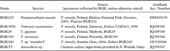
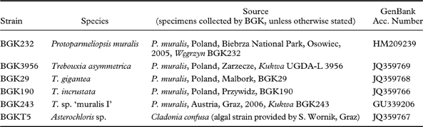
For the mycobiont isolation we used a specimen of Protoparmeliopsis muralis (BGK 232) collected in Osowiec, which is located in the Biebrza National Park (Poland). This particular specimen contained Trebouxia sp. ‘muralis I’ as the photobiont. Other specimens of P. muralis were used for photobiont isolation (as listed in Table 1). The following algae were used in resynthesis experiments: Trebouxia asymmetrica Friedl & Gärtner, T. gigantea (Hildreth & Ahmadjian) Gärtner, T. incrustata Ahmadjian & Gärtner, Trebouxia sp. ‘muralis I’, and Asterochloris sp. (isolated from Cladonia confusa R. Sant. and kindly provided by S. Wornik, Graz). The strains used in the experiments are deposited at the Department of Molecular Biology of the University of Gdańsk, and detailed information on these strains is given in Table 1.
List of mycobiont and algal species used in resynthesis experiments with details of the lichens specimens that were used for the isolation of the biont. GenBank accession numbers of the ITS rDNA sequences obtained in this study are provided.
Isolation and culture of photobionts
Prior to isolation, the lichens were examined under a stereomicroscope and pieces of thalli were washed in sterile water with Tween 80. Photobionts were isolated following the thallus fragmentation procedure of Yamamoto (Reference Yamamoto1990), modified according to Stocker-Wörgötter (Reference Stocker-Wörgötter, Kranner, Beckett and Varma2002). The fragments were inoculated on agar slants in tubes containing solid medium. After 4–6 weeks the algae were transferred to a sterile Petri dish containing solid medium with a sterile inoculation loop. The photobionts were sub-cultured on Bold's Basal Medium (BBM) (Deason & Bold Reference Deason and Bold1960; Bischoff & Bold Reference Bischoff and Bold1963) and Modified Bold's Basal Medium (MBB) (Friedl Reference Friedl1989) in order to provide optimal conditions for the growth of algae (from 2 to 5 replicates on each medium). The cultures were grown in a culture chamber under a light-dark cycle at 20°C for 14 h and 15°C for 10 h, and a light intensity of 50–100 µmol m–2 s–1 (standard conditions).
Isolation and culture of mycobiont
The mycobiont was isolated from the spores using the Ahmadjian method (Ahmadjian Reference Ahmadjian1993), with an additional pre-washing step of the apothecia in water (a small drop of Tween 80 detergent was added to bi-distilled water in a 50 ml beaker). In order to remove contaminations and dirt particles by rotations the beaker was placed on a magnetic stirrer.
The cultures obtained were multi-spore cultures. The mycobiont was sub-cultured on the following media: the modified Bold's Basal Medium with 1% glucose (Deason & Bold Reference Deason and Bold1960; Bischoff & Bold Reference Bischoff and Bold1963), Murashige-Skoog (Stocker-Wörgötter Reference Stocker-Wörgötter2001a ), G-LBM (Brunauer et al. Reference Brunauer, Hager, Grube, Türk and Stocker-Wörgötter2007) and Saboraud 2% glucose medium (Fluka N. 84086). The cultures were kept in the dark in the culture chamber at 20°C for 14 h and 10°C for 10 h. Well-developed mycelia were used for further resynthesis experiments.
Resynthesis of Protoparmeliopsis muralis with different photobionts
Axenically grown mycelia were used for the resynthesis experiments and different algal species were combined with the mycobiont. Fungal colonies were homogenized in sterile double-distilled water with a pestle in a mortar. Then a small amount of algal cells (about 1/3 of the volume of the mycobiont suspension) was taken from an axenic culture and added to the homogenized mycobiont. The suspension containing mixed bionts was transferred with a Pasteur pipette to a new Petri dish containing nutrient media. The following media were used for resynthesis experiments: Murashige-Skoog (Stocker-Wörgötter Reference Stocker-Wörgötter2001a ), G-LBM (Brunauer et al. Reference Brunauer, Hager, Grube, Türk and Stocker-Wörgötter2007), Saboraud 2% glucose medium (Fluka N. 84086) and BBM (Deason & Bold Reference Deason and Bold1960; Bischoff & Bold Reference Bischoff and Bold1963) enriched with 0·5% mannitol. The cultures were kept in the culture chamber under a day/night cycle at 20°C for 14 h and 10°C for 10 h and a light intensity of 50–100 µmol m–2 s–1 (standard conditions). The effect of the growth of the bionts in the resynthesis culture was observed with the stereomicroscope every week and assessed visually.
Microscopic investigations
In order to examine the identity of isolated photobionts, light microscopy studies were performed using a Nikon Eclipse E800 microscope. The identification of algae was carried out following a determination key by Ettl & Gärtner (Reference Ettl and Gärtner1995).
Scanning electron microscopy (SEM)
The sample, a small piece of the resynthesis culture, was attached to the holder, frozen in liquid nitrogen and sputtered with platinum. Afterwards, the selected specimens from the resynthesis experiments were investigated with a JEOL JSM-7401F Field Emission Scanning Electron Microscope (Biology Centre - Institute of Parasitology, Ceske Budejovice, Czech Republic) at an accelerating voltage of 3·0 kV.
Control samples, aposymbiotically grown Trebouxia sp. ‘muralis I’ and P. muralis, were cut from the agar medium and chemically fixed according to the following protocol: samples were fixed for 2 h at room temperature and then overnight at 4°C in the solution consisting of 5% glutaraldehyde and 0·5 M cacodylate buffer, pH 7·0. The samples were then washed twice with 0·5 M cacodylate buffer, pH 7·0, followed by washing in distilled water (twice for 15 min). In the next step the samples were gradually dehydrated in 10%, 20%, 30% ethanol (15 min each), then 50%, 75% ethanol (30 min each), followed by 96% and finally twice in anhydrous ethanol (1 h each). Dehydrated samples were subjected to critical point drying using EMITECH K850 at 35–40°C and a pressure of 73 Atm. The samples were then sputtered with gold. The selected control specimens were investigated using a Philips XL30 Scanning Electron Microscope (Laboratory of Electron Microscopy, University of Gdańsk, Gdańsk, Poland) at an accelerating voltage of 15·0 kV.
DNA analyses
Total genomic DNA was extracted directly from algal and fungal cultures using the CTAB method (Armaleo & Clerc Reference Armaleo and Clerc1995). DNA was re-suspended in sterile distilled water. PCR amplifications were performed using Tetrad MJ Research thermal cycler or GeneAmp 9700 PCR Thermal Cycler (Applied Biosystems). One unit of RedTaq polymerase (Sigma) was used for each 50 µl of master mix containing 5 µl of 10× Taq polymerase reaction buffer, 0·2 mM of each of the four dNTP's and 0·5 µM of each primer. The primers Al1500bf (Helms et al. Reference Helms, Friedl, Rambold and Mayrhofer2001) and LR3 (Friedl & Rokitta Reference Friedl and Rokitta1997) were used as PCR primers for amplification of algal nuclear Internal Transcribed Spacer (ITS) ribosomal DNA (rDNA). In several cases, a semi-nested-PCR was performed using Al1500bf (Helms et al. Reference Helms, Friedl, Rambold and Mayrhofer2001) and ITS4M (Guzow-Krzemińska Reference Guzow-Krzemińska2006) primers. For the fungal DNA, ITS1F (Gardes & Bruns Reference Gardes and Bruns1993) and ITS4 (White et al. Reference White, Bruns, Lee, Taylor, Innis, Gelfand, Sninsky and White1990) primers were used. After an initial denaturation step at 95°C for 10 min, the PCR ran for 35 cycles (95°C for 1 min, 51°C for 40 s, 72°C for 1 min) with a final extension step at 72°C for 10 min. PCR products were resolved on agarose gels in order to determine DNA fragment lengths and then purified using High Pure PCR Product Purification Kit (Roche) and sequenced using Macrogen (Korea) sequencing service (www.macrogen.com). For sequencing of the algal ITS region, the following primers were employed: ITS1 (White et al. Reference White, Bruns, Lee, Taylor, Innis, Gelfand, Sninsky and White1990) and ITS4M (Guzow-Krzemińska Reference Guzow-Krzemińska2006). For sequencing of fungal DNA, ITS1F (Gardes & Bruns Reference Gardes and Bruns1993) and ITS4 (White et al. Reference White, Bruns, Lee, Taylor, Innis, Gelfand, Sninsky and White1990) primers were used.
The newly determined ITS rDNA sequences from photobionts and mycobionts of P. muralis (Table 1) were compared to the sequences available in GenBank using BLAST (Altschul et al. Reference Altschul, Gish, Miller, Myers and Lipman1990).
Results
The symbionts
The following Trebouxia species were isolated from Protoparmeliopsis muralis thalli and grown aposymbiotically: T. asymmetrica, T. gigantea, T. incrustata and unknown Trebouxia sp. ‘muralis I’ (here named after Guzow-Krzemińska Reference Guzow-Krzemińska2006). Despite several attempts, we failed to isolate T. impressa from the thalli of P. muralis although it was previously reported to be associated with this lichen (Guzow-Krzemińska Reference Guzow-Krzemińska2006). However, this particular photobiont was identified in the thallus of P. muralis only once based on ITS rDNA sequencing. The specimen that was reported to contain T. impressa as photobiont was collected in 2003 (Guzow-Krzemińska Reference Guzow-Krzemińska2006), and during the present work it was too old to provide a source of the algal strain and it failed to grow in culture.
The algal cultures were identified under a light microscope by comparing morphological features and by using a determination key (Ettl & Gärtner Reference Ettl and Gärtner1995). Moreover, the algal ITS rDNA region was amplified, sequenced and the identity of the isolates was checked using BLAST search (Altschul et al. Reference Altschul, Gish, Miller, Myers and Lipman1990). ITS rDNA sequences of algal strains that were used for the resynthesis experiments were deposited in GenBank and their accession numbers are provided in Table 1.
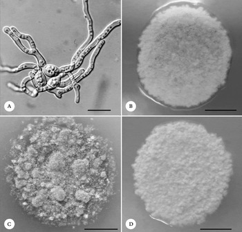
The mycobiont cultures obtained were poly-spore cultures as the mycelium was grown from multiple spores released from a single apothecium (Fig. 1A). The mycobiont investigated was associated in nature with Trebouxia sp. ‘muralis I’ that was identified in the thallus of this specimen based on ITS rDNA sequencing. Protoparmeliopsis muralis was successfully sub-cultured on Murashige-Skoog (Stocker-Wörgötter Reference Stocker-Wörgötter2001a ), G-LBM (Brunauer et al. Reference Brunauer, Hager, Grube, Türk and Stocker-Wörgötter2007) and BBM medium (Deason & Bold Reference Deason and Bold1960; Bischoff & Bold Reference Bischoff and Bold1963) enriched with glucose. The mycelia were well developed on these media (Fig. 1B–D), but the growth of the mycobiont was slightly inhibited on Saboraud 2% glucose medium. The identity of the sub-cultured mycobiont was confirmed by BLAST analysis of its nuclear ITS rDNA sequence (GenBank accession number HM209239).
Mycobiont isolated from lichen Protoparmeliopsis muralis. A, germinating fungal spores; B, mycelium grown on BBM medium enriched with 1% glucose after 3 months of incubation; C, mycelium grown on MS medium after 3 months of incubation; D, mycelium grown on G-LBM medium after 3 months of incubation. Scales: A=20 µm; B–D=1 cm.

Resynthesis experiments
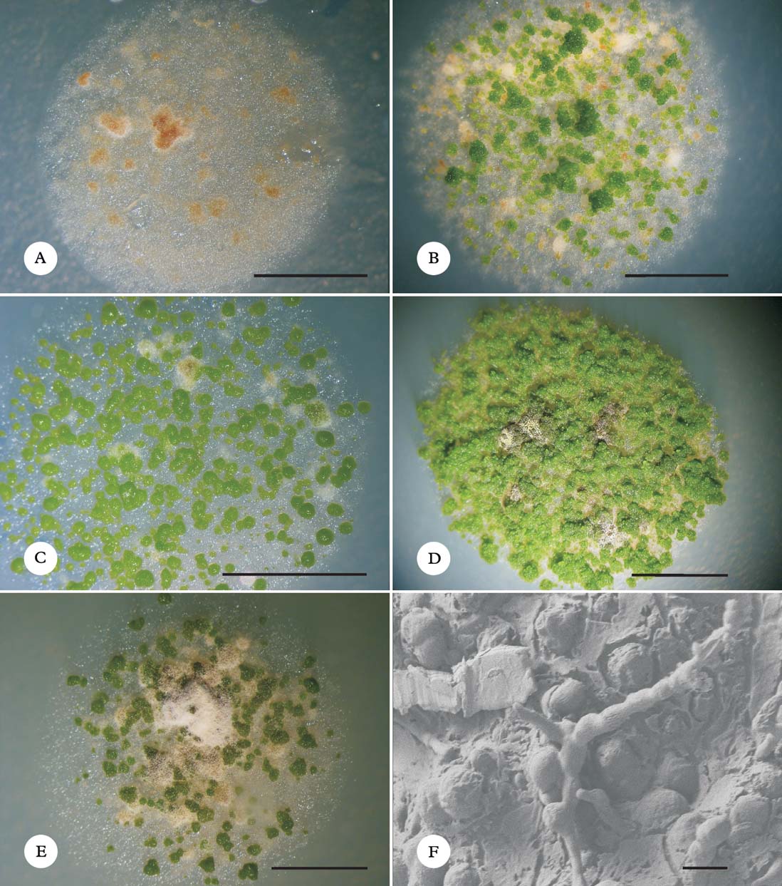
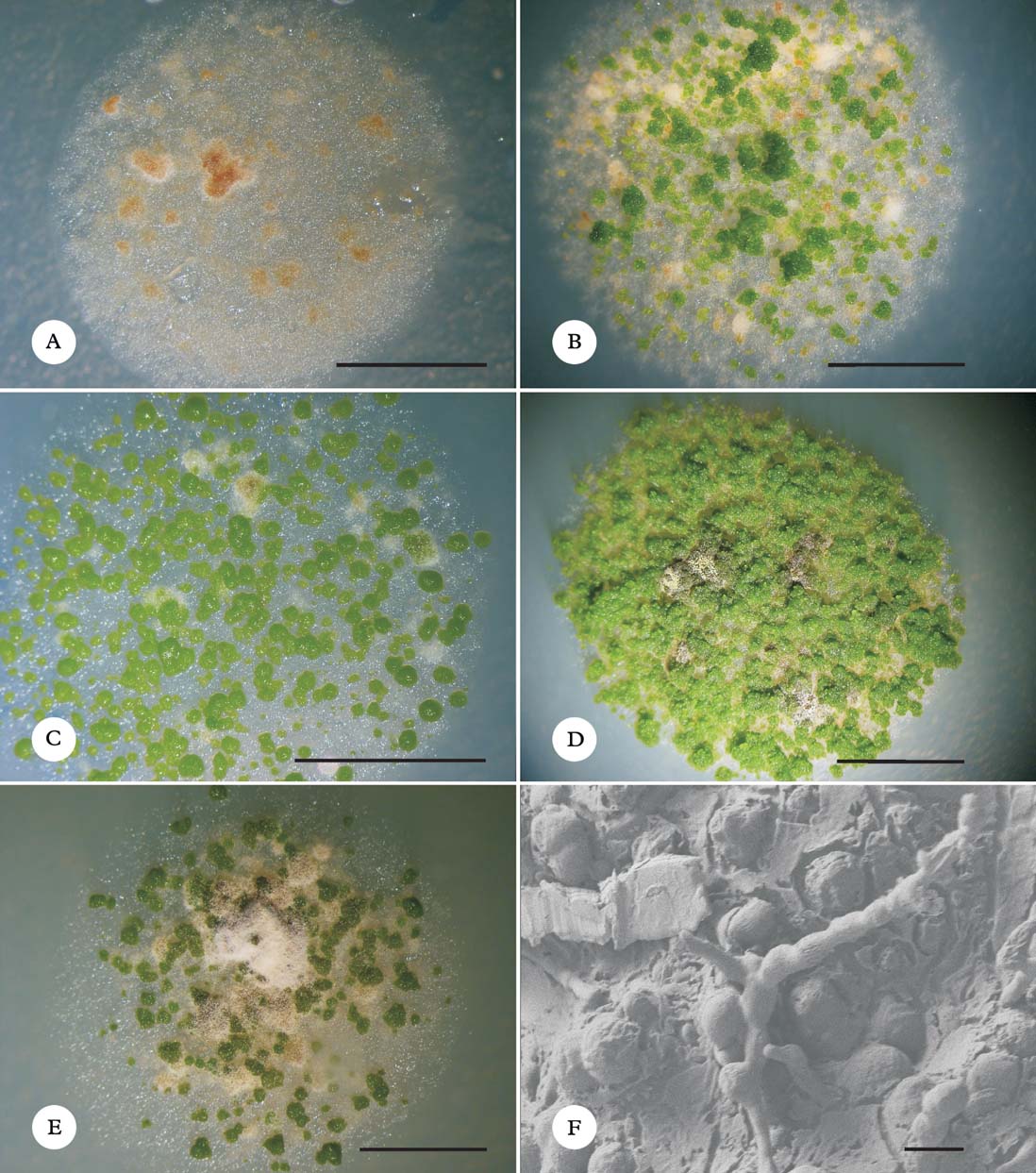
The compatibility of Protoparmeliopsis muralis with different algal species under sterile conditions was investigated using in vitro resynthesis experiments. The mycobiont used in our study was associated in nature with Trebouxia sp. ‘muralis I’. Different media were used for the resynthesis experiments and it was observed that the growth of some algae, such as T. asymmetrica (Fig. 2A), was inhibited on Saboraud 2% glucose medium. From the species analyzed, only Trebouxia gigantea and T. incrustata exhibited an increased growth on this medium (Fig. 2B). Other Trebouxia spp. stopped growing on this medium at the beginning of the resynthesis experiment, while the growth of Asterochloris sp. was comparable to that of the fungus until the third week of incubation, when it was observed that the growth of the mycobiont was faster, and after seven weeks the photobiont was finally overgrown by the mycobiont. On the other hand, on other media tested (i.e. Murashige-Skoog and G-LBM), the cultures were overgrown by the algae in most cases (Fig. 2C, D & E). We also observed using SEM that on these media, co-cultures of the fungus and the algae were covered with a gelatinous matrix as presented for G-LBM (Fig. 2F).
Cultures from resynthesis experiments of Protoparmeliopsis muralis with Trebouxia asymmetrica and T. gigantea grown on different media after 7 weeks of incubation. A, T. asymmetrica on Saboraud 2% medium; B, T. gigantea on Saboraud 2% medium; C, T. asymmetrica on Murashige-Skoog medium; D, T. gigantea on Murashige-Skoog medium; E, T. asymmetrica on G-LBM medium; F, scanning electron micrograph of the resynthesis of Protoparmeliopsis muralis with T. gigantea on G-LBM medium. Scales: A–E=1 cm; F=10 µm.
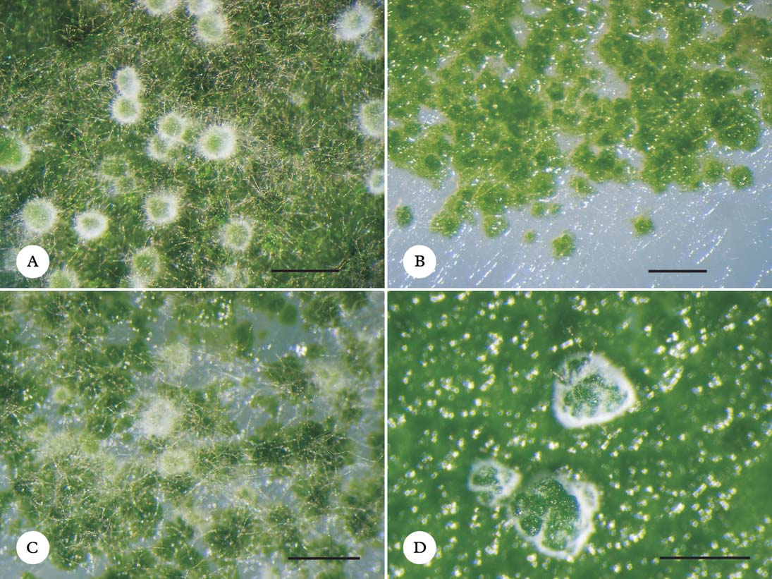
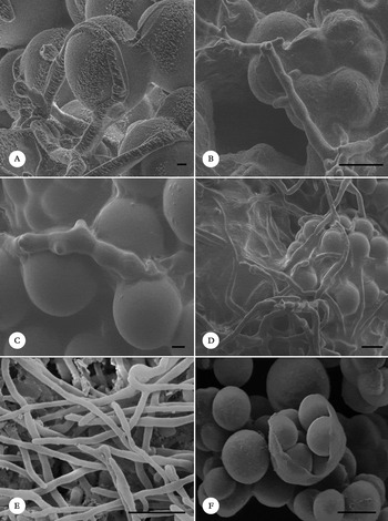
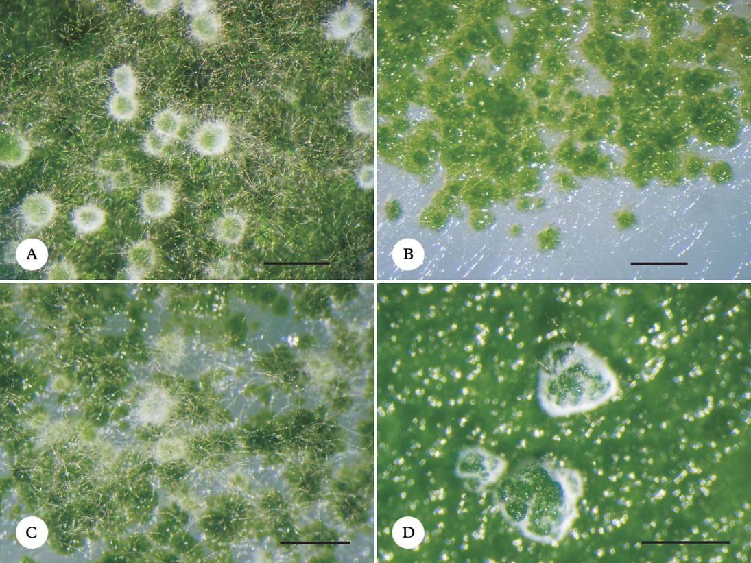
It was found that the modified BBM medium (enriched with mannitol) enabled the balanced growth of both bionts and the mycobiont formed numerous filamentous hyphae. We observed that the mycobiont was able to establish the primordial stages of lichenization with all Trebouxia photobionts investigated on this medium. After 7 weeks of incubation it was observed that fungal hyphae enveloped the algal cells, forming a dense network around the photobionts (Fig. 3A–C). Scanning electron microscopy (SEM) was used for studying the early contact between lichen bionts. SEM observations were performed after 4 and 10 months of incubation and showed a very close contact between hyphae and compatible photobionts in the resynthesis cultures (Fig. 4A & B). The hyphae enveloped even single cells and formed appressoria around compatible photobiont cells. Moreover, the surface of the cultures, at least partly, was covered with a gelatinous matrix. Algal cells were incorporated within this matrix but there was no thallus-like differentiation on agar media. Furthermore, SEM controls of aposymbiotically grown bionts (i.e. P. muralis and Trebouxia sp. ‘muralis I’) were also prepared (Fig. 4E & F). The presence of crystals of secondary metabolites was observed on the fungal hyphae but no such gelatinous matrix was noticed on either the algal or fungal cells.
Cultures from resynthesis experiments grown on BBM medium enriched with mannitol after 7 weeks of incubation. A, Trebouxia incrustata; B, Trebouxia sp. ‘muralis I’; C, Trebouxia asymmetrica; D, Asterochloris sp. Scale=1 mm.
Scanning electron microscope (SEM) micrographs. A, co-culture of Protoparmeliopsis muralis with Trebouxia gigantea; B, co-culture of P. muralis with Trebouxia sp. ‘muralis I’. ; C, co-culture of P. muralis with Asterochloris sp.; D, co-culture of P. muralis with Asterochloris sp. Scale bar; E, aposymbiotically grown P. muralis; F, aposymbiotically grown Trebouxia sp. ‘muralis I’. Scales: A & C=2 µm; B & D–F=10 µm.

The mycobiont was also able to establish primordial stages of lichenization with another photobiont, Asterochloris sp., regarded as an incompatible algal taxon. In this study, we observed that Protoparmeliopsis muralis interacted with Asterochloris sp., although the algal taxa of this genus have never been reported to form a lichen thallus with this mycobiont. However, it seemed that the growth of the algae was accelerated in comparison to the slow growth of the mycobiont. The algae formed cell aggregates enveloped by fungal hyphae (Fig. 3D). However, the fungal hyphae did not tightly cover all the algal cells, but were concentrated only in some parts of the resynthesis culture; in other parts, the hyphae were only growing on the algal cells without characteristic envelopment or close contact. The contact observed with the scanning electron microscope between P. muralis and Asterochloris sp. was close and the hyphae formed appressoria. Moreover, the algae and fungi were covered with a gelatinous matrix. Finally, some of the algal cell aggregates were embedded within this matrix which may have formed as a response to the close contact between the symbionts, and probably also as a reaction to environmental influences, such as protecting the algal cells against dryness on exposed surfaces (Fig. 4C & D).
Discussion
Different levels of specificity and selectivity have been reported from a considerable number of lichens, representing diverse growth-form types (e.g. Beck & Koop Reference Beck and Koop2001; Romeike et al Reference Romeike, Friedl, Helms and Ott2002; Blaha et al. Reference Blaha, Baloch and Grube2006; Guzow-Krzemińska Reference Guzow-Krzemińska2006; Wornik & Grube Reference Wornik and Grube2010). A previous study based on ITS rDNA sequencing of selected samples from the natural environment showed that several Trebouxia spp. were associated with Protoparmeliopsis muralis (Guzow-Krzemińska Reference Guzow-Krzemińska2006).
Although many papers have been published on in vitro resynthesis of lichens from their aposymbiotic bionts (e.g. Stocker-Wörgötter & Türk Reference Stocker-Wörgötter and Türk1991; Stocker-Wörgötter Reference Stocker-Wörgötter2001a , Reference Stocker-Wörgötter b , Reference Stocker-Wörgötter, Kranner, Beckett and Varma2002, Reference Stocker-Wörgötter2010; Brunauer et al. Reference Brunauer, Hager, Grube, Türk and Stocker-Wörgötter2007), our knowledge of the conditions needed for re-lichenization of many species still not investigated has to be complemented by novel approaches using new methodologies (e.g. Joneson et al. Reference Joneson, Armaleo and Lutzoni2011). The issue of ‘resynthesis’ of lichens from their partners is, in general, quite difficult to explore experimentally, because each time the experimental system has to be optimized de novo. However, we found here that the modified BBM medium (enriched with mannitol) enabled the balanced growth of both bionts. Joneson et al. (Reference Joneson, Armaleo and Lutzoni2011) also used BBM medium enriched with MY for Cladonia grayi resynthesis experiments. Therefore, it might be inferred that a slightly enriched BBM medium could be used as a general medium for resynthesis experiments for a variety of lichens in future studies.
Joneson & Lutzoni (Reference Joneson and Lutzoni2009) observed interactions of Cladonia grayi with its Asterochloris photobiont and other phototrophs, and showed that the morphological response of C. grayi was distinctive only in symbiotic growth with the compatible biont. Compatibility of lichen bionts has been discussed previously by many authors (e.g. Beck et al. Reference Beck, Kasalicky and Rambold2002; Joneson & Lutzoni Reference Joneson and Lutzoni2009). The non-compatible bionts do not interact to form any of the initial stages of lichen development. On the other hand, compatible bionts enter into stage two of the development, that is the envelopment of the alga by the mycobiont hyphae. At the same time, gene expression was also studied and several proteins were found to play an important role in an early contact of the bionts (Meessen & Ott Reference Meessen and Ott2010; Joneson et al. Reference Joneson, Armaleo and Lutzoni2011). Moreover, the differential gene expression observed in the C. grayi model system suggested that mycobionts and photobionts communicate before as well as during cellular contact (Joneson et al. Reference Joneson, Armaleo and Lutzoni2011). Such findings could explain the process of recognition of the compatible bionts during re-lichenization.
Although several papers on in vitro re-lichenization have been published to date, in most cases the morphology of the resynthesis culture differs from the original lichen thallus as far as they are grown on agar substrata. However, the limited number of reports showed the ability of forming thallus lobes or even fruiting bodies in in vitro resynthesis experiments, but in most cases the use of soil substratum seems to be crucial for the development of more differentiated structures (e.g. Bubrick & Galun Reference Bubrick and Galun1986; Stocker-Wörgötter & Türk Reference Stocker-Wörgötter and Türk1991, Reference Stocker-Wörgötter and Türk1993; Stocker-Wörgötter Reference Stocker-Wörgötter2001a , Reference Stocker-Wörgötter b ; Stenroos et al. Reference Stenroos, Stocker-Wörgötter, Yoshimura, Myllys, Thell and Hyvönen2003; Stocker-Wörgötter & Elix Reference Stocker-Wörgötter and Elix2006). In our short-term study (regarding the slow growth of the P. muralis lichen fungus), we did not obtain any of the more differentiated structures. However, the formation of the primordial stages of lichenization was observed for all Trebouxia spp. investigated here, including Trebouxia asymmetrica, T. gigantea, T. incrustata and an unidentified lineage of Trebouxia sp. ‘muralis I’ that were previously found to be compatible photobionts for this lichen-forming fungus. Moreover, P. muralis was able to relichenize with Asterochloris sp., regarded as an incompatible photobiont, although the contact between fungal and algal cells was not as tight as with Trebouxia photobionts. In the case of Asterochloris, the algal aggregates were enveloped by fungal hyphae, but in some parts of the culture the hyphae were only growing next to algal cells without characteristic close contact of both bionts. Moreover, no thallus-like structures were formed. Related findings were presented by Schaper & Ott (Reference Schaper and Ott2003) for Fulgensia bracteata, which was able to establish a symbiotic association with less compatible algal partners. Asterochloris spp. are known to be mainly associated with Cladoniaceae and Stereocaulaceae, but they have also been reported from other lichen families, such as Parmeliaceae (summarized in Skaloud & Peksa Reference Skaloud and Peksa2010). However, in some lineages of Asterochloris the specificity to the mycobiont is very low as the algae were found to associate with about 20 fungal species from different genera (Skaloud & Peksa Reference Skaloud and Peksa2010). Our findings suggest that Asterochloris could also be accepted by the Protoparmeliopsis muralis fungus as a temporary photobiont, until a more suitable algal symbiont (Trebouxia) is available, and a final switch to its ‘preferred’ algal partner may occur. This could be advantageous for survival of the mycobiont, especially on substrata where the lichen fungus acts as a pioneer, successfully colonizing new habitats.
A part of this study was financially supported by the Marie Curie Fellowship within the 6th European Community Framework Programme, project no. 24206, to BGK (fellow) and EST-W (leader of the project) and performed at the University of Salzburg. BGK also acknowledges the financial support of the Marie Curie European Reintegration Grant within the 7th European Community Framework Programme, project no. 239343. EST-W acknowledges the support of FWF projects 18210 and 20887. We are grateful to the Austrian Science Foundation FWF for supporting Project P23570 (ESt-W). The teachers and instructors from EMBO Electron Microscopy and Stereology course held in České Budejovice (Czech Republic in 2008) are acknowledged for advice and help with sample preparations and assistance with electron microscopy investigations, especially Martina Tesařová and Jana Nebesářová. Dorota Łuszczek (University of Gdańsk) is acknowledged for help with control sample preparation for SEM investigations. BGK also thanks Grzegorz Węgrzyn, Andreas Beck, Georg Brunauer, Armin Hager and Sabine Wornik for helpful discussion, and is grateful to the collectors named in Table 1 for providing fresh material. Hans Peter Comes is acknowledged for making his laboratory facilities available. We are grateful to anonymous reviewers for their valuable comments and suggestions to improve the manuscript.