


Introduction
Fully grown mammalian oocytes are arrested at the prophase stage of the first meiotic division. This stage is known as the germinal vesicle (GV) stage. Resuming meiosis, the oocytes undergo GV breakdown (GVBD), extrude the first polar body and progress to metaphase of the second meiotic division (MII), which is then arrested again until fertilization or parthenogenetic activation. The physiological trigger for meiotic resumption is the preovulatory gonadotrophin surge. This signal is transduced within the germ cell via activation and deactivation of specific proteins, especially kinases. The activation and deactivation is caused by phosphorylation and/or dephosphorylation of these kinases, which is considered to be the most important mechanism regulating cell-cycle progression.
Mature oocytes have elevated levels of maturation promoting factor (MPF) and mitogen-activated protein kinase (MAPK) activity. MPF is a heterodimeric protein kinase composed of a catalytic subunit, p34cdc2 kinase, and a regulatory subunit, cyclin B (Lohka et al., Reference Lohka, Hayes and Maller1988). Activation of MPF induces entry into metaphase in eukaryotes (Gautier et al., Reference Gautier, Norbury, Lohka, Nurse and Maller1988). MAPK is also called extracellular signal regulated kinase (ERK) and functions as another main regulator of oocyte maturation. MAPK appears to facilitate meiotic resumption, maintain the normal morphology of the meiotic spindle, inhibit interphase transition between the two meiotic divisions and prevent the release from MII arrest (Verlhac et al., Reference Verlhac, Kubiak, Clarke and Maro1994; Su et al., Reference Su, Rubinstein, Luria, Lax and Breitbart2001). Among the substrates of the MAPK are the kinases MNK (MAPK signal-interacting kinase) and MSK (MAPK/stress-activated protein kinase (SAPK)-activated kinase (Rubinfeld & Seger, Reference Rubinfeld and Seger2004). Upstream kinases known as as Raf and MEK (MAP/ERK kinase) are also substrates of MAPK, supporting the hypothesis of a feedback loop regulating this protein kinase cascade (Davis, Reference Davis1993; Rubinfeld & Seger, Reference Rubinfeld and Seger2004).
The first found and best known physiological substrate of MAPK in oocytes is p90rsk (RSK). This protein kinase belongs to a family of Ser/Thr kinases that was originally identified as phosphorylating the S6 protein of the 40S ribosomal subunit in maturing Xenopus oocytes (Erikson & Maller, Reference Erikson and Maller1985). Therefore, this family of 90 kDa ribosomal S6 kinases is also known as RSK, p90rsk or MAPK-activated protein kinase-1. The RSK family belongs to the serine–threonine kinases, but it is unique in having a special structure. It consists of two functional kinase domains that are each similar to distinct protein kinases (Jones et al., Reference Jones, Erikson, Blenis, Maller and Erikson1988). Phosphorylation of p90rsk substrate is mediated via the N-terminal domain, whereas the C-terminal domain is necessary for full activation of the molecule (Frödin & Gammeltoft, Reference Frödin and Gammeltoft1999).
Another S6 kinase of 70 kDa (p70S6K) was identified that shows substantial sequence homology to the N-terminal kinase of RSK (Bannerjee et al., Reference Bannerjee, Ahmad, Grove, Kozlosky, Price and Avruch1990; Kozma et al., Reference Kozma, Ferarri, Bassand, Siegmann, Trotty and Thomas1990). p70S6K is thought to be the major physiological S6 kinase (Ballou et al., Reference Ballou, Luther and Thomas1991; Chung et al., Reference Chung, Kuo, Crabtree and Blenis1992; von Manteuffel et al., Reference Von Manteuffel, Dennis, Pullen, Gingras, Sonnenberg and Thomas1997), but it is not phosphorylated and subsequently activated during oocytes maturation and, therefore, probably has no function in the process of meiotic resumption (mouse: Gavin & Schorderet-Slatkine, Reference Gavin and Schorderet-Slatkine1997; Xenopus: Schwab et al., Reference Schwab, Kim, Terade, Edfäll, Kozma, Thomas and Maller1999).
RSK is located downstream of the Raf–MEK–MAPK signalling cascade and evidence from Xenopus oocytes suggests that p90rsk mediates most of the MAPK function in regulating meiotic cell-cycle progression (Bhatt & Ferrell, Reference Bhatt and Ferrell Jr1999; Gross et al., Reference Gross, Schwab, Lewellyn and Maller1999, Reference Gross, Schwab, Taieb, Lewellyn, Qian and Maller2000). After its first appearance, concomitantly with or shortly after GVBD, the activity of p90rsk increases with progression of meiosis, with a peak at the MI stage and remains high in MII-arrested mammalian oocytes (mouse: Kalab et al., Reference Kalab, Kubiak, Verlhac, Colledge and Maro1996; Tong et al., Reference Tong, Fan, Chen, Song, Schatten and Sun2003; rat: Tan et al., Reference Tan, Chen, Yang, Wang, Li, Schatten and Sun2001; Lu et al., Reference Lu, Smith, Chen, Yang, Han, Schatten and Sun2001; rabbit: Yu et al., Reference Yu, Bou, Chen and Sun2002; pig: Sugiura et al., Reference Sugiura, Iwamori, Kagii, Goto, Naruoka, Yada, Yamanouchi and Tojo2002; Fan et al., Reference Fan, Tong, Lian, Li, Gao, Cheng, Chen, Schatten and Sun2003). RSK slowly becomes dephosphorylated in activated eggs shortly before pronuclear formation.
In mouse, as well as in porcine oocytes, p90rsk was observed partially phosphorylated, but inactive, in oocytes immediately following recovery from the follicles in the absence of MAPK phosphorylation (Kalab et al., Reference Kalab, Kubiak, Verlhac, Colledge and Maro1996; Fan et al., Reference Fan, Tong, Lian, Li, Gao, Cheng, Chen, Schatten and Sun2003).
Studies analysing p90rsk in in vivo matured Xenopus oocytes gave different results. One group (Erikson & Maller, Reference Erikson and Maller1989) detected the molecule at around 90 kDa by immunoblotting, whereas another group (Hsiao et al., Reference Hsiao, Chou, Shih and Ferell1994) found p90rsk being complexed with MAPK in a heterodimer around 110 kDa.
The aim of the present study was to compare the phosphorylation pattern of p90rsk from in vitro and in vivo matured porcine oocytes. Additionally, we wanted to show the activity profile of the kinase. We report our results using a commercially available kinase assay kit analysing S6 kinase activity.
Materials and Methods
Unless otherwise stated, chemicals and reagents were obtained from Sigma–Aldrich. Porcine FSH and LH were obtained from the National Hormone and Peptide Program of the Harbor–UCLA Medical Center. Ovaries were collected from peripubertal gilts (aged 6–7 months, bodyweight: 90–120 kg) at a local slaughterhouse and were transported in prewarmed (30–33 °C) physiological salt solution to the laboratory within 1 h of slaughter of the animals. The cumulus–oocyte complexes (COCs) for in vitro maturation were obtained by slitting individual antral follicles (3–5 mm in diameter) and flushing each antrum with prewarmed (39 °C) phosphate buffered saline (PBS) containing 1% (v/v) heat-inactivated fetal calf serum (FCS). Only COCs with at least three compact and homogenous layers of cumulus cells were selected and stored at 39 °C in 2 ml PBS supplemented with FCS (collection medium) for up to 45 min. The COCs were cultured in groups of around 25 oocytes for different time periods, from 22 h up to 46 h in 4 h intervals, in TCM containing 20% (v/v) FCS, 0.02 mg/ml insulin, 0.08 mg/ml l-glutamine, 50 μg/ml gentamycin, 5.0 μg/ml LH and 2.5 μg/ml FSH (culture medium). For the inhibition experiment, the MEK-specific inhibitor U0126 was used. The inhibitor was dissolved in dimethylsulphoxide (DMSO) and added to the culture medium in a concentration of 10 μM. To exclude an influence of the solvent on the maturation process, the solvent was added to the culture medium instead of the inhibitor. A concentration of 0.2% (v/v) of DMSO was used in the culture medium. A stock solution containing 5 mM U0126 was prepared. From this stock solution, 2 μ1 were added to 998 μ1 culture medium or collection medium to make a final concentration of 10 μM U0126.
Incubation was carried out in 1 ml medium in 4-well multidishes at 39 °C, in an atmosphere of 5% CO2 in air and saturated humidity. At the end of culture, COCs were denuded by treatment with 0.25% (w/v) hyaluronidase (bovine testes) followed by repeated pipetting to remove the remaining cumulus cells. The denuded oocytes and separated cumulus cells were then washed three times in protein-free PBS. Samples of 20 oocytes per time point and cumulus cells from 10 oocytes, respectively, were frozen in about 4 μ1 of this protein-free PBS at −80 °C until electrophoresis was performed.
For in vivo maturation, 25 peripubertal hybrid gilts aged of 7 to 8.5 months and weighing between 90–110 kg were kept in groups of two to four animals without direct contact to a boar. The animals were injected with 600–800 IU equine chorionic gonadotrophin (eCG, Pregmagon®) at the start of transport from the farmer to the Department of Reproductive Biology. Equine chorionic gonadotrophin was injected to stimulate follicle development and to avoid uncontrolled induction of transport heat. After 72 h the gilts were tested for signs of heat in the presence of a boar. If they showed no signs of heat, they were injected with 500 IU human chorionic gonadotrophin (hCG, Ovogest®) to mimic the LH surge and, therefore, induce oocyte maturation (Hunter & Polge, Reference Hunter and Polge1966, Ratky et al., Reference Ratky, Rath and Brüssow2003a, Reference Ratky, Brüssow, Torner and Egerszegib).
Ovariectomy was carried out via midventral laparotomy following routine operation procedures. To prevent any influence of tissue hypoxia, particular care was taken to remove the ovaries before the blood vessels were ligated. The animals were ovariectomized at selected time points according to the in vitro maturation intervals. The time points chosen were 0 h (at the regular time of hCG injection, nine animals), 22 h, (nine animals) and 30 h (seven animals) after hCG injection corresponding to 0, 22 h and 30 h of in vitro maturation. Twenty oocytes for one sample per time point were necessary for characterization of the protein processed by the in vitro matured oocytes, using electrophoresis, immunoblot and enhanced chemiluminescence. In both groups, in vitro and in vivo, the nuclear status of the oocytes was assessed. Oocytes were fixed for at least 24 h in a freshly prepared solution containing 25% acetic acid (v/v) and 75% ethanol and stained with a 2% (w/v) orcein solution in acetic acid. During in vitro maturation, at least 80 oocytes per time point (0, 22, 26, 30, 34, 46 h) were fixed, stained and their nuclear status was assessed and for the in vivo matured oocytes a total number of at least 20 oocytes from each of the three time points (0, 22 and 30 h) chosen, were fixed, stained and their nuclear status was assessed. The nuclear status of the oocytes during the maturation process, with an addition of the inhibitor as well as with the addition of 0.2% (v/v) of DMSO, was controlled by samples at the same time points (0, 22, 26, 30, 34, 46 h) as the in vitro maturation but without the addition of an inhibitor. Again at least 80 oocytes were fixed, stained and their nuclear status was assessed. The assessment of the nuclear status was carried out under phase-contrast microscopy with 200–400 fold magnification, according to the protocol by Motlik and Fulka (Reference Motlik and Fulka1976).
In order to prepare the samples for electrophoresis, 10–20 μ1 of modified Laemmli buffer (without mercaptoethanol) was added to 20 denuded oocytes or cumulus cells from 10 COCs in order to extract the proteins. The samples were boiled for 5 min, put on ice immediately, centrifuged for at least 1 min at 9980 g and then put back on ice until they were filled into the gel pockets. The proteins were separated by discontinuous SDS-PAGE with a 5% stacking and a 9% separation gel for 10 min at 100 V and 1 h at 200 V.
After SDS-PAGE, proteins were transferred electrophoretically onto nitrocellulose membranes for 2 h with a constant current of 1 mA/cm2 at room temperature. The membrane was blocked (TBS/Tween 0.1%, 5% teleostean gelatine) for at least 2 h at room temperature, washed three times for 10 min (TBS, 1% Tween) and incubated in washing solution containing 1:200 diluted polyclonal rabbit anti-human antibody for RSK (Rsk-1 (C-21): sc-231; Santa Cruz Biotechnology) overnight at RT. After three washes of 10 min (TBS, 1% Tween), membranes were incubated in diluted conjugated probe antibody (1:500 up to 1:2000) for 1 h at room temperature. This second antibody was a horseradish peroxidase-conjugated goat anti-rabbit antibody (goat anti-rabbit HRP-conjugated, Perbio Science). For visualization of the biotinylated standard (high molecular weight protein standard), horseradish peroxidase-conjugated streptavidin was used.
The membranes were washed five times for 10 min (TBS, 1% Tween) and then processed using an enhanced chemiluminescence detection system (Perbio Science).
For detection of MAPK, the membranes were incubated in stripping solution (100 mM β-mercaptoethanol, 2% SDS, 62.5 mM Tris, pH 6.7) for 2 h at room temperature, washed three times (TBS, 1% Tween) and reprobed with 1:200 diluted polyclonal rabbit anti-rat antibody (ERK 1 (K-23): sc-94; Santa Cruz Biotechnology). After three washes for 10 min. (TBS, 1% Tween), they were then incubated with HRP-conjugated goat anti-rabbit antibody and submitted to chemiluminescence.
For analysis of the activation pattern of p90rsk in the in vitro matured oocytes, a commercially available kinase assay kit was used (S6 Kinase Assay Kit, Upstate). The assay kit is designed to measure the phosphotransferase activity of S6 kinase in immunoprecipitates and column fractions; crude cell lysates may also be used. It is based on phosphorylation of a specific substrate modelled after the major phosphorylation sites in S6 kinase using the transfer of radiolabeled phosphate [γ-32P]-ATP by S6 kinase. The phosphorylated substrate is separated from the residual [γ-32P]-ATP using P81 phosphocellulose paper and quantified using a scintillation counter. The S6 kinase assay kit was performed according to the manufacturer's instructions.
Statistical analysis
All experiments were repeated at least three times. Nuclear maturation results were analysed statistically (http://faculty.vassar.edu/lowry/VassarStats.html) using chi-squared analysis. Data of kinase assays were analysed by one way analysis of variance followed by Tukey HSD test. A probability of p < 0.01 was considered to be statistically significant.
Results
Almost all of the oocytes were in the GV stage at 0 h of in vitro and in vivo maturation (99.2% and 100%). After 22 h the majority of oocytes was still in the GV stage (in vitro: 59.1%, in vivo: 84%) and after 30 h most of the oocytes were in the MI stage (in vitro: 80.3%, in vivo: 80.0%) (Tables 1 and 2).
State of meiotic maturation of pig oocytes after in vitro maturation for indicated times

GV, germinal vesicle stage; Di/MI, diakinesis/metaphase I; AI–MII, anaphase I–metaphase II. The percentages are rounded to one decimal place, therefore the rows not always add up to 100%. Values of rows with different superscripts (a–f) differ significantly (p < 0.01).
State of meiotic maturation of pig oocytes after treatment with hCG
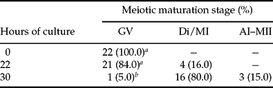
GV, germinal vesicle stage; Di/MI, diakinesis/metaphase I; AI–MII, anaphase I–metaphase II. The percentages are rounded to one decimal place, therefore the rows not always add up to 100%. Values of rows with different superscripts (a, b) differ significantly (p < 0.01).
At all selected maturation time points up to 46 h of maturation with the inhibitor, over 90% of the oocytes remained in the GV stage (Table 3), whereas over 90% of the oocytes cultured without the addition of the inhibitor had reached the MII stage after 46 h of in vitro maturation (cf. Table 2). Statistical analysis of the data showed that there was a significant (p < 0.01) difference in frequency distribution after 0 and 30, 26 and 30 and 30 and 46 h of in vitro maturation with the inhibitor. At all these time points, over 90% of the oocytes remained in the GV stage, but at 30 h of in vitro maturation a higher percentage of oocytes was found in the MII stage (30 h = 6.7%), whereas all the other time points showed no or only very low percentages of MII stage oocytes.
State of meiotic maturation of pig oocytes after in vitro maturation in medium supplemented with U0126 for indicated times

GV, germinal vesicle stage; Di/MI, diakinesis/metaphase I; AI–MII, anaphase I–metaphase II. The percentages are rounded to one decimal place, therefore the rows not always add up to 100%. Values of rows do not differ significantly (p > 0.01).
A comparison of the results of the oocytes matured without the inhibitor and those with an addition of the inhibitor shows that at all time points the two groups are significantly different (p < 0.01) except for the time point 0 h, which shows no significant difference from all time points during in vitro maturation with an addition of the inhibitor (cf. Table 2 and 3). To exclude any effect of adding DMSO, the inhibitor solvent, on the in vitro maturation process, controls (46 h) were carried out with addition of DMSO (0.2%,v/v) but without U0126. Under these culture conditions, 6.0% (7/115) of the oocytes remained in the GV stage whereas 7.8% (9/115) and 86.1% (99/115) of the oocytes reached the Di/MI and AI–MII stage, respectively. These maturation rates were not significantly different from the maturation results following culture of COCs in the absence of DMSO for 46 h (cf. Table 1). Therefore any influence of DMSO on the maturation rates can be disregarded.
Phosphorylation of p90rsk in oocytes during IVM. Proteins of 20 oocytes were loaded per lane and the membrane was incubated with the anti-RSK antibody. From 22 to 46 h of IVM, p90rsk bands became of a lower mobility, i.e. the degree of phosphorylation increased during IVM.

Electrophoresis, immunoblot and subsequent chemiluminescence analysis revealed a stepwise phosphorylation pattern in the in vitro matured oocytes at the expected molecular weight of around 90 kDa. These steps are visible as bands of p90rsk, which showed different mobility in electrophoresis (Fig. 1). In oocytes immediately following recovery from the follicles until 22 h of culture, p90rsk is observed as double bands. Proceeding with maturation at first, the highest mobility band becomes less and less intense before it disappears whereas the second high mobility band at first becomes more intense before it starts to fade. Around 34 h of maturation, a lower mobility band appears and is seen here until the end of the culture period.
Analysis of the in vivo matured oocytes detected p90rsk at a molecular weight of slightly less than 200 kDa (Figure 2a, b). Although the same number of oocytes per time point was analysed, the received bands in the immunoblots show very different intensity. Therefore different exposure times of the same immunoblot are depicted. In all replicates, three bands of different mobility as seen in the in vitro matured oocytes and corresponding to the phosphorylation of the molecule were visible and at 0 h p90rsk was detected as a double band (Fig. 2b). Reprobing the immunoblots of the in vivo matured oocytes with a MAPK-specific antibody detected no bands in the molecular range 70 to 200 kDa.
Phosphorylation of p90rsk in oocytes during in vivo maturation. Proteins of 20 oocytes were loaded per lane and the anti-RSK antibody was used. The same immunoblot was visualized by an exposure time to the X-ray film of 1 (a) and 5 (b) min. The p90rsk was detected at a molecular weight of about 200 kDa. The phosphorylation degree of p90rsk increased during in vivo maturation at the chosen time points (0 h, 22 h, 30 h).

Figure 3 shows the observed phosphorylation pattern of p90rsk during in vitro maturation carried out with an addition of the MEK inhibitor U0126 to the culture medium. In the immunoblot picture, one can clearly see that the two high mobility bands were already present during culture conditions without the inhibitor from 18 until 22 h. Proceeding with the culture, no lower mobility bands become visible; only the highest mobility band, corresponding to the non-phosphorylated form of p90rsk, becomes prominent.
Phosphorylation of p90rsk in oocytes during IVM of COCs in medium supplemented with U0126. Proteins of 20 oocytes were loaded per lane and the membrane was incubated with the anti-RSK antibody. The MEK inhibitor U0126 (10 μM) prevented an increase of the phosphorylation of p90rsk in oocytes. No mobility shift of the bands could be observed.

Phosphorylation of p90rsk in cumulus cells during IVM. Proteins from cumulus cells of five COCs were loaded per lane and the anti-RSK antibody was used. In comparison to cumulus cells obtained immediately following recovery from the follicles (0 h), the phosphorylation of p90rsk in cumulus cells had not increased after either 24 h of IVM or 48 h of IVM.

Analysing, via electrophoresis and immunoblot, samples of cumulus cells from 10 in vitro matured COCs cultured for 0, 24 and 48 h, demonstrated the existence of p90rsk in porcine cumulus cells. In contrast what was found for the oocytes during maturation culture, increasing phosphorylation of the molecule, did not detect the corresponding varying mobility bands (Fig. 4).
In cumulus cells from in vivo matured oocytes, p90rsk was found at around 90 kDa (Fig. 5). At 0 h of in vivo maturation the kinase can only be detected as a single band. Longer exposure time did not reveal any further bands. At 22 and 30 h of in vivo maturation a double band that corresponded to the kinase was visible. A shift, as found in the oocytes, was not observed.
Phosphorylation of p90rsk in cumulus cells during in vivo maturation. Proteins of cumulus cells of 10 COCs were loaded per lane and the anti-RSK antibody was used. As in cumulus cells cultured in vitro, no mobility shift of the p90rsk bands could be observed. In contrast to the p90rsk of in vivo matured oocytes (Fig. 2) in cumulus cells of in vivo matured COCs, the p90rsk bands appeared at around 90 kDa.

To determine the enzyme activity of p90rsk, samples of 20 oocytes from the six time points analysed previously via electrophoresis and immunoblot (0, 22, 26, 30, 34 and 46 h IVM) were submitted to the kinase assay kit procedure. The highest activity was measured at 0 h, before the start of maturation. A decrease in the activity was found at all other time points (22, 26, 30, 34, 46 h) during the following maturation process (Fig. 6). Statistical analysis of the data revealed no significant differences between the activities at the start of maturation (0 h) and all other time points during maturation. In a second step, samples of 20 oocytes, each matured with an addition of the inhibitor U0126 to the culture medium, were analysed. The same time points (0, 22, 26, 30, 34, 46 h) that were already analysed via electrophoresis and immunoblot were submitted to the kinase assay kit. Again, the highest activity was measured at 0 h, before the start of maturation. A decrease in the activity was found at all other time points (22, 26, 30, 34, 46 h) during the following maturation process (Fig. 6). Statistical analysis of the data revealed a significant difference between activities at the start of maturation (0 h) and at all other time points during maturation.
Discussion
In the present in vitro maturation system with porcine oocytes, GVBD took place from 22 to 30 h of culture in more than 85% of the oocytes. Most of the oocytes were in the MI stage after 30 h of IVM and the percentage of MII stage oocytes increased significantly after 34 h of culture, and reached this stage in over 90% of the oocytes after 46 h of IVM. These detected kinetics are in agreement with previous findings (Wehrend, Reference Wehrend and Meinecke2001; Sugiura et al., Reference Sugiura, Iwamori, Kagii, Goto, Naruoka, Yada, Yamanouchi and Tojo2002; Fan et al., Reference Fan, Tong, Lian, Li, Gao, Cheng, Chen, Schatten and Sun2003). In the present investigation, the kinetics of the in vivo maturation process following hCG application to eCG primed gilts corresponded well to previous observations (Hunter & Polge, Reference Hunter and Polge1966) and to the present in vitro data. Earlier studies comparing the in vivo and in vitro maturation kinetics in porcine oocytes observed a delay in in vivo matured oocytes of 4 h compared to in vitro matured ones. This difference appeared at the beginning of nuclear maturation and remained unchanged throughout the whole period studied (24 h) (Motlik & Fulka, Reference Motlik and Fulka1976).
Activity of the assumed p90rsk in pig oocytes cultured in vitro without or with the MEK inhibitor U0126 for indicated times. Values of columns with different superscripts (a, b) differ significantly (p < 0.01). Values of the putative p90rsk of oocytes cultured without U0126 do not differ significantly (p > 0.01).

During IVM with medium containing 10 μM U0126, a specific MEK inhibitor, GVBD was significantly inhibited. More than 90% of the oocytes remained in the GV stage at all time points investigated. The higher percentage of MII stage oocytes after 30 h of IVM is probably due to selection of oocytes that had already resumed meiosis at the start of the in vitro maturation period. Fan et al. (Reference Fan, Tong, Lian, Li, Gao, Cheng, Chen, Schatten and Sun2003) also found a significant inhibition of GVBD with the same concentration of the inhibitor in the medium, although still 23% of the oocytes underwent GVBD after 44 h of IVM. This difference might be due to the different composition of the culture medium. Kagii et al. (Reference Kagii, Naito, Sugiura, Ohashi, Goto, Yamanouchi and Tojo2000) in their experiments observed 42% of the oocytes undergoing GVBD. As these authors used medium containing 100 μM U0126, this difference in GVBD rate is not due to inhibitor concentration. Differences might be due to the selection of oocytes and/or the use of an inhibitor-free collection medium before the start of culture. On the basis of the present results, it can be concluded that the used concentration of the inhibitor predictably inhibits the in vitro maturation process under the applied conditions.
In oocytes immediately following recovery from the follicles (0 h) p90rsk is already observed as double bands, the first phosphorylation step has already taken place. These two bands with the highest mobility remain until about 22 h of in vitro maturation. After 22 h, the intensity of the band corresponding to the first phosphorylation step increases, and after 26 h the third and lowest mobility band appears. This lowest mobility band increases in intensity until the end of maturation whereas the two other high mobility bands disappear. p90rsk phosphorylation, therefore, starts around the time of GVBD. This observation is in agreement with the findings by other authors in porcine oocytes (Sugiura et al., Reference Sugiura, Iwamori, Kagii, Goto, Naruoka, Yada, Yamanouchi and Tojo2002; Fan et al., Reference Fan, Tong, Lian, Li, Gao, Cheng, Chen, Schatten and Sun2003). Grove et al. (Reference Grove, Price, Banerjee, Balasubramanyam, Ahmad and Avruch1993) proposed that this first phosphorylation step might be caused by autophosphorylation and that the partially phosphorylated p90rsk is not an active kinase. In mouse and rat oocytes, p90rsk is also observed as double bands during the GV stage (mouse: Kalab et al., Reference Kalab, Kubiak, Verlhac, Colledge and Maro1996; rat: Tan et al., Reference Tan, Chen, Yang, Wang, Li, Schatten and Sun2001). In these species, a phosphorylation of p90rsk, visible as a shift of the bands, takes place after GVBD.
Phosphorylation of p90rsk, as well as nuclear maturation under the present in vitro maturation conditions with medium containing 10 μM U0126, was completely inhibited. Only the two high mobility bands that are visible from the start of IVM remain until the end and become more and more intense. Fan et al. (Reference Fan, Tong, Lian, Li, Gao, Cheng, Chen, Schatten and Sun2003) also reported an inhibition of p90rsk phosphorylation using U0126 in the same concentration. In their studies this inhibition of p90rsk phosphorylation and significant inhibition of GVBD was only observed in COCs. In denuded oocytes, they still observed the inhibition of phosphorylation, but GVBD occurred in the same percentage of cells as in COCs or denuded oocytes cultured in the inhibitor-free medium. Kagii et al. (Reference Kagii, Naito, Sugiura, Ohashi, Goto, Yamanouchi and Tojo2000) also examined porcine oocytes cultured with an addition of U0126. They came to same conclusion: that the inhibitory effects of U0126 needed the presence of cumulus cells.
Two studies exist on p90rskin vivo matured oocytes in Xenopus (Erikson & Maller, Reference Erikson and Maller1989; Hsiao et al., Reference Hsiao, Chou, Shih and Ferell1994). Erikson & Maller (Reference Erikson and Maller1989) detected the molecule at around 90 kDa by immunoblotting. As they only describe that the kinase was purified from unfertilized eggs, it is difficult to assign the results to the maturation process and, therefore, to compare them with the results from Hsiao et al. (Reference Hsiao, Chou, Shih and Ferell1994). Hsiao et al. (Reference Hsiao, Chou, Shih and Ferell1994) found p90rsk being complexed with MAPK in a heterodimer. It was shown that ERK2 when phosphorylated and active is monomeric, but when it is non-phosphorylated and inactive about half of it is monomeric and half is a component of a 110 kDa complex. The other part of the complex was identified as Rsk, and much or all of the cell's Rsk was coimmunoprecipitated with ERK2. Phosphorylation and activation of MAPK correlated with the release of ERK2 from the complex. This study also reports MAPK being part of a similar sized complex in murine somatic cells. The present observations instead indicate that p90rsk exists as a dimer in in vivo matured oocytes. This dimer, as well as the monomeric form that is seen in in vitro matured oocytes, undergoes a mobility shift during the maturation process due to phosphorylation of the molecule. MAPK being part of the complex can be excluded, as a probe with the MAPK-specific antibody revealed no bands in the immunoblot picture. Philipova & Whitaker (Reference Philipova and Whitaker2005) reported that they found ERK1 capable of dimerization, both in vivo and in vitro, in sea urchin embryos and human cells. They differentiated between three forms of the ERK1 molecule: (i) monomers that are mainly unphosphorylated and inactive in vivo; (ii) monophosphodimers consisting of one phosphorylated and one unphosphorylated ERK1 molecule and which generate the basal ERK1 activity; and (iii) bisphosphodimers formed from two phosphorylated ERK1 molecules being the predominant form of highly active ERK1 in vivo.
In the cumulus cells from the in vivo matured oocytes, p90rsk was observed at the expected molecular weight of around 90 kDa. Earlier studies of cumulus cells from in vitro matured oocytes showed that p90rsk phosphorylation occurs earlier in cumulus cells compared to the oocytes, already beginning after 12 h of in vitro maturation and culminating at 24 h (Fan et al., Reference Fan, Tong, Lian, Li, Gao, Cheng, Chen, Schatten and Sun2003). Analysis of p90rsk phosphorylation of cumulus cells after in vitro maturation in our laboratory, consistent with our results in the in vivo matured cumulus cells, revealed no mobility shift of p90rsk during the in vitro maturation period (Ebeling et al., Reference Ebeling, Bösebeck and Meinecke2005). These differences in phosphorylation might be due to different maturation conditions, especially the different culture medium additives.
Activity of p90rsk in in vitro matured porcine oocytes was analysed with a commercially available kinase assay. This kit was used in another study analysing p90rsk in porcine oocytes (Sugiura et al., Reference Sugiura, Iwamori, Kagii, Goto, Naruoka, Yada, Yamanouchi and Tojo2002). The kit was applied according to the manufacturer's instructions and results revealed an activity course during in vitro maturation very different from what was expected. The kit uses an S6 kinase-specific substrate, not p90rsk specific. As there are two major S6 kinases in vivo, p90rsk and p70S6K (Bannerjee et al., Reference Bannerjee, Ahmad, Grove, Kozlosky, Price and Avruch1990), one has to rule out the possibility of measuring the wrong kinase. Sugiura et al. (Reference Sugiura, Iwamori, Kagii, Goto, Naruoka, Yada, Yamanouchi and Tojo2002) discuss that biochemical and molecular studies indicated that RSK is the protein kinase responsible for S6 phosphorylation during meiotic maturation of Xenopus oocytes (Erikson & Maller, Reference Erikson and Maller1985, Reference Erikson and Maller1986, Reference Erikson and Maller1989) and, furthermore, that p70S6K was inactive throughout the maturation period of Xenopus and mouse oocytes (Gavin & Schorderet-Slatkine, Reference Gavin and Schorderet-Slatkine1997; Schwab et al., Reference Schwab, Kim, Terade, Edfäll, Kozma, Thomas and Maller1999). Therefore the present and measured S6 kinase activity should be the activity of RSK. On the basis of the species-specific differences that are already distinct, comparing the phosphorylation pattern of a kinase, this argument is to be judged critically.
In addition to this, the company renamed the kinase assay kit during the course of the studies from ‘S6 kinase assay kit’ to ‘p70S6K assay kit.’ An inquiry at the company confirmed this renaming, but they also confirmed that it could still be used for analysis of both S6 kinases.
As the present results, with the activity being high at the start of maturation and declining during the progress of maturation and also the same picture in in vitro matured oocytes with an addition of the inhibitor U0126, resemble the measured activity for p70S6K in Xenopus oocytes (Schwab et al., Reference Schwab, Kim, Terade, Edfäll, Kozma, Thomas and Maller1999) we conclude that the measured activity in our studies was not p90rsk but p70S6K. Schwab et al. (Reference Schwab, Kim, Terade, Edfäll, Kozma, Thomas and Maller1999) observed that p70S6K activity is present in oocytes before induction of maturation by progesterone and saw a decrease in activity upon the induction.
As an overall conclusion, the present investigation confirmed the reported phosphorylation pattern of p90rsk in in vitro matured porcine oocytes and revealed a similar stepwise phosphorylation in in vivo matured porcine oocytes. Interestingly the kinase was detected at a molecular weight of approximately 200 kDa in the in vivo matured oocytes, indicating that the molecule exists here as a dimer.
The commercially available kinase assay kit used was found to be unsuitable for measuring p90rsk activity, instead detecting the p70S6K. Future efforts should focus on suitable methods for kinase activity assessment (e.g. in-gel kinase assay).
Acknowledgements
The study was supported by a grant of the H. Wilhelm Schaumann Stiftung, Hamburg, Germany.



