Introduction
In recent years, mines in the Red Canyon portion of the White Canyon district in south-eastern Utah have yielded many new minerals. Our investigation of the secondary mineralization at the Blue Lizard mine has already resulted in the description of sixteen new minerals, most of which are Na uranyl sulfates. As we have expanded our efforts to other nearby uranium mines in Red Canyon, additional new species are being revealed. The new Na–Mg uranyl carbonate leószilárdite (Olds et al., Reference Olds, Sadergaski, Plášil, Kampf, Burns, Steele and Marty2016) was recently described from the Markey mine and, herein, we describe another new uranyl carbonate, markeyite, from this mine.
Markeyite (/ma:r 'ki: ait/) is named for the locality, the Markey mine. The new mineral and name were approved by the International Mineralogical Association Commission on New Minerals, Nomenclature and Classification (IMA 2016-090). After the initial approval of the mineral and its publication in the CNMNC Newsletter No. 35 (Kampf et al., Reference Kampf, Plášil, Kasatkin, Marty and Čejka2017), a change in the formula from Ca9(UO2)4(CO3)12(OH)2·28H2O to Ca9(UO2)4(CO3)13·28H2O was approved by the officers of the CNMNC (Hålenius et al., Reference Hålenius, Hatert, Pasero and Mills2017). The description is based on one holotype and five cotype specimens. The holotype and four cotypes are deposited in the collections of the Natural History Museum of Los Angeles County, 900 Exposition Boulevard, Los Angeles, CA 90007, USA, catalogue numbers 67091 (holotype), 67092, 67093, 67094 and 69095, respectively. One cotype specimen is housed in the collections of the Fersman Mineralogical Museum of the Russian Academy of Sciences, Moscow, Russia, with registration number 4932/1.
Occurrence
Markeyite was found underground in the Markey mine, Red Canyon, White Canyon District, San Juan County, Utah, USA (37°32′57″N, 110°18′08″W). The Markey mine is located ~1 km southwest of the Blue Lizard mine, on the east-facing side of Red Canyon, ~72 km west of the town of Blanding, Utah, and ~22 km southeast of Good Hope Bay on Lake Powell. The geology of the Markey Mine is quite similar to that of the Blue Lizard mine (Chenoweth, Reference Chenoweth1993; Kampf et al., Reference Kampf, Plášil, Kasatkin, Marty and Čejka2016), although the secondary mineralogy of the Markey mine is notably richer in carbonate phases. The information following is taken largely from Chenoweth (Reference Chenoweth1993).
Jim Rigg of Grand Junction, Colorado began staking claims in Red Canyon in March of 1949. The Markey group of claims, staked by Rigg and others, was purchased by the Anaconda Copper Mining Company on June 1, 1951. After limited exploration and production, the mine closed in 1955. The mine was subsequently acquired from Anaconda by Calvin Black of Blanding, Utah under whose ownership the mine operated from 1960 to 1982 and was a leading producer in the district for nearly that entire period.
The uranium deposits in Red Canyon occur within the Shinarump member of the Upper Triassic Chinle Formation, in channels incised into the reddish-brown siltstones of the underlying Lower Triassic Moenkopi Formation. The Shinarump member consists of medium- to coarse-grained sandstone, conglomeratic sandstone beds and thick siltstone lenses. Ore minerals were deposited as replacements of wood and other organic material and as disseminations in the enclosing sandstone. Since the mine closed in 1982, oxidation of primary ores in the humid underground environment has produced a variety of secondary minerals, mainly carbonates and sulfates, as efflorescent crusts on the surfaces of mine walls.
Markeyite is a rare mineral in the secondary mineral assemblage. It occurs on asphaltum in association with calcite, gypsum and natrozippeite. Other secondary minerals in the general assemblage include: ammoniozippeite, andersonite, anglesite, aragonite, arsenuranospathite, atacamaite, bayleyite, bluelizardite, bobcookite, brochantite, čejkaite, chalcanthite, chalconatronite, chinleite-(Y), covellite, cuprosklodowskite, cyanotrichite, deliensite, devilline, erythrite, eugsterite, fermiite, jarosite, johannite, klaprothite, leószilárdite, leydetite, magnesioleydetite, mahnertite, malachite, marécottite, melanterite, metakahlerite, metasideronatrite, natrojarosite, plášilite, posnjakite, pseudojohannite, redcanyonite, römerite, sabugalite, schröckingerite, sideronatrite, sulfur, thénardite, thérèsemagnanite, uramarsite, uranospathite, wetherillite, zippeite and other potentially new minerals currently under investigation.
Physical and optical properties
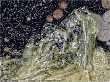
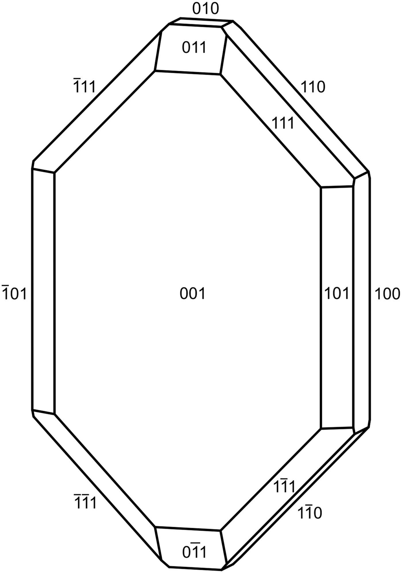
Markeyite crystals are blades and tablets (Fig. 1) up to ~1 mm in maximum dimension, flattened on {001} and elongate on [010]. Measurements on a Huber Reflection Goniometer 302 confirmed the crystal forms {100}, {010}, {001}, {110}, {101}, {011} and {111} (Fig. 2). No twinning was observed.
Markeyite (blades in centre) with similar new Ca uranyl carbonate (tapering crystals along bottom) and calcite (brown and grey balls) on asphaltum; field of view: 1.6 mm across.

Crystal drawing of markeyite; clinographic projection in nonstandard orientation, [010] vertical.

Crystals are pale yellowish green and transparent with vitreous lustre. The streak is white. The mineral fluoresces bright bluish white under a 405 nm laser. The Mohs hardness is between 1½ and 2, based upon scratch tests. Crystals are brittle with irregular fracture and three cleavages: perfect on {001}, good on {100} and {010}. At room temperature, the mineral dissolves very slowly in H2O (minutes) and dissolves immediately with effervescence in dilute HCl. The density measured by flotation in a mixture of methylene iodide and toluene is 2.68(2) g/cm3. The calculated density based on the empirical formula and unit-cell parameters obtained from single-crystal X-ray diffraction data is 2.699 g/cm3.
Optically, markeyite is biaxial (–), with α = 1.538(1), β = 1.542(1) and γ = 1.545(1) (measured in white light). The 2V measured directly on a spindle-stage is 81(2)°; the calculated 2V is 81.6°. Dispersion is r < v, weak. The mineral is weakly pleochroic: X = light greenish yellow, Y ≈ Z = light yellow; X > Y ≈ Z. The optical orientation is X = c, Y = b, Z = a.
Raman spectroscopy
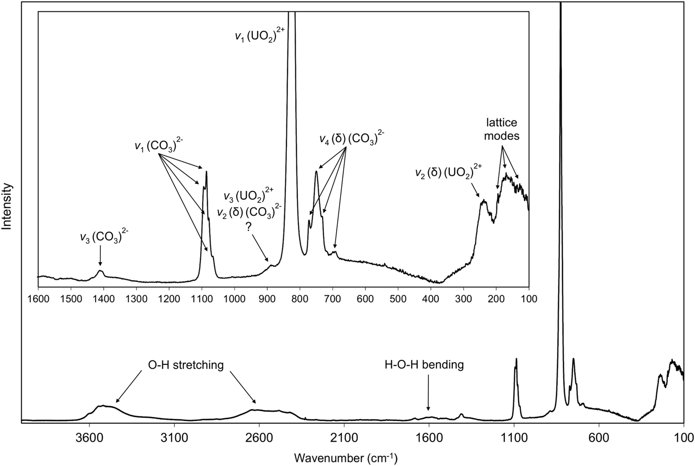
Raman spectroscopy was conducted on a Horiba XploRA PLUS using a 532 nm diode laser. A background correction was applied using the Horiba software. The Raman spectrum of markeyite is shown in Fig. 3.
The Raman spectrum of markeyite.

The broad multiple bands in the 3700–3300 and 2800–2300 cm–1 ranges are attributed to ν O–H stretching vibrations of structurally nonequivalent/symmetrically distinct hydrogen-bonded OH and H2O groups. The 3700–3300 cm–1 range corresponds to weak hydrogen bonds and the 2800–2300 cm–1 range to strong hydrogen bonds (Libowitzky, Reference Libowitzky1999). A weak, broad band centred at ~1600 cm–1 is attributed to the ν2 (δ) bending vibrations of H2O.
The aforementioned H2O band partly overlaps with a very weak broad band associated with the split doubly degenerate ν3 (CO3)2– antisymmetric stretching vibrations of the (CO3)2– units, with a more distinct weak band at 1412 cm–1. Medium to strong bands at 1095, 1086, 1078 and 1067 cm–1 are connected with the ν1 (CO3)2– symmetric stretching vibrations. These bands are consistent with the presence of four structurally nonequivalent carbonate units (Koglin et al., Reference Koglin, Schenk and Schwochau1979; Anderson et al., Reference Anderson, Chieh, Irish and Tong1980; Čejka, Reference Čejka, Burns and Finch1999 and Reference Čejka2005; and references therein), but do not preclude the presence of a fifth carbonate unit.
A weak band at 882 cm–1 may be due to the ν2 (δ) (CO3)2– bending vibrations or to the ν3 (UO2)2+ antisymmetric stretching vibration corresponding with the U–O bond length in uranyl at ~1.80 Å; an overlap/coincidence of these two bands is possible. A very strong band at 825 cm–1 is assigned to the ν1 (UO2)2+ symmetric stretching vibrations and provides an inferred U–O bond length of ~1.79 Å (Bartlett and Cooney, Reference Bartlett and Cooney1989). Also a coincidence of the ν2 (δ) (CO3)2– bending vibration and ν1 (UO2)2+ symmetric stretching vibration is likely.
Weak to strong bands at 772, 751, 733 and 694 cm–1 are assigned to the doubly degenerate ν4 (δ) (CO3)2– bending vibrations. A medium broad band at 238 cm–1 is assigned to the split doubly degenerate ν2 (δ) (UO2)2+ bending vibrations and weak to medium bands at 170, 155 and 128 cm–1 to the lattice modes (Koglin et al., Reference Koglin, Schenk and Schwochau1979; Anderson et al., Reference Anderson, Chieh, Irish and Tong1980; Čejka, Reference Čejka, Burns and Finch1999 and Reference Čejka2005).
Chemical composition
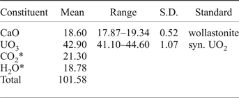
Chemical analyses (nine) were performed using a CamScan 4D electron microprobe in energy-dispersive spectroscopy mode (20 kV, 5 nA and 3 µm beam diameter). Attempts to use wavelength-dispersive spectroscopy with a higher beam current were made, but resulted in partial dehydration and totals significantly higher than 100 wt.%. H2O and CO2 were not determined directly because of extreme paucity of material. The H2O and CO2 contents were calculated by stoichiometry on the basis of 75 O atoms per formula unit and confirmed by the crystal-structure refinement and Raman spectroscopy. No other elements with atomic numbers higher than 8 were observed. Analytical data are given in Table 1.
Chemical composition (in wt.%) for markeyite.

* Based on the structure.
S.D. – Standard deviation.
The empirical formula is Ca8.91(U1.01O2)4(CO3)13·28H2O. The ideal formula is Ca9(UO2)4(CO3)13·28H2O which requires CaO 18.52, UO3 41.98, CO2 20.99 and H2O 18.51, total 100 wt.%. The Gladstone-Dale compatibility index 1 – (KP/KC) for the empirical formula is –0.027, in the excellent range (Mandarino, Reference Mandarino2007), using k(UO3) = 0.118, as provided by Mandarino (Reference Mandarino1976).
X-ray crystallography and structure refinement
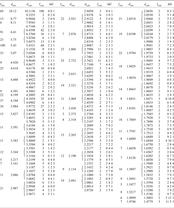
Powder X-ray studies were done using a Rigaku R-Axis Rapid II curved imaging plate microdiffractometer, with monochromatized MoKα radiation (λ = 0.71075 Å). A Gandolfi-like motion on the ϕ and ω axes was used to randomize the sample and observed d values and intensities were derived by profile fitting using JADE 2010 software (Materials Data, Inc.). The powder data presented in Table 2 show good agreement with the pattern calculated from the structure determination. Unit-cell parameters refined from the powder data using JADE 2010 with whole pattern fitting are: a = 17.9688(13), b = 18.4705(6), c = 10.1136(4) Å and V = 3356.6(3) Å3.
Powder X-ray diffraction data (d in Å) for markeyite. Only calculated lines with I ≥ 2 are listed.

The single-crystal structure data were collected at room temperature using the same diffractometer and radiation noted above. The data were processed using the Rigaku CrystalClear software package and an empirical (multi-scan) absorption correction was applied using the ABSCOR program (Higashi, Reference Higashi2001) in the CrystalClear software suite. The structure was solved by direct methods using SIR2011 (Burla et al., Reference Burla, Caliandro, Camalli, Carrozzini, Cascarano, Giacovazzo, Mallamo, Mazzone, Polidori and Spagna2012). SHELXL-2013 (Sheldrick, Reference Sheldrick2015) was used for the refinement of the structure.
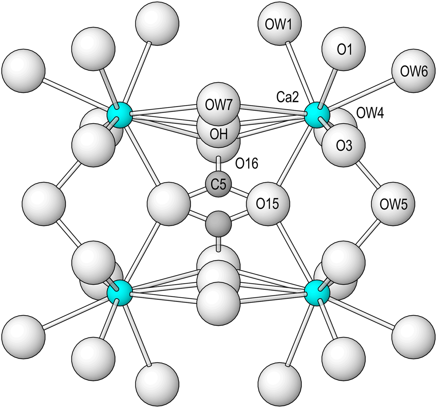
Determining the locations of most atoms was straightforward. The initial structure determination showed that the O15 site, nearly fully occupied by O, is only 2.28 Å from an equivalent O15 site. We first thought that the short O15–O15 distance could be indicative of a very strong hydrogen bond; however, the distance is typical for an O–O edge of a CO3 group. Closer examination of difference-Fourier maps revealed that two partially-occupied CO3 groups (centred by C5) share this O15–O15 edge and are completed by a partially occupied O site, O16. In subsequent refinements, the occupancies of the C5 and O16 sites were refined as equivalent. Another partially occupied O site (OH) was located 1.43 Å from the O16 site and 1.31 Å from OW7 on the opposite side. The OH site cannot be occupied when either the O16 or OW7 site is occupied. In subsequent refinements, the occupancies of the OW7 and OH sites were refined with a combined occupancy of 1.0. The aforementioned sites are shown in Fig. 4. There are three other partially occupied O sites, OW10, OW11 and OW12. The OW11 and OW12 sites are separated by only 1.41 Å; consequently, their occupancies were refined with a combined occupancy of 1.0. In the final refinement, soft distance restraints were placed on the distances between the atoms in the C5O3 group, which slightly improved the refinement and bond-valence sums. All sites, except the low-occupancy OW12 site, were refined with anisotropic displacement parameters.
The grouping of Ca2 polyhedra in markeyite, viewed down [001], showing the linkage by the partially occupied C5 carbonate group, OH and OW7.

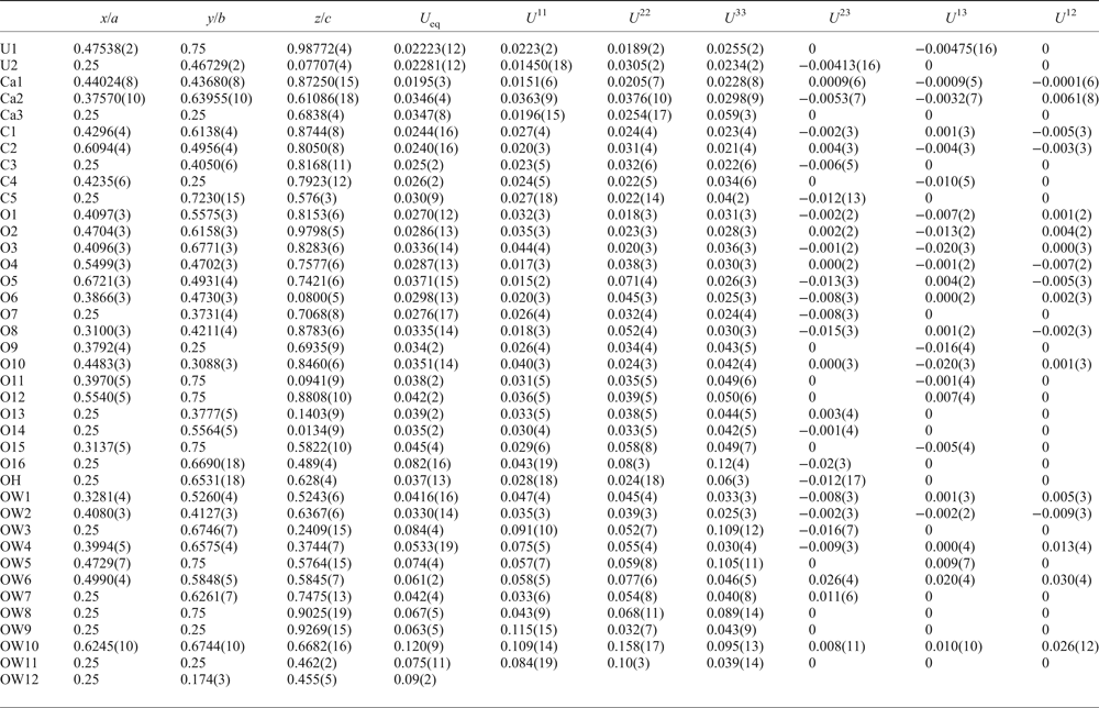
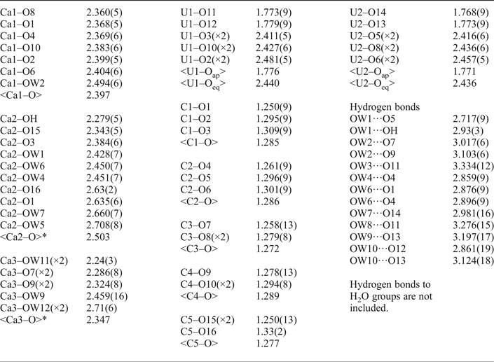
Data collection and refinement details are given in Table 3, atom coordinates and displacement parameters in Table 4, selected bond distances in Table 5 and a bond-valence analysis in Table 6. The crystallographic information files have been deposited with the Principal Editor of Mineralogical Magazine and are available as Supplementary material (see below).
Data collection and structure refinement details for markeyite.*
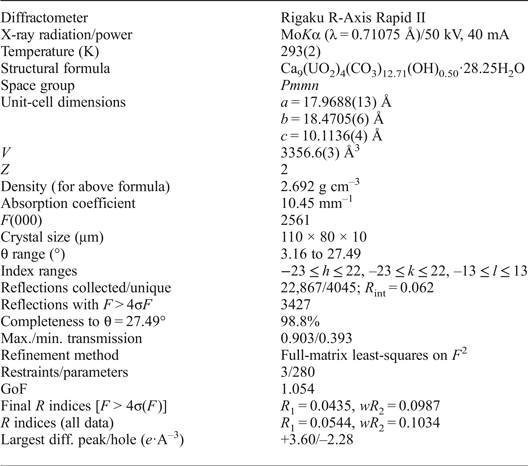
*R int = Σ|F o2–F o2(mean)|/Σ[F o2]. GoF = S = {Σ[w(F o2–F c2)2]/(n–p)}1/2. R 1 = Σ||F o|–|F c||/Σ|F o|. wR 2 = {Σ[w(F o2–F c2)2]/Σ[w(F o2)2]}1/2; w = 1/[σ2(F o2)+(aP)2+bP] where a = 0.0420, b = 40.5434 and P = [2F c2+Max(F o2,0)]/3.
Atom coordinates and displacement parameters (Å2) for markeyite.

* Occupancies: C5/O16: 0.35(3); O15: 0.93(4); OH/OW7: 0.25/0.75(3); OW10: 0.75(3); OW11/OW12: 0.71/0.29(5).
Selected bond distances (Å) for markeyite.

* Weighted average based on partial occupancies of OH, O15, O16, OW7, OW11 and OW12 sites; the effective coordination of Ca2 is 8.27 and that of Ca3 is 7.00.
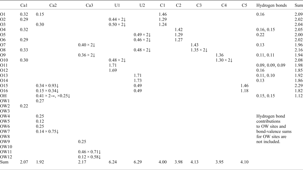
Bond-valence analysis for markeyite. Values are expressed in valence units.*

* Multiplicity is indicated by ×↓→. Bond strength contributions to Ca sites from partially occupied O sites are adjusted for their occupancies. Ca2+–O and C4+–O bond-valence parameters from Brown and Altermatt (Reference Brown and Altermatt1985); U6+–O bond valence parameters from Burns et al. (Reference Burns, Ewing and Hawthorne1997); hydrogen-bond strengths based on O–O bond lengths from Ferraris and Ivaldi (Reference Ferraris and Ivaldi1988).
Description and discussion of the structure
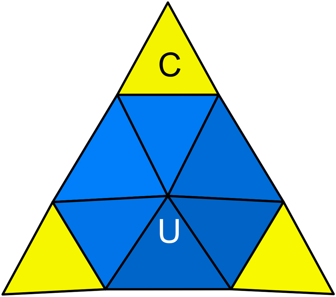
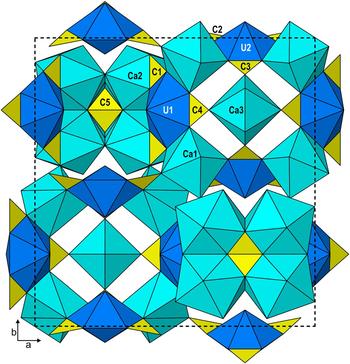
Two U sites (U1 and U2) in the structure of markeyite are each surrounded by eight O atoms forming a squat UO8 hexagonal bipyramid. These bipyramids are each chelated by three CO3 groups, forming uranyl tricarbonate clusters (UTC) of formula [(UO2)(CO3)3]4– (Burns Reference Burns2005; Fig. 5). There are three different Ca–O polyhedra in the structure: Ca1 bonds to seven fully occupied O sites; Ca2 bonds to five fully occupied and five partially occupied O sites (Fig. 4) for a total effective coordination of 8.27; and Ca3 bonds to five fully occupied and four partially occupied O sites for a total effective coordination of 7. The three Ca–O polyhedra share edges and corners with the UTCs in very different ways. The Ca1 polyhedra share edges with U1 and U2 bipyramids and corners with C1 and C2 triangles in different UTCs. Pairs of Ca2 polyhedra share an edge to form a dimer, which is linked to a second dimer through the partially occupied C5 triangles and OH and OW7 sites. The group of four Ca2 polyhedra (and C5 triangle) is linked to two U1 UTCs by edge sharing between Ca2 polyhedra and C1 triangles. The Ca3 polyhedra share corners with two C3 and two C4 triangles, each being in a different UTC. The linkages between UTCs and Ca polyhedra form thick corrugated heteropolyhedral layers parallel to {010} (Fig. 5) and these layers link to one another and to interlayer H2O groups (OW3, OW8 and OW10) only via hydrogen bonds (Fig. 6).
The uranyl tricarbonate cluster (UTC) of formula [(UO2)(CO3)3]4–.

The heteropolyhedral layer in markeyite.

The formula based upon the refined structure is Ca9(UO2)4(CO3)12.71(OH)0.50·28.25H2O. The ideal formula assumes full occupancy of the O15 site and half occupancy of the C5 and O16 sites, providing one CO3 group per formula unit. The OH site, with an occupancy of 0.25, is combined with the nearby OW7 site, with a refined occupancy of 0.75. The resultant ideal formula is Ca9(UO2)4(CO3)13·28H2O.
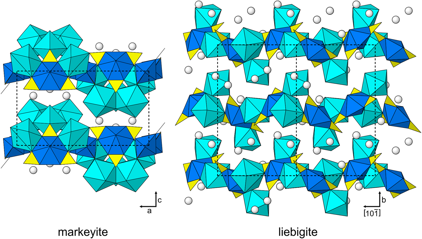
The structure of liebigite, Ca2(UO2)(CO3)3·11H2O (Mereiter, Reference Mereiter1982), contains the same structural components as that of markeyite. The same types of polyhedral linkages occur in both structures. As in the structure of markeyite, the Ca–O polyhedra link the UTCs in the structure of liebigite forming thick corrugated heteropolyhedral layers and these layers link to one another and to interlayer H2O groups only via hydrogen bonds (Fig. 7). However, topologies of the two structures are quite different. Of particular note, in the liebigite structure, one of the Ca–O polyhedra shares an edge with a CO3 group of the UTC, but there is no such linkage in the markeyite structure.
The structures of markeyite and liebigite viewed parallel to the heteropolyhedral layers. UO8 hexagonal bipyramids are dark blue, CO3 triangles are yellow and Ca–O polyhedra are light blue; O atoms of isolated H2O groups are white balls. Unit cell outlines are shown as dashed black lines.

Acknowledgements
Igor Pekov and an anonymous reviewer are thanked for their constructive comments on the manuscript. An anonymous CNMNC member is thanked for pointing out the likelihood that the short O15–O15 distance corresponds to the edge of an additional CO3 group. A portion of this study was funded by the John Jago Trelawney Endowment to the Mineral Sciences Department of the Natural History Museum of Los Angeles County. This research was also financially supported by GACR post-doctoral Grant no. 13-31276P to J.P. and by the long-term project DKRVO 2016-02 of the Ministry of Culture of the Czech Republic (National Museum 00023272) to J.Č.
Supplementary material
To view supplementary material for this article, please visit https://doi.org/10.1180/minmag.2017.081.085