Significant Outcomes
∙ A decline in cognitive function was associated with decreased regional cerebral blood flow (rCBF) among late-onset Alzheimer’s disease (AD) patients with low vascular risk, but not among those with high vascular risk.
∙ Hypertension and diabetes mellitus have distinct influences on the relationships between cognitive function and rCBF in late-onset AD patients.
Limitations
∙ The severity of each metabolic disease and dose of medications used to treat these diseases were not evaluated.
∙ The sample size was small.
Introduction
In its typical course, AD begins with episodic memory dysfunction, followed by additional cognitive impairment (1,Reference McKhann, Drachman, Folstein, Katzman, Price and Stadlan2). Single-photon emission computed tomography (SPECT) studies show that rCBF in the parietal-temporal lobes is significantly correlated with global cognitive function as measured by the Mini-Mental State Examination (MMSE) and the Alzheimer’s Disease Assessment Scale-cognitive subscale (ADAS-cog) (Reference Lampl, Sadeh, Laker and Lorberboym3–5). It is likely that the pattern of rCBF reduction depends on the age of AD onset. Patients who develop the disease after age 65 (late-onset) show topographic patterns of brain grey matter atrophy in the medial temporal lobe as well as hippocampal atrophy; whereas, early-onset AD shows atrophy in the occipital and parietal lobes, including the precuneus (6–9). In our recent study of patients with late-onset AD, we revealed that rCBF was reduced in the frontal, temporal, and limbic lobes, and to a lesser degree in the parietal and occipital lobes (Reference Takahashi, Oda, Okubo and Shirayama10). Moreover, these patients’ scores on the ADAS-cog were significantly correlated with rCBF in some gyri of the frontal, parietal, and limbic lobes (Reference Takahashi, Oda, Okubo and Shirayama10). Many genetic association studies have investigated genetic risk factors associated with late-onset AD risk, including apolipoprotein E heterozygosity (Reference Reitz and Mayeux11). A recent study showed that a late-onset AD polygenic risk profile score predicts hippocampal function (12).
Compared with early-onset AD patients, late-onset AD patients are more likely to exhibit comorbid hypertension and/or hypercholesterolaemia (Reference Panegyres and Chen13). These finding suggests that the cognitive impairment of late-onset AD patients has a greater association with cerebrovascular disease risk factors, which are thought to contribute to a faster rCBF regulation in AD (14). As reviewed by Kisler et al. (Reference Kisler, Nelson, Montagne and Zlokovic15), neurovascular dysfunction may influence rCBF regulations in AD. Notably, cognitive impairment in AD patients is affected by cerebrovascular disease risk factors (Reference Richard and Pasquier16), including hypertension (Reference Bellew, Pigeon, Stang, Fleischman, Gardner and Baker17–Reference Razay, Williams, King, Smith and Wilcock19), hyperlipidaemia (Reference Hajjar, Schumpert, Hirth, Wieland and Eleazer20–22), and diabetes mellitus (21,Reference Sato, Hanyu, Hirao, Kanetaka, Sakurai and Iwamoto23). These risk factors reportedly have both short-term and long-term impacts on cognitive decline among elderly people without dementia, and on AD onset and subsequent deterioration of cognitive function (24–Reference Sato and Morishita26). Compared with AD patients without cerebrovascular risk factors, those with cerebrovascular risk factors show reduced brain perfusion in broader regions and a more severe decrease in global cognitive function measured by the MMSE (14). Importantly, treatment of vascular risk factors has been associated with slower progression of global cognitive decline among AD patients without cerebral vascular disease (Reference Deschaintre, Richard, Leys and Pasquier27).
We analysed brain perfusion using three-dimensional stereotactic surface projection (3D-SSP) and the stereotactic extraction estimation method (SEE) level 3. The 3D-SSP can more accurately measure quantitative data and detect the localisation of metabolic abnormalities using stereotactic coordinates (Reference Minoshima, Frey, Koeppe, Foster and Kuhl28). Compared with statistical parametric mapping, this method is less affected by brain atrophy and by partial volume effects (29). Furthermore, using the SEE method in combination with 3D-SSP enables a more objective evaluation of rCBF (30).
In our present study, we aimed to examine how cerebrovascular risk factors, assessed using the Hachinski Ischemic Score (HIS), might influence the rCBF evaluated by SPECT and its relationship with cognitive impairment evaluated with the ADAS-cog in 40 patients with late-onset AD. We additionally studied how these relationships were affected by the metabolic diseases, hypertension, hyperlipidaemia, and diabetes mellitus, in various regions.
Materials and methods
Patients
This study included 40 drug-naïve patients with late-onset AD who were enrolled from the outpatient clinic of Teikyo University Chiba Medical Center. AD was diagnosed following the DSM-IV-TR criteria for dementia of the Alzheimer’s type (31), and the enrolled patients fulfilled the NINCDS-ADRDA criteria for probable or possible AD (Reference McKhann, Drachman, Folstein, Katzman, Price and Stadlan2). To select patients with early-stage disease, participants were required to have MMSE scores of 26 or below (Reference Folstein, Folstein and McHugh32,Reference Kukull, Larson, Teri, Bowen, McCormick and Pfanschmidt33). Magnetic resonance imaging or computed tomography was performed when needed, for example, in cases of normal-pressure hydrocephalus. Patients were also examined with regards to thyroid function and vitamin levels to rule out hypothyroidism or other types of dementia, such as vascular dementia, frontotemporal dementia, or dementia with Lewy bodies. Patients were excluded from this study if they had received medical treatment for AD, since acetylcholinesterase inhibitors significantly influence rCBF, cognitive function, and their relationship in AD patients (34). Other criteria for exclusion were a history of cerebral vascular disease (including indicating a of history of stroke on the HIS); history of head trauma; seizures or other neurological disorders; mental retardation; alcohol or substance abuse; schizophrenia; major depressive disorder; bipolar disorder; and cardiac, pulmonary, vascular, or haematological conditions or other illnesses of sufficient severity to adversely affect cognition or functioning. The severity of functional impairment was evaluated using the Functional Assessment Stating scale (35). This study was approved by the ethics committee of Teikyo University Chiba Medical Center (study number 11-17), and was performed in accordance with the Helsinki Declaration of 1975, as revised in 2008. After a full explanation of all study procedures, patients and their closest caregivers gave written informed consent.
Assessment of cognitive function and vascular risk factors
The severity of AD and cognitive impairment was assessed using the ADAS-cog (Reference Rosen, Mohs and Davis36), a common rating instrument for assessing cognitive dysfunction in AD. The ADAS-cog comprises 11 components for measuring cognitive function – including word recall, word recognition, constructional praxis, orientation, naming, commands, ideational praxis, remembering test instructions, spoken language ability, word finding, and comprehension. Total scores on the ADAS-cog range from 0 to 70, with higher total scores indicating poorer cognitive performance.
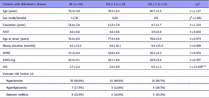
The HIS (37,38) was used to evaluate the degree of vascular risk. The HIS comprises of 13 items, but we omitted the item regarding ‘history of stroke’. Possible scores range from 0 to 18. Patients were divided into two groups: those with high vascular risk (HIS value of ≥5) and those with low vascular risk (HIS ≤4). The HIS cut-off values were selected based on the bimodal distribution of HIS scores in the present study (Table 1).
Demographic characteristics

ADAS-cog, Alzheimer’s Disease Assessment Scale-cognitive subscale; FAST, Functional Assessment Stating scale; HIS, Hachinski Ischemic Score; MMSE, Mini-Mental State Examination; n, number.
Values are reported as mean±SD.
***p<0.001.
SPECT imaging
In all subjects, cerebral blood flow was examined by brain perfusion SPECT. Twenty minutes before imaging, patients received an intravenous injection of 222 MBq of N-isopropyl-p-123I-iodoamphetamine. Image scanning was performed using a dual-head rotating gamma camera (Millennium MG, GE Healthcare, Milwaukee, WI, USA) with a parallel beam collimator, permitting spatial resolution of 10 mm full width, at half maximum. Continuous images were captured in 32 steps (64 projections), and each collected step counted for 30 s. Image reconstruction was performed by filtered backprojection, using Butterworth and Ramp filters with attenuation correction (Chang, 0.11 per cm). SPECT images had a matrix size of 64×64 mm and slice thickness of 6.78 mm.
Image analysis
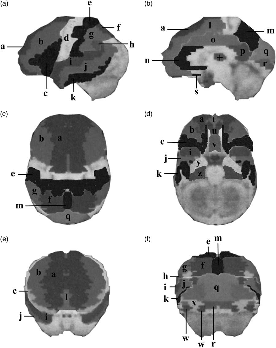
SPECT image data were analysed using the 3D-SSP programmed in Neurological Statistical Image Analyze Software (NEUROSTAT) (Reference Minoshima, Frey, Koeppe, Foster and Kuhl28). To evaluate the spatial distribution of abnormal cerebral blood flow, the original data were first realigned to the bicommissural (anterior commissure-posterior commissure) line and then transferred into the stereotactic standard atlas after rotation and centring (Fig. 1). Next, maximum cortical activity was projected onto the brain surface pixel. Brain activity data sets were normalised to mean cortical activity. The pixel values from each individual’s image data were compared with the normal database generated from 18 normal subjects (age range, 60–81 years). We then calculated pixel-by-pixel z-scores, representing the degree of rCBF reduction. Additionally, pixel-by-pixel data were used to divide the whole brain into segments at classified gyrus levels using SEE level 3 (14,30).
Brain surface images. (a) Lateral, (b) medial, (c) superior, (d) inferior, (e) anterior, (f) posterior. a, Superior frontal gyrus; b, middle frontal gyrus; c, inferior frontal gyrus; d, precentral gyrus; e, postcentral gyrus; f, superior parietal lobule; g, inferior parietal lobule; h, supramarginal gyrus; i, superior temporal gyrus; j, middle temporal gyrus; k, inferior temporal gyrus; l, medial frontal gyrus; m, precuneus; n, anterior cingulate; o, cingulate gyrus; p, posterior cingulate; q, cuneus; r, lingual gyrus; s, subcallosal gyrus; t, medial frontal gyrus; u, orbital gyrus; v, rectal gyrus; w, fusiform gyrus; x, inferior occipital gyrus; y, uncus; z, parahippocampal gyrus; +, thalamus.

Statistical analysis
Values are expressed as the mean±SD. Statistical analysis was performed using the Student’s t-test for parametric data, and the χ2 test for categorical data. Correlations between rCBF and cognitive function were examined using Pearson’s correlation coefficient. Differences between groups and correlations were considered significant when p value was<0.05.
Results
Patient demographics
We divided all patients into two groups, based on the HIS: those with high vascular risk factors (HIS≥5) and those with low vascular risk factors (HIS≤4). The two groups did not significantly differ in demographic data with the exception of HIS values denoting vascular risk (Table 1). The two groups did not significantly differ with regards to ADAS or MMSE scores.
Reduced hypoperfusion among AD patients regardless of their HIS
Patients with HIS ≤4 and ≥5 showed apparent reductions of rCBF, with z-scores of >1.5, in both sides of the inferior frontal, orbital, rectal, and subcallosal gyri of the frontal lobe, and the anterior cingulate of the limbic lobe (Table 2, bold). Both groups showed hypoperfusion on one side of the inferior temporal gyrus of the temporal lobe, the fusiform of the occipital lobe, and the uncus of the limbic lobe (Table 2). Overall, the patterns and degree of brain hypoperfusion were almost overlapping between groups, with some differences. Compared with AD patients with HIS ≥5, those with HIS ≤4 showed significantly lower rCBF, with z-scores>1.5, in the right side of the superior parietal lobule, inferior parietal lobule, and angular gyrus of the parietal lobe (Table 2). Patients with HIS ≥5 showed significantly lower rCBF in the right side of the transverse temporal gyrus of the temporal lobe, and a stronger degree of rCBF reduction, with z-scores around 1.5, in the medial frontal gyrus of the frontal lobe and the parahippocampal gyrus of the limbic lobe than patients with HIS ≤4 (Table 2).
Reduction of regional cerebral blood flow (rCBF) in Alzheimer’s disease (AD) patients with Hachinski Ischemic Score (HIS) values of ≤4 versus ≥5

* p<0.05, ** p<0.01, compared to the group of AD patients without vascular disease risk factors (HIS≤4).
Effects of vascular risk factors on the relationship between rCBF and cognitive function
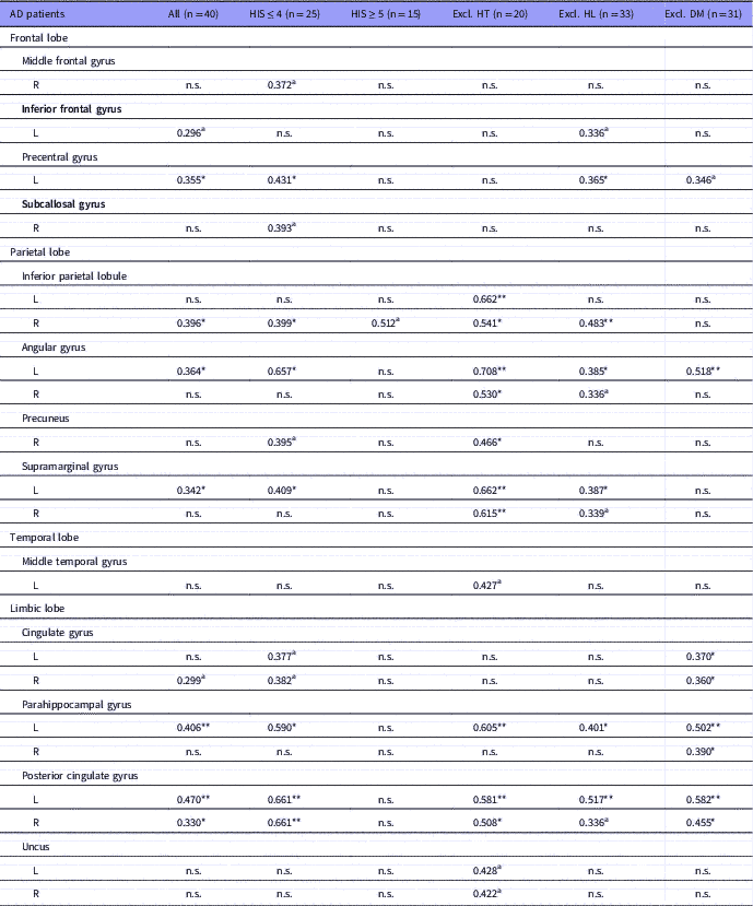
In all late-onset AD patients, ADAS-cog scores were significantly correlated with rCBF decreases in the precentral gyrus of the frontal lobe, the inferior parietal lobule, the angular, and supramarginal gyri of the parietal lobe, and the parahippocampal gyrus and posterior cingulate gyrus of the limbic lobe (Table 3). Compared with the full patient cohort, AD patients with low vascular risk (HIS≤4) showed a stronger association between hypoperfusion and ADAS-cog scores in the precentral gyrus of the frontal lobe, the inferior parietal lobe, angular gyrus, and supramarginal gyrus of the parietal lobe, and the parahippocampal and posterior cingulate gyri of the limbic lobe (Table 3). In contrast, among AD patients with high vascular risks (HIS≥5), we detected only one relationship (at the trend level) between the ADAS-cog and rCBF in the right inferior frontal gyrus (Table 3).
Relationships between regional cerebral blood flow (rCBF) and Alzheimer’s Disease Assessment Scale-cognitive subscale (ADAS-cog) total scores in various groups of patients with Alzheimer’s disease (AD)

DM, diabetes mellitus; Excl., excluding; HIS, Hachinski Ischemic Score; HL, hyperlipidaemia; HT, hypertension; L, left; R, right; n.s., not significant.
*p<0.05, **p<0.01, significant correlation.
a p<0.07, a trend for weak correlation.
Effects of metabolic diseases on the relationship between rCBF and cognitive function
We examined how metabolic diseases, namely hypertension, diabetes mellitus, and hyperlipidaemia, impacted the relationship between rCBF and the ADAS-cog. When patients with hypertension were removed from the analysis, we observed a stronger relationship between the ADAS-cog and rCBF in the inferior parietal lobule, the angular, precuneus, and supramarginal gyri of the parietal lobe, and the parahippocampal gyrus and posterior cingulate of the limbic lobe (Table 3). Omitting patients with hyperlipidaemia from our analysis also led to enhancement of the relationships between ADAS-cog and rCBF in the precentral gyrus of the frontal lobe, the inferior parietal lobule, the angular and supramarginal gyri of the parietal lobe, and the parahippocampal gyrus and posterior cingulate of the limbic lobe (Table 3). Removing patients with diabetes mellitus from our analysis led to enhancement of the relationship between the ADAS-cog total scores and rCBF in the angular gyrus of the parietal lobe, and the cingulate, parahippocampal, and posterior cingulate gyri of the limbic lobe (Table 3).
Discussion
Late-onset AD patients with HIS≤4 or ≥5 showed nearly overlapping degrees and regions of hypoperfusion. As shown in Table 2, prominent hypoperfusion was noted in both sides of the inferior frontal, orbital, rectal, and subcallosal gyri of the frontal lobe, and in the anterior cingulate of the limbic lobe in both groups. Hypoperfusion in the right-side inferior temporal gyri of the temporal lobe, the right-side fusiform of the occipital lobe, and the left-side uncus of the limbic lobe may be noteworthy in late-onset AD (Table 2). However, late-onset AD patients with HIS values≤4 presented more severe hypoperfusion in the right side of the superior parietal lobule, inferior parietal lobule, and angular gyrus of the parietal lobe than patients with HIS≥5 (Table 2). This finding suggests that factors other than hypoperfusion in the parietal lobe contribute to high vascular risk. In addition there were no differences in ADAS-cog scores between the two groups (Table 1).
There were different relationships between rCBF and ADAS-cog scores in late-onset AD patients with HIS values of ≤4 versus ≥5. When compared with the total cohort of AD patients, the subgroup with HIS≤4 showed a strong relationship between rCBF and ADAS-cog scores in the precentral gyrus of the frontal lobe, the inferior parietal lobe, angular gyrus, and supramarginal gyrus of the parietal lobe, and the parahippocampal and posterior cingulate gyri of the limbic lobe (Table 3). Among these regions, the inferior parietal lobe, angular gyrus and supramarginal gyrus of the parietal lobe were related to changes in the ADAS-cog and rCBF during 18 months of follow-up (39). Dementia levels evaluated with the ADAS-cog can be explained, at least in part, by hypoperfusion in these regions.
On the other hand, AD patients with HIS≥5 showed no significant relationship between rCBF and ADAS-cog scores. Although this may have been due to the small sample size, this suggests that vascular risk factors affect the relationship between rCBF and ADAS-cog scores. Moreover, it is well documented that vascular risk factors promote cognitive dysfunction (Reference Skoog, Kakaria and Breteler40) and increase the risk of AD (Reference Luchsinger, Reitz, Honig, Tang, Shea and Mayeux41). Thus, the existence of factors other than hypoperfusion may worsen the ADAS-cog scores and lessen the correlation between hypoperfusion and ADAS-cog scores.
Further analysis was done to elucidate how vascular factors influence rCBF in late-onset AD. To examine how metabolic disease impacted rCBF, we separately analysed data sets, excluding data from patients with each candidate disease, one by one. This approach is based on the assumption that if a significant relationship emerges after excluding a specific metabolic disease from correlation analysis, the excluded disease may be an important factor in disturbing the relationship between rCBF and cognitive function.
Excluding subjects with hypertension from the analysis revealed a strong relationship between ADAS-cog and hypoperfusion in the inferior parietal lobule, angular gyrus, precuneus, and supramarginal gyrus of the parietal lobe, and the parahippocampal and posterior cingulate gyri of the limbic lobe (Table 3). Thus, it appeared that hypertension affected the parietal lobe and, to a lesser degree, the limbic lobe. Supporting this finding, a published review indicated that hypertension is a strong predictor of memory impairment (Reference Skoog, Kakaria and Breteler40). Moreover, hypertension impairs hippocampal neurogenesis and long-term memory (42). Thus, in cases with hypertension, factors other than hypoperfusion could lead to worse ADAS-cog scores and loss of the simple correlation between hypoperfusion and ADAS-cog scores.
Omitting hyperlipidaemia from analysis had less impact on the relationship between hypoperfusion and cognitive impairment in late-onset AD patients (Table 3). The relationship between ADAS-cog and rCBF when excluding hyperlipidaemia was almost the same as when all patients were considered, suggesting that hyperlipidaemia does not affect the relationship between hypoperfusion and cognitive function. Further investigation of hyperlipidaemia in late-onset AD is necessary.
The exclusion of patients with diabetes mellitus revealed a strong relationship between ADAS-cog and rCBF in the angular gyrus of the parietal lobe and in the cingulate, parahippocampal, and posterior cingulate gyri of the limbic lobe (Table 3). Thus, diabetes mellitus had some effects on the limbic lobe according to SPECT data. Diabetes mellitus is reportedly a risk factor for mild cognitive impairment among elderly subjects (43) and is associated with abnormalities in structural imaging markers, including hippocampal atrophy, brain volume reduction, and white matter hyperintensity (44,Reference Fotuhi, Do and Jack45). Therefore, in patients with diabetes mellitus, factors other than hypoperfusion might worsen the ADAS-cog scores and result in the loss of the simple correlation between hypoperfusion and ADAS-cog scores.
There are some limitations to the present study. First, severity of each metabolic disease and dose of medication to treat these diseases were not evaluated; although, these factors may affect the ADAS-cog scores and rCBF. In addition the sample size was small.
In conclusion, we found that cognitive function according to the ADAS-cog was significantly associated with rCBF in late-onset AD patients with low vascular risk (HIS≤4), but not those with high vascular risk (HIS≥5), indicating that reductions in rCBF are the main cause of cognitive deficits in late-onset AD. We also observed that metabolic diseases, namely hypertension and diabetes mellitus, influenced and disrupted the relationship between hypoperfusion and cognitive impairments. Thus, factors other than hypoperfusion, such as hypertension and diabetes mellitus, could be involved in the cognitive dysfunction of late-onset AD patients with high vascular risk.
Financial Support
This research did not receive any specific grant from any funding agencies in the public, commercial, or not-for-profit sectors.
Conflicts of Interest
Y.S. has received research support from Eisai, MSD, Pfizer, Takeda and Mitsubishi-Tanabe. M.T., Y.O., and Y.S. have no competing interest.