INTRODUCTION
Neospora caninum is a tissue cyst-forming coccidian, closely related to Toxoplasma gondii, that causes reproductive failure in cattle (Dubey et al. Reference Dubey, Schares and Ortega-Mora2007). Currently, N. caninum is considered to be a major cause of infectious bovine abortion worldwide, causing important economic losses to the cattle industry (Dubey et al. Reference Dubey, Schares and Ortega-Mora2007). Dogs and coyotes act as definitive hosts in the life cycle of this parasite, and cattle and a wide range of other ungulates can act as intermediate hosts (Dubey and Schares, Reference Dubey and Schares2006).
N. caninum infection may occur by horizontal transmission when cattle ingest sporulated oocysts shed by the definitive host, although several reports have shown the importance of endogenous congenital transmission from a persistently infected dam to a foetus (Trees and Williams, Reference Trees and Williams2005). Infection of a pregnant animal may result in abortion or the birth of a weak calf, although in the majority of cases, the calf is born clinically healthy but persistently infected. Different host factors are involved in the outcome of neosporosis during pregnancy (Innes et al. Reference Innes, Wright, Bartley, Maley, Macaldowie, Esteban-Redondo and Buxton2005; Collantes-Fernández et al. Reference Collantes-Fernandez, Rodriguez-Bertos, Arnaiz-Seco, Moreno, Aduriz and Ortega-Mora2006b; Dubey et al. Reference Dubey, Buxton and Wouda2006), but other factors, such as isolate virulence, may play an important role. Thus, determining the influence of intra-species diversity on N. caninum infection is important for understanding the epidemiology and pathogenicity of this parasite.
The broad host range of N. caninum, its global distribution and the possibility of sexual recombination in its definitive host suggest significant biological diversity. N. caninum isolates have shown extensive genetic diversity (Schock et al. Reference Schock, Innes, Yamane, Latham and Wastling2001; Regidor-Cerrillo et al. Reference Regidor-Cerrillo, Pedraza-Díaz, Gómez-Bautista and Ortega-Mora2006, Reference Regidor-Cerrillo, Gómez-Bautista, Pereira-Bueno, Aduriz, Navarro-Lozano, Risco-Castillo, Fernández-García, Pedraza-Diaz and Ortega-Mora2008; Basso et al. Reference Basso, Schares, Bärwald, Herrmann, Conraths, Pantchev, Vrhovec and Schares2009), as well as significant variations in their in vitro behaviour (Schock et al. Reference Schock, Innes, Yamane, Latham and Wastling2001; Pérez-Zaballos et al. Reference Pérez-Zaballos, Ortega-Mora, Álvarez-García, Collantes-Fernández, Navarro-Lozano, García-Villada and Costas2005) and pathogenicity in murine models (Lindsay et al. Reference Lindsay, Lenz, Cole, Dubey and Blagburn1995; Atkinson et al. Reference Atkinson, Harper, Ryce, Morrison and Ellis1999; Miller et al. Reference Miller, Quinn, Windsor and Ellis2002; Collantes-Fernández et al. Reference Collantes-Fernández, López-Pérez, Álvarez-García and Ortega-Mora2006a; Rojo-Montejo et al. Reference Rojo-Montejo, Collantes-Fernández, Regidor-Cerrillo, Álvarez-García, Marugan-Hernández, Pedraza-Díaz, Blanco-Murcia, Prenafeta and Ortega-Mora2009; Pereira García-Melo et al. Reference Pereira García-Melo, Regidor-Cerrillo, Ortega-Mora, Collantes-Fernández, de Oliveira, de Oliveira and Caetano da Silva2009). Nevertheless, the number of isolates studied is limited, and most of these studies have been carried out with isolates obtained from animals with clinical signs of infection. Thus, the analysed population could be biased towards the most virulent isolates.
Here we studied the variability of 8 N. caninum isolates using a well-established experimental mouse model. These isolates were obtained from asymptomatic but congenitally infected calves in Spain, and they have been genetically characterized using microsatellite markers (Regidor-Cerrillo et al. Reference Regidor-Cerrillo, Gómez-Bautista, Pereira-Bueno, Aduriz, Navarro-Lozano, Risco-Castillo, Fernández-García, Pedraza-Diaz and Ortega-Mora2008). This work was approached with the aim of investigating the pathogenic diversity of isolates of this species, at least of those isolated from healthy but congenitally infected cattle.
MATERIALS AND METHODS
Culture and preparation of bovine N. caninum isolates for BALB/c mouse inoculation
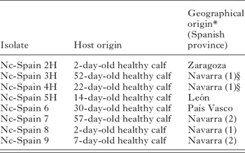
The isolates of N. caninum used in this study are shown in Table 1. These isolates were maintained in monolayer cultures of the monkey kidney cell line MARC-145, as previously described (Regidor-Cerrillo et al. Reference Regidor-Cerrillo, Gómez-Bautista, Pereira-Bueno, Aduriz, Navarro-Lozano, Risco-Castillo, Fernández-García, Pedraza-Diaz and Ortega-Mora2008).
Summary of name, host and geographical origin of the isolates included in this study

* Calves from Navarra originated from 2 dairy herds. The number in parentheses identifies the dairy herd.
§ Nc-Spain 3H and Nc-Spain 4H are genetically identical by multilocus microsatellite analysis. They were isolated from calves with different dams.
The tachyzoites used in the mouse inoculations were recovered from 3-day growth cultures when the greater part of them was still intracellular (at least 80% of undisrupted parasite vacuoles in the cell monolayer). Tachyzoite cultures were scraped with a plastic cell scraper, passed through 21-gauge needles, harvested by centrifugation at 1350 g for 10 min and suspended in sterile phosphate-buffered saline (PBS). Tachyzoite number was determined by trypan blue exclusion followed by counting in a Neubauer chamber, and the organisms were resuspended in PBS at the required dose of 106 tachyzoites in a final volume of 200 μl per mouse. Mice were inoculated within 1 h of tachyzoite collection. All isolates used were passaged in cell culture between 11 and 25 times.
Murine model and sample collection
Pathogenicity studies were performed using a BALB/c mouse model as described previously by Collantes-Fernández et al. (Reference Collantes-Fernández, López-Pérez, Álvarez-García and Ortega-Mora2006a). Female inbred BALB/c mice weighing 18–22 g were purchased from a commercial supplier (Harlan Iberica, Barcelona, Spain). Mice were housed in groups of 10 each in a controlled environment, with 12-h light and 12-h dark cycles and provided with rodent feed and water ad libitum. The uninfected control group was intraperitoneally injected with sterile PBS, and the other 8 groups were intraperitoneally inoculated with 106 tachyzoites per mouse of each isolate (day 0) (Table 1). Mice were examined daily for clinical signs compatible with neosporosis (rough hair coats, inactivity, anorexia and neurological signs consisting of head tilting, walking in circles, ataxia, pelvic limb weakness and paralysis). Five mice from N. caninum-inoculated groups and 3 mice from the control group were randomly sacrificed by CO2 inhalation on days 1, 2, 4, 8, 16, 32 and 64 post-inoculation (p.i.), except for the Nc-Spain 5H-infected group, for which day 64 p.i. was not performed. In the Nc-Spain 4H, Nc-Spain 6, Nc-Spain 8 and Nc-Spain 9 inoculated groups, 10 instead of 5 mice were evaluated on day 32 p.i. All animal-handling procedures complied with EU legislation.
Blood samples were collected by cardiac puncture at necropsy and recovered in EDTA tubes. Plasma and peripheral blood cells were obtained by centrifugation at 2000 g for 10 min. Plasma was aliquoted and preserved at −80°C for ELISA, and peripheral blood cells were stored at 4°C for less than 24 h until genomic DNA extraction was carried out. Mouse brains and lungs were also collected under aseptic conditions and portions were frozen at −80°C until DNA was extracted. Tissue specimens from brain, lungs, heart and liver were fixed in 10% neutral buffered formalin for processing by routine histological methods.
DNA extraction and nested-PCR
Genomic DNA was extracted from 20 mg of brain and lung tissues using the Real Pure Extracción DNA Genómico kit (Durviz, Valencia, Spain) and from blood cells using the Real Pure Extracción DNA SSS kit (Durviz, Valencia, Spain), according to the manufacturer's instructions. The concentration of DNA was determined by spectrophotometry and adjusted to 50 ng/μl. A total of 5 μl was used for PCR amplification. Detection of parasite DNA was performed by a nested-PCR on the ITS-1 region of N. caninum as described by Buxton et al. (Reference Buxton, Maley, Wright, Thomson, Rae and Innes1998). To avoid false-positive reactions, DNA extraction, PCR sample preparation and electrophoresis were performed in separate rooms with different sets of instruments, and aerosol barrier tips and disposable gloves were employed. A secondary amplification product was visualized as a 249 bp band by 1·5% agarose gel electrophoresis in 0·5× TBE buffer and ethidium bromide staining. DNA extracted from tachyzoites maintained in cell culture and H2O was also included as a PCR control in each batch of reactions.
Real-time PCR
Positive samples from lung and brain containing parasite DNA, as assessed by nested-PCR, were quantified by real-time PCR. We used primer pairs from the N. caninum Nc-5 sequence to quantify parasites, and primers from the 28S rRNA gene to quantify host DNA (Collantes-Fernández et al. Reference Collantes-Fernández, Zaballos, Alvarez-García and Ortega-Mora2002). The N. caninum number was calculated by interpolation of the corresponding Ct values (cycle threshold: the fractional cycle number reflecting a positive PCR result) on a standard curve generated in each real-time PCR run by assaying serial 10-fold dilutions of parasite DNA equivalent to 10−1–104 tachyzoites. The amount of DNA per sample was normalised by quantification of the 28S rRNA gene, and a standard curve was generated with 5-fold serial dilutions of mouse brain DNA quantified by UV spectrophotometry. Parasite number in tissue samples (parasite load) was expressed as parasite number/μg host DNA.
Histopathological analysis
Samples of brain, heart, liver and lung from mice were processed by routine histological methods. Tissues were fixed in 10% neutral buffered formalin and dehydrated through graded alcohols before being embedded in paraffin wax, sectioned at 4 μm and stained with haematoxylin and eosin (H&E). Analysis was based on the observation of lesions characteristic of or consistent with N. caninum infection (Lindsay and Dubey, Reference Lindsay and Dubey1989; Collantes-Fenandez et al. Reference Collantes-Fernandez, Alvarez-Garcia, Perez-Perez, Pereira-Bueno and Ortega-Mora2004) in the brain: meningitis, perivascular cuffing, gliosis, glial nodules and granulomas with or without necrosis; in the heart: myocarditis; in the liver: periportal or random hepatitis and hepatocellular necrosis; and in the lungs: interstitial pneumonia. Lesions were assessed according to the severity of inflammation and the extension of affected tissue in each section, and lesion scores were assigned on a 3-point scale according to the following schemes. The lesions in brain tissues were scored based on the following criteria: no lesion (=0); mild meningitis, perivascular cuffing and gliosis (=1); moderate meningitis, perivascular cuffing, gliosis, mild glial nodules and focal granulomas (=2); or severe lesions including meningitis, perivascular cuffing, gliosis, glial nodules and multifocal granulomas (=3). The liver lesion scoring was based on the following criteria: no lesion (=0); mild portal or perisinusoidal hepatitis (=1); moderate hepatitis and minimal hepatocellular necrosis (=2); or severe hepatitis and moderate hepatocellular necrosis (=3). Lesions in lung and heart were assigned according to the severity of interstitial pneumonia and myocarditis, respectively: no lesion (=0); mild (=1); moderate (=2); or severe lesion (=3). The average of these values was determined for each animal, and a median score corresponding to the phase of infection for each group and organ was assessed. Representative lesion photomicrographs from target organs that yielded marked differences are depicted in Fig. 1.
Histopathological lesions in sections of examined organs stained with H&E from BALB/c mice inoculated with Neospora caninum isolates. (A) Mild focal meningoencephalitis in the brain from a mouse infected with the Nc-Spain 2H isolate and sacrificed at day 64 p.i. (200×). (B) Focal meningoencephalitis, perivascular cuffing, gliosis and granulomatous inflammation surrounding a necrotic focus in the brain from a mouse infected with the Nc-Spain 7 isolate and sacrificed at day 38 p.i. with clinical signs (200×). (C) Foci of minimal interstitial pneumonia in the lung from a mouse infected with the Nc-Spain 5H isolate and sacrificed at day 32 p.i. (200×). (D) Moderate interstitial pneumonia in the lung from a mouse infected with the Nc-Spain 5H isolate and sacrificed at day 8 p.i. (200×). (E) Mild focal granulomatous hepatitis in the liver from a mouse infected with the Nc-Spain 9 isolate and sacrificed at day 2 p.i. (200×). (F) Multifocal hepatitis with necrosis from a mouse infected with the Nc-Spain 4H isolate and sacrificed at day 2 p.i.(200×). Scale bars=100 μm.

Serum IgG1 and IgG2a analysis
Neospora-specific IgG2a and IgG1 isotypes were determined by ELISA using soluble N. caninum tachyzoite antigen, as described by Collantes-Fernández et al. (Reference Collantes-Fernández, López-Pérez, Álvarez-García and Ortega-Mora2006a). Briefly, an ELISA was developed using 96-well plates coated with soluble N. caninum tachyzoite antigen (0·5 μg in 100 μl/well), diluted serum samples (1:100) and anti-mouse IgG2a or IgG1 antibody (1:5000; Southern Biotechnology, Birmingham, AL, USA). The serum isotype balance was evaluated by the IgG1/IgG2a ratio.
Statistical analysis of data
Frequencies of parasite DNA detection, parasite burdens and severity of lesion scores were analysed by grouping the information from days 1 to 8 p.i. (early phase of infection) and from days 16 to 64 p.i. (late phase of infection). Differences in PCR detection of parasite DNA were evaluated using the Χ2 or Fisher exact F-test. Variations in parasite burdens and the severity of lesions in target tissues among inoculated mouse groups were analysed using the Kruskal-Wallis test. When statistically significant differences were found by these tests, a nonparametric multiple-comparison test value was applied to examine all possible pairwise comparisons. A P value of less than 0·05/k was considered statistically significant, where k is the number of groups analysed. Finally, a one-way ANOVA test followed by Duncan's Multiple Range test was employed to compare serum anti-N. caninum antibody values and the IgG1:IgG2a ratio. A value of P<0·05 was considered statistically significant. Statistical analyses were carried out using SAS 8.02 (SAS Institute, Cary, NC), and graphics were generated in GraphPad Prism 5 Demo, v. 5.00 (GraphPad, San Diego, CA) software.
RESULTS
Morbidity and mortality
Clinical signs compatible with neosporosis, such as emaciation, rough hair coats, ataxia and paralysis, were only observed in 1 mouse infected with the Nc-Spain 7 isolate from day 31 p.i. This mouse was euthanized at day 38 p.i. due to the severity of the signs.
Parasite DNA detection and parasite burden determination in target organs
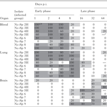
Parasite DNA was detected in the target organs of mice from all infected groups. During the early phase of infection, parasite DNA was detected in all infected groups, in the blood between days 1 and 4 p.i. and in the lung tissue until day 8 p.i. (Table 2). During the late phase of infection, the parasite was detected in the brain tissue, although in a lower percentage of mice compared to the blood and lungs during the early phase of infection (Table 2). Mice from the uninfected group were PCR negative in all target organs analysed.
Detection of N. caninum DNA by nested-PCR in the blood, lung and brain from BALB/c mice inoculated with 106 tachyzoites of each isolate

a Percentage detection.
b Grey intensity also indicates the frequency of N. caninum detection by PCR.
c Not evaluated at day 64 p.i.
The statistical analysis showed significant differences in parasite detection frequency and parasite burden among groups (P<0·007 by the Fisher exact test; P<0·0001 by the Kruskal-Wallis H-test). Mice infected with the Nc-Spain 7 isolate exhibited the lowest frequency of parasite detection in the blood and lung tissue (P<0·004; Fisher exact test). There was also a low frequency of detection in lung tissue from the Nc-Spain 5H infected mice, and significant differences were found when both infected groups (Nc-Spain 5H and Nc-Spain 7) were compared to those that showed the highest frequencies of detection (Nc-Spain 3H and Nc-Spain 6) (P<0·004; Fisher exact test). The infected groups with the lowest frequencies of detection (Nc-Spain 5H and Nc-Spain 7) also showed the lowest parasite burdens (Fig. 2A). However, pairwise comparisons indicated that only the parasite burdens in the lungs of mice infected with the Nc-Spain 7 isolate were significantly lower than those determined for the other infected groups (P<0·001, by the nonparametric multiple-comparison test).
Box-plot graph representing the maximum and minimum values, lower and upper quartiles and median values of parasite burden in BALB/c Neospora caninum-infected mice. (A) Parasite burden in lung tissue in the early phase of infection (from days 1 to 8 p.i.). (B) Parasite burden in brain tissue in the late phase of infection (from days 16 to 64 p.i.). According to the Kruskal-Wallis H-test and the nonparametric multiple-comparison test, the differences between N. caninum-infected groups were significant (*P<0·0055; **P<0·0015). Taking into account that the N. caninum detection limit by real-time PCR is 10−1 parasites (Collantes-Fernández et al. Reference Collantes-Fernández, Zaballos, Alvarez-García and Ortega-Mora2002), all positive samples had ⩾0·1 parasites, and negative samples (0 parasites) were represented on the log scale as <0·1 parasites.

During the late phase of infection, mice infected with the isolates Nc-Spain 5H and Nc-Spain 7, as well as the Nc-Spain 9 displayed the highest frequencies of N. caninum-DNA detection and parasite burdens in the brain. In contrast, the Nc-Spain 3H, Nc-Spain 6 and Nc-Spain 2H infected groups displayed the lowest (Table 2, Fig. 2B). However, only those frequencies and burdens detected in the Nc-Spain 5H and Nc-Spain 9 groups were significantly higher when compared to the group with the lowest frequencies and burdens in the brain (Nc-Spain 3H) (P<0·003; Fisher exact test).
Histopathological lesion analysis
A statistical analysis comparing the severity of lesions in the two phases of infection showed significant differences among inoculated mouse groups in lung and liver tissues for both phases, in heart tissue exclusively for the early phase and in brain tissue for the late phase (P<0·05, Kruskal–Wallis H-test).
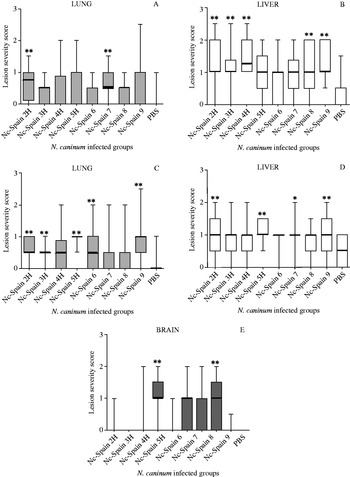
In the early phase, median lesion scores for the lungs ranged from nil to mild in all infected groups (Fig. 3A), but only the lesions observed in mice infected with the Nc-Spain 7 and Nc-Spain 2H isolates were significantly more severe than the control group (P<0·002, by the nonparametric multiple-comparison test). In the liver, the lesion scores were usually assessed as mild to moderate in all infected groups (Fig. 3B) and most of them were significantly more affected than the control group (P<0·005 by the nonparametric multiple-comparison test). No significant differences in the heart were detected among inoculated mice (data not shown).
Box-plot graph representing the maximum and minimum values, the lower and upper quartiles and median lesion severity score (nil=0, minimal=1, moderate=2 and severe=3) in the early phase of infection in lung (A) and liver (B) tissues and in the late phase in lung (C), liver (D) and brain (E) tissues. According to the Kruskal-Wallis H-test and the nonparametric multiple-comparison test, the differences between Neospora caninum-infected mouse groups were significant (*P<0·0055; **P<0·0015).

In the late phase of infection, lesion scores for the lungs remained absent to mild, although in contrast to the early phase, lesions were more severe than in the control group in more infected groups (5) (Nc-Spain 2H, Nc-Spain 3H, Nc-Spain 5H, Nc-Spain 6 and Nc-Spain 9). Furthermore, lung tissues in mice infected with the Nc-Spain 5H and Nc-Spain 9 isolates were also significantly more affected than in the Nc-Spain 8 infected mice (P<0·005 by the nonparametric multiple-comparison test) (Fig. 3C). In the liver, lesion scores were described as mild for all infected groups, and only 4 infected groups (Nc-Spain 2H, Nc-Spain 5H, Nc-Spain 7 and Nc-Spain 9) were significantly more affected than the control group (P<0·005 by the nonparametric multiple-comparison test) (Fig. 3D).
Finally, the median lesion scores for brain tissue increased from none during the early phase to mild during the late phase in some infected mouse groups. Thus, during the late phase the most severe brain lesions were caused by the Nc-Spain 5H, Nc-Spain 7 and Nc-Spain 9 isolates, whereas inflammation was absent in mice infected with the Nc-Spain 2H, Nc-Spain 3H and Nc-Spain 6 isolates. However, the differences were only significant in mice infected with the Nc-Spain 5H and Nc-Spain 9 isolates compared to the Nc-Spain 3H, Nc-Spain 6 and control groups (P<0·005 by the nonparametric multiple-comparison test) (Fig. 3E).
Humoral immune response analysis in Neospora caninum-infected BALB/c mice
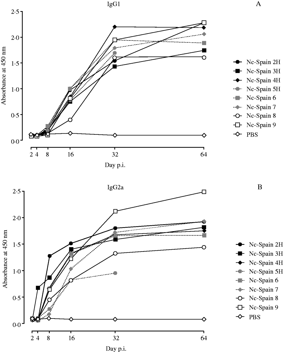
Specific anti-N. caninum IgG1 antibodies were detected at day 16 p.i. onwards in sera from all infected groups (Fig. 4A), although in the Nc-Spain 8 group, the increase was not significant. The highest IgG1 response was detected in mice infected with the Nc-Spain 4H and Nc-Spain 9 isolates from day 32 p.i. onwards, but IgG1 levels were only significantly higher than those detected in mice infected by the Nc-Spain 3H and Nc-Spain 8 isolates with the lowest levels (P<0·004, one-way ANOVA, followed by Duncan's test).
Kinetics of anti-Neospora caninum IgG1 (A) and IgG2a (B) isotypes from BALB/c mice inoculated with N. caninum isolates from day 2 p.i. onwards (see legend). Each data point represents the average absorbance at 450 nm.
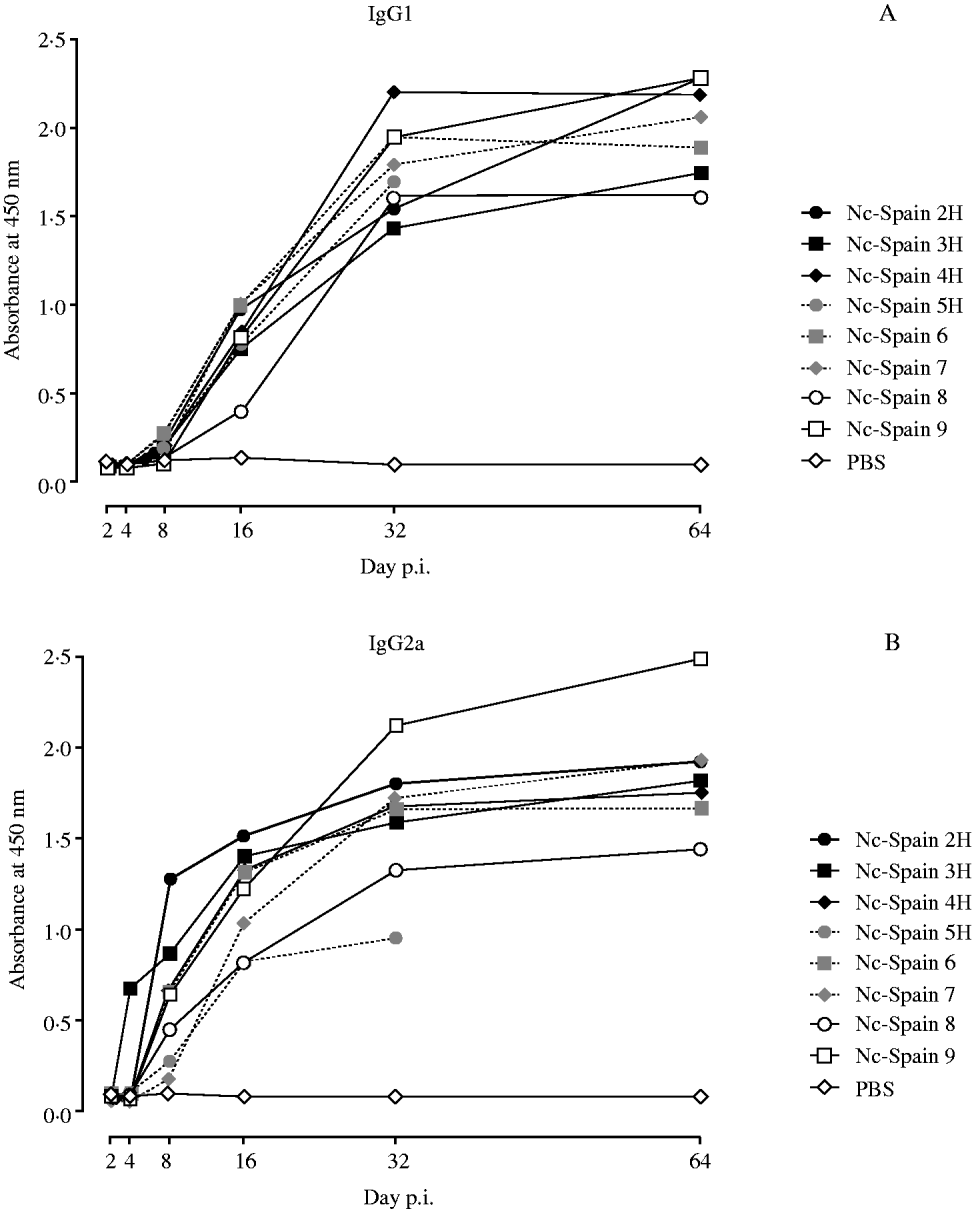
An early increase in N. caninum-specific IgG2a antibodies was detected in mice from all infected groups (Fig. 4B). The IgG2a response increased significantly in mice infected with the Nc-Spain 3H isolate from day 4 p.i., in mice infected with the Nc-Spain 2H, Nc-Spain 4H, Nc-Spain 6 and Nc-Spain 9 isolates from day 8 p.i. and in all infected groups from day 16 p.i. onwards. In the early phase (up to day 16 p.i.), the highest IgG2a levels were detected in mice infected with the Nc-Spain 2H and Nc-Spain 3H isolates, whereas in the late phase of infection (at days 32 and 64 p.i.) the highest levels were detected in mice infected with the Nc-Spain 9 isolate. The lowest IgG2a levels were observed in mice infected with the Nc-Spain 7 isolate until day 16 p.i., together with those detected in mice infected with the Nc-Spain 5H and Nc-Spain 8 isolates throughout the experimental period (P<0·0001, one-way ANOVA, Duncan's test).
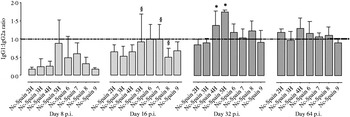
The IgG1:IgG2a ratio followed a similar pattern in all infected groups (Fig. 5). The mice had a mixed IgG2a:IgG1 response, with a slight predominance of IgG2a at days 8–16 p.i. (IgG1:IgG2a<1), followed by a tendency toward an IgG1 bias from day 32 p.i. onwards (IgG1:IgG2a>1). However, the IgG2a response predominated in mice infected with the Nc-Spain 2H, Nc-Spain 3H and Nc-Spain 9 isolates until day 32 p.i. Significant differences among N. caninum-infected mouse groups were only detected at day 32 p.i., when the IgG1:IgG2a ratio was higher in mice infected with Nc-Spain 5H or Nc-Spain 4H compared to those infected with the Nc-Spain 2H, Nc-Spain 3H or Nc-Spain 9 isolates (P<0·0001, one-way ANOVA, followed by Duncan's test).
Bars represent the average of anti-Neospora caninum IgG1:IgG2a isotype ratios from BALB/c mice infected with N. caninum isolates on days 8, 16, 32 and 64 p.i. (§ symbol indicates no significant detectable IgG1 or IgG2a levels; *P<0·0001). Error bars represent±S.D. The discontinuous line marks identical IgG1 and IgG2a levels (IgG1:IgG2a=1). Data from days 1, 2 and 4 p.i. are not plotted because anti-N. caninum IgG1 and IgG2a responses were not detected in any N. caninum-infected mouse groups, with the exception of IgG2a levels from the Nc-Spain 3H group at day 4 p.i.

DISCUSSION
Mice have been broadly used as laboratory models for studies of parasite infection in mammals, including the protozoan N. caninum. Although immunocompetent mice are relatively resistant to N. caninum infection, inbred BALB/c mice have been found to be susceptible to clinical neosporosis, and they have been broadly used to examine the immune response to and pathogenicity of N. caninum infections as well as the virulence of N. caninum isolates (Lindsay et al. Reference Lindsay, Lenz, Cole, Dubey and Blagburn1995; Atkinson et al. Reference Atkinson, Harper, Ryce, Morrison and Ellis1999; Shibahara et al. Reference Shibahara, Kokuho, Eto, Haritani, Hamaoka, Shimura, Nakamura, Yokomizo and Yamane1999; Miller et al. Reference Miller, Quinn, Windsor and Ellis2002; Collantes-Fernández et al. Reference Collantes-Fernández, López-Pérez, Álvarez-García and Ortega-Mora2006a; Rojo-Montejo et al. Reference Rojo-Montejo, Collantes-Fernández, Regidor-Cerrillo, Álvarez-García, Marugan-Hernández, Pedraza-Díaz, Blanco-Murcia, Prenafeta and Ortega-Mora2009; Pereira García-Melo et al. 2008). Nevertheless, the number of isolates bioassayed using this BALB/c model is small (10), and most of those studies were performed with N. caninum isolates obtained from clinically affected animals. Thus, the isolate populations characterized in vivo may not be representative of the N. caninum populations circulating in herds. Furthermore, the culture conditions used to maintain N. caninum isolates prior to experimental infection are relevant, as an attenuation of virulence in vivo by prolonged in vitro passage has been reported (Bartley et al. Reference Bartley, Wright, Sales, Chianini, Buxton and Innes2006). For these reasons, our goal in this study was the in vivo characterization of 8 N. caninum isolates, recently obtained from congenitally infected but healthy calves (Regidor-Cerrillo et al. Reference Regidor-Cerrillo, Gómez-Bautista, Pereira-Bueno, Aduriz, Navarro-Lozano, Risco-Castillo, Fernández-García, Pedraza-Diaz and Ortega-Mora2008), using a well-established BALB/c mouse model (Collantes-Fernández et al. Reference Collantes-Fernández, López-Pérez, Álvarez-García and Ortega-Mora2006a).
Almost none of the BALB/c mice inoculated with these isolates displayed clinical signs of infection, although Neospora infection was confirmed by parasite DNA detection in target organs and specific antibody detection in all N. caninum-infected groups. Furthermore, organic lesions were also observed. Similar results were seen when this mouse model was inoculated with the JPA1 (Shibahara et al. Reference Shibahara, Kokuho, Eto, Haritani, Hamaoka, Shimura, Nakamura, Yokomizo and Yamane1999), Nc-Spain 1H (Rojo-Montejo et al. Reference Rojo-Montejo, Collantes-Fernández, Regidor-Cerrillo, Álvarez-García, Marugan-Hernández, Pedraza-Díaz, Blanco-Murcia, Prenafeta and Ortega-Mora2009), Nc-Goiás 1 (Pereira García-Melo et al. Reference Pereira García-Melo, Regidor-Cerrillo, Ortega-Mora, Collantes-Fernández, de Oliveira, de Oliveira and Caetano da Silva2009) or Nc-Nowra (Miller et al. Reference Miller, Quinn, Windsor and Ellis2002) bovine isolates. In contrast, previous studies have demonstrated that BALB/c mice inoculated with N. caninum Nc-1 (Lindsay et al. Reference Lindsay, Lenz, Cole, Dubey and Blagburn1995, Reference Lindsay, Lenz, Blagburn and Brake1999; Collantes-Fernández et al. Reference Collantes-Fernández, López-Pérez, Álvarez-García and Ortega-Mora2006a), Nc-Liv or Nc-SweB1 (Atkinson et al. Reference Atkinson, Harper, Ryce, Morrison and Ellis1999; Collantes-Fernández et al. 2004, Reference Collantes-Fernández, López-Pérez, Álvarez-García and Ortega-Mora2006a) isolates exhibited nervous signs of neosporosis.
The temporal tissue dissemination of the parasite observed in infected mice followed a pattern similar to that described for the Nc-1 and Nc-Liv isolates in this animal model. Parasite distribution was characterized by an early phase of infection with parasitaemia and DNA detection in the lungs and a late phase of infection with parasite localization in the brain (Collantes-Fernández et al. Reference Collantes-Fernández, López-Pérez, Álvarez-García and Ortega-Mora2006a). However, some differences from this common profile could be detected depending on the isolate.
The spread of the parasite to target organs is not well understood, and 2 dissemination routes have been proposed. Some researchers have reported the possibility that tachyzoites may reach the brain of intraperitoneally Neospora-infected mice via the cerebrospinal fluid (Sawada et al. Reference Sawada, Park, Morita, Shimada, Umemura and Haritani1997). However, the results obtained in this and previous studies regarding parasitaemia and quick spread to other organs, such as the lungs (Collantes-Fernández et al. Reference Collantes-Fernández, López-Pérez, Álvarez-García and Ortega-Mora2006a; Pinitkiatisakul et al. Reference Pinitkiatisakul, Mattsson and Lundén2008; Rojo-Montejo et al. Reference Rojo-Montejo, Collantes-Fernández, Regidor-Cerrillo, Álvarez-García, Marugan-Hernández, Pedraza-Díaz, Blanco-Murcia, Prenafeta and Ortega-Mora2009; Pereira García-Melo et al. Reference Pereira García-Melo, Regidor-Cerrillo, Ortega-Mora, Collantes-Fernández, de Oliveira, de Oliveira and Caetano da Silva2009), may indicate that the bloodstream is the main route of parasite dissemination. This idea was also suggested in a previous study by Shibahara et al. (Reference Shibahara, Kokuho, Eto, Haritani, Hamaoka, Shimura, Nakamura, Yokomizo and Yamane1999) because N. caninum tachyzoites were mainly identified in the endothelial cells of blood vessels in infected nude mice. Nevertheless, these dissemination routes might not be mutually exclusive, and both may be used by the parasite.
Reduced parasite detection frequencies and parasite burdens were seen in the lungs of mice infected with the Nc-Spain 5H and Nc-Spain 7 isolates during the early phase of infection, whereas parasite frequencies and burdens detected in the brain were elevated for both isolates during the late phase of infection. In contrast, the detection pattern observed in groups infected with the Nc-Spain 2H, Nc-Spain 3H and Nc-Spain 6 isolates was characterized by a high frequency of detection and high parasite burdens in the blood and lungs during early infection but low levels in the brain during the late phase of infection. Previous in vitro studies have reported differences in parasite growth (Schock et al. Reference Schock, Innes, Yamane, Latham and Wastling2001; Pérez-Zaballos et al. Reference Pérez-Zaballos, Ortega-Mora, Álvarez-García, Collantes-Fernández, Navarro-Lozano, García-Villada and Costas2005) and tachyzoite-bradyzoite conversion rates (Vonlaufen et al. Reference Vonlaufen, Muller, Keller, Naguleswaran, Bohne, McAllister, Bjorkman, Muller, Caldelari and Hemphill2002; Risco-Castillo et al. Reference Risco-Castillo, Fernández-García and Ortega-Mora2004) among different N. caninum isolates in cell culture. In addition, a lower tachyzoite yield and slower tachyzoite-bradyzoite conversion in vitro have recently been related to the low virulence of the Nc-Spain 1H isolate, which was not detected in the brain during the chronic phase of infection, even in mice infected with a high dose of tachyzoites (107) (Rojo-Montejo et al. Reference Rojo-Montejo, Collantes-Fernández, Regidor-Cerrillo, Álvarez-García, Marugan-Hernández, Pedraza-Díaz, Blanco-Murcia, Prenafeta and Ortega-Mora2009). Thus, for mice infected with the Nc-Spain 5H or Nc-Spain 7 isolates, the high parasite burdens detected during the late phase of infection could be explained by better parasite dissemination, a higher capacity of growth in these immunoprivileged tissues or better evasion of the host immune response. However, further experiments will be needed to determine whether differences in their migratory capacity and parasite motility, invasion and proliferation rates or tachyzoite-bradyzoite conversion rates can explain the variability in detection frequencies and parasite burdens shown by these isolates, including the reduced presence of the parasite in the brains of mice infected with the Nc-Spain 2H, Nc-Spain 3H or Nc-Spain 6 isolates during the late phase of infection.
In addition to clinical signs and mortality, assessments of the histopathological effects of N. caninum have been routinely carried out in experimental infections to describe the severity of the infection; however, there are limits to this technique, which could influence the results, including the number of samples evaluated, the criteria used and the experience of the examiner. In previous studies of N. caninum-infected but not clinically affected BALB/c mice, the severity grade of brain lesions was assessed as nil to moderate, whereas moderate or severe lesions were described in clinically affected mice (Lindsay et al. Reference Lindsay, Lenz, Cole, Dubey and Blagburn1995, Reference Lindsay, Lenz, Blagburn and Brake1999; Shibahara et al. Reference Shibahara, Kokuho, Eto, Haritani, Hamaoka, Shimura, Nakamura, Yokomizo and Yamane1999; Miller et al. Reference Miller, Quinn, Windsor and Ellis2002; Collantes-Fernández et al. Reference Collantes-Fernandez, Alvarez-Garcia, Perez-Perez, Pereira-Bueno and Ortega-Mora2004). In this study, histological lesion analysis for some target organs was also conducted, and lesions were categorized as absent to moderate in all tissues analysed from N. caninum-infected mice. In addition, a correlation between the lesion severity grade and parasite burden in the brain could also be discerned, as has been previously reported (Collantes-Fernández et al. Reference Collantes-Fernandez, Alvarez-Garcia, Perez-Perez, Pereira-Bueno and Ortega-Mora2004). Thus, those N. caninum-infected mice with higher parasite burdens (produced by the Nc-Spain 5H, Nc-Spain 7 and Nc-Spain 9 isolates) had the most severe brain lesions in the late phase of infection, and those with lower parasite burdens (produced by the Nc-Spain 2H, Nc-Spain 3H and Nc-Spain 6 isolates) had the least severe lesions.
A relationship between parasite number and lesion severity could also be established in the lungs during the late phase of infection, when the majority of the groups showed a higher presence or severity of lesions. More affected lungs were observed in those infected groups (Nc-Spain 2H, Nc-Spain 5H, Nc-Spain 6 and Nc-Spain 9) in which the parasite DNA was detected during the late phase of infection and higher median parasite burdens were quantified (data not shown), although the parasite detection percentages were drastically diminished compared to the early phase of infection. A difference in parasite behaviour that is dependent upon the tissue reached may explain these results. In the brain, N. caninum tachyzoites can convert to the bradyzoite stage inside tissue cysts, where the parasite can persist for a long time. Tissue cysts in the lungs have not yet been described in either natural or experimental infection, and the parasite may proliferate as tachyzoites in the infected tissues until the immune response is able to control parasite multiplication and eliminate it, although previously generated histological lesions may persist.
The specific humoral immune response was also evaluated for mice infected with the different isolates. Cytokines such as interferon-γ (IFN-γ) and interleukin 4 (IL-4), related to a Th1 and Th2-type immune response, respectively, play important roles in the control of N. caninum multiplication and the susceptibility of BALB/c mice to neosporosis (Long et al. Reference Long, Baszler and Mathison1998; Baszler et al. Reference Baszler, Long, McElwain and Mathison1999). Here N. caninum-specific serum isotypes IgG2a and IgG1, also related to the Th1/Th2-type immune response balance, were determined to discern the cytokine and type immune response bias throughout the infection. In previous studies, variations in the IgG1 and IgG2a immune responses of mice related to the isolate, the dose administered and the route of inoculation used have been reported (Quinn et al. Reference Quinn, Miller, Ryce, Windsor and Ellis2002; Rojo-Montejo et al. Reference Rojo-Montejo, Collantes-Fernández, Regidor-Cerrillo, Álvarez-García, Marugan-Hernández, Pedraza-Díaz, Blanco-Murcia, Prenafeta and Ortega-Mora2009). In this study, antibody production for both isotypes showed a similar profile to that reported by Collantes-Fernández et al. (Reference Collantes-Fernández, López-Pérez, Álvarez-García and Ortega-Mora2006a), with IgG2a predominating in the early phase of infection, and IgG1 increasing in the late phase, although different kinetics for the IgG1:IgG2a balance were observed, depending on the inoculated isolate. The earlier IgG2a response stimulated by the Nc-Spain 2H and Nc-Spain 3H isolates could indicate a predominance of the Th1-type response, which could be related to the better control of parasite dissemination and the reduced parasite burdens and lesions seen in the brain during the late phase of infection. Similarly, the delayed and minor IgG2a response observed in mice infected with the Nc-Spain 5H and Nc-Spain 7 isolates, which were only significantly increased from day 16 p.i. onwards, may suggest poor immune control of parasite dissemination, since both mouse groups presented elevated parasite burdens and higher brain lesion severity in the late phase of infection.
Interestingly, mice infected with the Nc-Spain 3H and Nc-Spain 4H isolates, which displayed the same microsatellite profile and were obtained from different calves from the same herd (Regidor-Cerrillo et al. Reference Regidor-Cerrillo, Gómez-Bautista, Pereira-Bueno, Aduriz, Navarro-Lozano, Risco-Castillo, Fernández-García, Pedraza-Diaz and Ortega-Mora2008), showed significant variations in IgG1 and IgG2a kinetics. However, there were no statistical differences in morbidity, parasite distribution, parasite burden and the severity of lesions caused by these two isolates in mice. These results indicate that the isolates have different immunogenic capacities, although further studies are needed to evaluate the existence of other differences in their biological characteristics.
In conclusion, this paper describes the pathogenic characterization of 8 isolates obtained from asymptomatic calves in a BALB/c mouse model. Because the results obtained in mice may not be applicable to the natural host, further studies are needed to determine the inherent virulence of these isolates, as well as other epidemiological features such as variations in the vertical transmission rate using pregnant murine and bovine models. Nevertheless, this work confirms the intra-species diversity in virulence of N. caninum isolates, which may be related to the differences observed in the presentation of neosporosis.
ACKNOWLEDGEMENTS
This work was financed by the projects PETRI 95-0777.OP from the Spanish Government and Hipra Laboratories S.A. and RTA04-047-C2 from the INIA. Debora García-Melo was financed by Coordenação de Aperfeiçoamento de Pessoal de Nível Superior (CAPES)- Ministério da educação of Brazil. We would like to thank Ricardo García de la Mata for performing the statistical analyses and Vanesa Navarro (SALUVET, UCM) for her excellent technical assistance.