Millions of endoscopic procedures are performed in US hospitals every year.Reference Rutala and Weber 1 Over the last decade, reports have implicated endoscopes as a source of infectious disease outbreaks and pseudo-outbreaks.Reference Srinivasan, Wolfenden and Song 2 – Reference Marsh, Krauland and Nelson 6 Recently, an increasing number of US hospitals have reported multidrug-resistant (MDR) organism clusters linked to endoscopes.Reference Epstein, Hunter and Arwady 7 – Reference Gastmeier and Vonberg 9
In November of 2014, a cluster of patients with bronchoscopically obtained clinical cultures positive for carbapenem-resistant Klebsiella pneumoniae (CR-KP) and gentamicin-resistant MDR Pseudomonas aeruginosa (MDR-PA) was identified in the medical intensive care unit (MICU) at University of Pittsburgh Medical Center-Presbyterian Hospital (UPMC-P), a 757-bed urban teaching hospital in Pittsburgh, Pennsylvania. Each of the patients in this cluster had no prior history of isolation of CR-KP or MDR-PA from clinical cultures. An epidemiologic investigation was conducted under the auspices of the infection prevention department’s role in patient safety to identify a common infection source.
Methods
Epidemiologic investigation
In November 2014, CR-KP and MDR-PA isolates were identified in a bronchoalveolar lavage (BAL) specimen from a MICU patient without recent health care. This finding was unexpected. Over the next 3 weeks, clinical cultures taken either during or after a bronchoscopy procedure from an additional 9 MICU patients grew CR-KP and/or MDR-PA, triggering an epidemiologic investigation. House-wide CR-KP incident isolates were identified, and potential commonalities were investigated. Analysis by patient care area identified the 32-bed MICU as accounting for 17 of 21 (81%) of the newly identified CR KP cases, and MDR-PA was also isolated from a subset of cases. During the period from July 2014 to December 2014, case patients were retrospectively identified as persons with incident clinical cultures positive for (1) CR-KP resistant to ertapenem; (2) MDR-PA nonsusceptible to gentamicin, tobramycin, ciprofloxacin, levofloxacin, meropenem, aztreonam, and cefepime and susceptible to piperacillin and piperacillin/tazobactam; or (3) both organisms. Analyses of antibiotic susceptibility of all isolates were performed using minimal inhibitory concentration (MIC) and/or disc diffusion methods. These results were interpreted using Clinical and Laboratory Standards (CLSI) criteria.
During the cluster’s peak period of October–November 2014, 25 unique case patients were identified, 19 (76%) of whom were exposed to the same bronchoscope (named B1; Olympus BF-160 Video Bronchoscope, Olympus, Tokyo, Japan). This finding prompted investigation of the potential association of B1 exposure with the cluster. All 9 MICU-circulating bronchoscopes were immediately taken out of service, were cultured according to previously published methods,Reference Srinivasan, Wolfenden and Song 2 and underwent borescopy to visualize the internal lumen and to identify structural defects and/or deposits of biological material. Of 9 bronchoscopes, 8 were negative for bacterial growth and were returned to service. The B1 bronchoscope was positive for both CR-KP and MDR-PA. Bronchoscope reprocessing, including manual, enzymatic precleaning (Steris, Mentor, OH), and automated high-level disinfection (Custom Ultrasonics, Ivyland, PA) was assessed by UPMC infection prevention personnel to ensure compliance with current guidelines.Reference Rutala and Weber 10 No breaches in bronchoscopy technique or bronchoscope precleaning were detected. High-level disinfection (HLD) records were reviewed, and no failures were identified.
Bacterial genomics
PFGE was performed on 3 CR-KP B1 isolates and a subset of clinical CR-KP isolates according to published methods.Reference Marsh, Krauland and Nelson 6 , Reference Vimont, Mnif, Fevre and Brisse 11 Subsequent whole-genome sequencing (WGS) was performed on an expanded set of 28 isolates (14 CR-KP and 14 MDR-PA), including 4 isolates cultured from the B1 bronchoscope (3 CR-KP and 1 MDR-PA), to strengthen evidence for horizontal transmission. Genomic DNA samples were extracted using the Qiagen DNeasy Kit (Qiagen, Venlo, Netherlands) according to the manufacturer’s instructions. DNA samples were quantified by Qubit fluorimetry (Qubit fluorimeter, Thermo Fisher Scientific, Waltham, MA), and genomic libraries were prepared using a modified Illumina Nextera protocol (Illumina, San Diego, CA).Reference Baym, Kryazhimskiy, Lieberman, Chung, Desai and Kishony 12 Libraries were sized using an Agilent bioanalyzer (Agilent, Santa Clara, CA), normalized, pooled, and sequenced using an Illumina NextSeq500.
FastQ reads were quality filtered and assembled using SPAdes version 3.10.1 software.Reference Inouye, Dashnow and Raven 13 Assemblies were annotated using Prokka version 0.1.1 software.Reference Seemann 14 Reads had a sequencing depth ranging from 47X to 256X, and assemblies had a median number of 75 contigs per sample, with an average assembly length of 7.1 Mbp for MDR-PA and 5.5 Mbp for CR-Kp. Sequence types were identified using SRST2.Reference Inouye, Dashnow and Raven 13 Reads were aligned to reference assembly, CR-KP112 and MDR-PA012 for K. pneumonia and P. aeruginosa genomes, respectively, using BWA-MEM version 0.7.12-r1039 software (http://bio-bwa.sourceforge.net/). Single-nucleotide polymorphisms (SNPs) were identified using Snippy genome software with default parameters. Snippy uses Freebayes as the variant caller and filters out SNPs with <10 reads and 90% alternate allele proportion. A phylogenetic tree based on the aligned core SNPs was generated using RAxML version 8.2.9 software by running 100 bootstrap replicates under the generalized time-reversible model (GTRCAT) and Lewis correction for ascertainment bias.Reference Stamatakis 15 Phylogenies were visualized using the python package ETE3. Isolates with <30 SNP differences were considered genetically related.
Clinical data
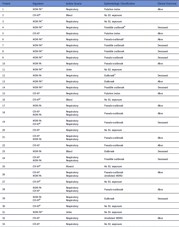
The electronic medical records of case patients were reviewed to assess clinical outcomes and epidemiology. Patients were classified epidemiologically as “outbreak,” “possible outbreak,” or “pseudo-outbreak.” The “outbreak” classification was assigned to patients with isolates recovered from a clinical culture ≥1 day after B1 exposure (Table 1). The “pseudo-outbreak” classification included patients with isolates obtained from culture during exposure to B1 who had no subsequent recovery of outbreak-related isolates. The “possible outbreak” classification was assigned to patients whose death occurred within 10 days of B1 exposure such that subsequent recovery and epidemiologic classification of isolates from clinical cultures could not be assessed. Genetically unrelated MDR-PA or CR-KP isolates were regarded as isolates unlikely to belong to a B1-related cluster.
Isolate Source of 33 Case Patients Along With Epidemiologic Classification and Clinical Outcome of 23 Patients With Bronchoscope B1 Exposure

Note. MDR-PA, multidrug-resistant Pseudomonas aeruginosa; CR-KP, carbapenem-resistant Klebsiella pneumoniae.
a Isolate unavailable for molecular analysis.
b Possible outbreak: patients whose death occurred within 10 days of bronchoscope B1 exposure such that subsequent recovery and epidemiologic classification of isolates from clinical cultures could not be assessed.
c Pseudo-outbreak: patients with isolates obtained from culture during exposure to bronchoscope B1 who had no subsequent recovery of outbreak-related isolates.
d Outbreak: patients with isolates recovered from a clinical culture ≥1 day after bronchoscope B1 exposure.
Results
Epidemiology
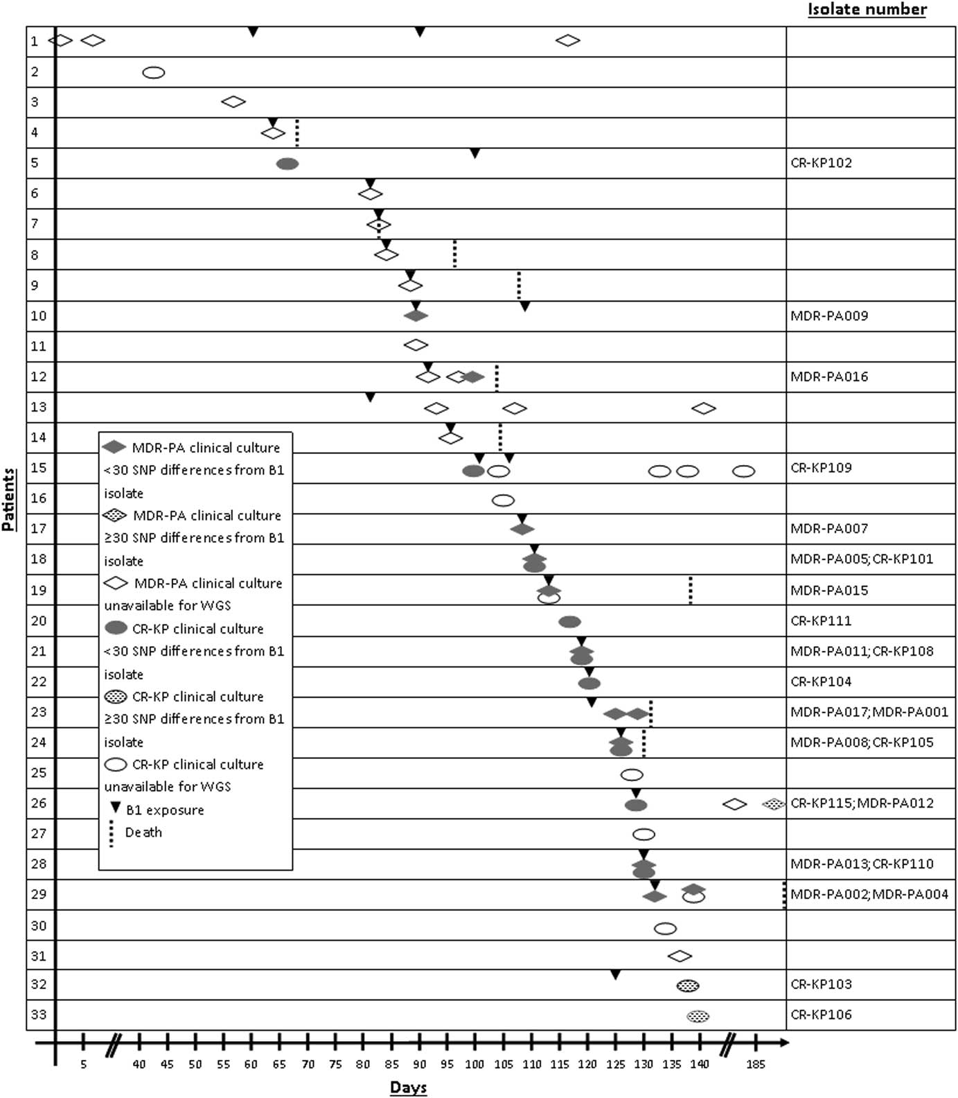
A total of 33 case patients with cultures positive for CR-KP and/or MDR-PA from predominantly respiratory sources collected between July and December 2014 (Table 1) were identified and plotted over time (Fig. 1). Of these 33 case patients, 23 (69.7%) were exposed to bronchoscope B1. Of these 23 patients, 19 (82.6%) had cultures positive for MDR-PA and 11 (47.8%) had cultures positive for CR-KP. Upon removal of B1 from service, the number of patients with newly detected CR-KP or MDR-PA decreased.
Timeline depicting the relative chronology of bronchoscope B1 exposures and CR-KP (n = 22) and MDR-PA (n = 31) positive cultures among 33 case patients over a 185-day period (July–December 2014). Diamonds represent multidrug-resistant Pseudomonas aeruginosa (MDR-PA) detection in case patient clinical specimens. Ovals represent carbapenem-resistant Klebsiella pneumonia (CR-KP) detection in case patient clinical specimens. Shading represents genetic relatedness. Triangles represent B1 exposure. Dashed lines represent patient deaths.

MDR-PA epidemiology
Patient 1, who had clinical cultures positive for MDR-PA prior to B1 exposure, was identified as a potential transmission source (Fig. 1). Patient 1 was exposed to bronchoscope B1 3 days prior to the bronchoscope procedure of patient 4, who became culture positive for MDR-PA on the day of B1 exposure. Subsequently, 18 case patients with exposure to bronchoscope B1 developed MDR-PA positive cultures (Fig. 1).
CR-KP epidemiology
Patients 5 and 15 were considered putative index patients for contamination of B1 with CR-KP. Patient 5 was culture positive for CR-KP 100 days prior to B1 exposure, whereas patient 15 was culture positive for CR-KP 7 hours prior to B1 exposure. Both patients 5 and 15 had a bronchoscopy with B1 performed 11 days prior to patient 18—the first case patient who became culture positive for CR-KP on the same day of B1 exposure. Subsequently, 9 patients with B1 exposure developed CR-KP–positive cultures (Fig. 1).
B1 borescopy and culture
Borescopy revealed that the lumen of B1 was physically defective and contained proteinaceous debris. Culture of bronchoscope B1 in December 2014 identified 3 CR-KP and 1 MDR-PA isolates.
Bacterial genotyping
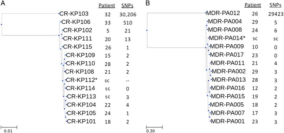
PFGE demonstrated that the 3 CR-KP B1 scope isolates and the subset of CR-KP isolates were genetically related (data not shown). Of the 33 case patients, 16 patients did not have MDR-PA or CR-KP isolates available for WGS (Fig. 1). Phylogenetic analysis of the 24 isolates available for genomic comparison from 17 case patients revealed clustering of patient- and bronchoscope-derived isolates that was concordant with the epidemiology. Overall, 7 CR-KP patient isolates were highly related to the B1 isolate CR-KP112 with ≤4 SNP differences identified (Fig. 2A). Of these patients, 6 had exposure to bronchoscope B1 prior to becoming CR-KP culture positive. One patient (patient 15) was exposed to B1 after CR-KP culture positivity, supporting patient 15 as a possible index patient. The B1 isolates CR-KP113 and CR-KP114 were nearly identical to CR-KP112, with ≤3 SNP differences. Isolate CR-KP106 collected from patient 33 who lacked B1 exposure showed 510 SNP differences from B1 isolate CR-KP112 (Fig. 2A). Isolate CR-KP103, collected 12 days after B1 exposure from patient 32, had 30,206 SNP differences from B1 isolate CR-KP112, suggesting that B1 was not the source of this patient’s CR-KP infection.
Phylogenies of CR-KP (A) and MDR-PA (B) genomes obtained from patient isolates. Genetic distance is based upon the number of SNP differences relative to the reference genome. Note. * = reference genome; sc = scope isolate. The scale indicates the number of nucleotide substitutions per site.

Moreover, 12 MDR-PA patient isolates clustered with B1 isolate MDR-PA114 on the phylogenetic tree with ≤6 SNP differences (Fig. 2B). One isolate from patient 26 (MDR-PA012), collected 56 days after B1 exposure, had >29,000 SNP differences from isolate MDR-PA014, indicating that B1 was an unlikely source of this patient’s MDR-PA–positive culture.
In addition, 7 patients with B1 exposure had clinical cultures positive for both CR-KP and MDR-PA (Fig. 1, patients 18, 19, 21, 24, 26, 28, and 29). Of 5 patients with both MDR-PA and CR-KP isolates available for WGS, 4 were genetically related to isolates recovered from bronchoscope B1 (Fig. 2A and B, patients 18, 21, 24, and 28). Together, these genomic data suggest transmission to patients of CR-KP and MDR-PA from bronchoscope B1.
We identified 6 case patients without B1 exposure during the cluster’s peak period, including 1 patient whose isolate (CR-KP111) was genetically related to the outbreak (13 SNP differences) (Figs. 1 and 2A). No epidemiologic link to the MICU cluster was identified, suggesting an undetected horizontal transmission route.
Clinical histories and epidemiologic designation
Of the 23 patients with B1 exposure, only 1 patient (patient 32) was considered unrelated to the outbreak because the clinical CR-KP isolate was genetically distinct from the CR-KP isolate recovered from the B1 bronchoscope (Table 1; Fig. 2). Three patients (patients 1, 5, and 15) were considered possible index patients. Of the remaining 19 patients with exposure to the B1 bronchoscope, 4 patients with MDR-PA or CR-KP recovered from follow-up respiratory cultures were classified as “outbreak” cases. However, 5 patients died within 10 days with no other clinical specimens classified as “possible outbreak.” Furthermore, 9 patients whose follow-up respiratory cultures were MDRO negative and 1 patient without follow-up respiratory cultures were characterized as “pseudo-outbreak” cases. Isolates from 8 B1-exposed patients were unavailable for WGS. In all, 10 B1-exposed patients died: by definition, all 5 patients classified as “possible outbreak” died; 3 of 4 patients classified as “outbreak” died; and 2 patients classified as “pseudo-outbreak” died. Our investigation did not include an analysis of attributable mortality.
Discussion
We have described the contamination of a single bronchoscope with 2 different MDR pathogens and an outbreak and pseudo-outbreak that involved at least 19 patients. We presume that horizontal transmission from a contaminated bronchoscope to patients undergoing bronchoscopy was the source of infection among outbreak and possible outbreak cases, and we hypothesize that the pseudo-outbreak occurred due to contamination of patient specimens as they were being withdrawn from the bronchoscope, a phenomenon that has been previously described.Reference Weinstein and Stamm 16 However, we cannot exclude transient colonization. Although pseudo-outbreaks are not considered true infections, they can nonetheless lead to adverse patient outcomes due to receipt of unnecessary antimicrobial therapy or other treatments.
Our investigation revealed 2 putative CR-KP index patients (patients 5 and 15). The temporal proximity of patient 15’s infection with subsequent B1-related CR-KP infections suggests that this patient was the likely index case. However, the 21 SNP differences between CR-KP isolates obtained from patient 5 (CR-KP102) and B1 (CR-KP112) do not rule out the possibility that patient 5 was the index case (Fig. 2A).
After detecting microbial contamination of B1, UPMC-P instituted bedside precleaning of bronchoscopes with an enzymatic solution with a goal of reducing the likelihood of luminal biofilm formation. More recently, bronchoscope storage was reconfigured so the flexible portion of each device would hang vertically without coiling. Additionally, periodic culturing of clinical endoscopes was planned. Surveillance culturing of endoscopes is a proposed strategy to detect contamination, and while the practice is not currently part of US guidelines, the Centers for Disease Control and Prevention (CDC) endorses surveillance culturing of duodenoscopes as a supplement to recommended endoscope reprocessing procedures. Thus, surveillance culture may expand to bronchoscopes and other types of endoscopes in the future. However, the optimal frequency of culturing and the sensitivity of surveillance endoscope culturing for detection of contamination are unknown. Moreover, these measures are not sufficient to detect device-associated outbreaks in a timely fashion.
The bronchoscope model implicated in this case is commonly used in many hospitals, and the outbreak we describe involved one of many Olympus BF-160 bronchoscopes in our hospital. Borescopy revealed a luminal defect and, despite compliance with manufacturer’s recommended reprocessing procedures, accumulation of proteinaceous debris in bronchoscope B1 that may have contributed to the establishment of a biofilm and subsequent contamination with MDR-PA and CR-KP. Several other published reports implicate defective bronchoscopes as the cause of polymicrobial contamination that was linked to outbreaks and pseudo-outbreaks.Reference Kirschke, Jones and Craig 17 – Reference Guy, Vanhems and Dananche 19 Our report, along with others linking bacterial contamination to defective equipment,Reference Kirschke, Jones and Craig 17 , Reference Cetre, Nicolle and Salord 18 , Reference Srinivasan, Wolfenden and Song 20 gives credence to the notion that routine maintenance of bronchoscopes should include visual luminal inspection. Close scrutiny by the FDA of newly marketed medical devices is also necessary to identify engineering vulnerabilities that may engender persistent microbial contamination.
The outbreak and pseudo-outbreak we report largely came to the attention of practitioners because it involved MDROs. Hospital infection prevention departments should consider dedicated surveillance of all endoscopes. Samples that yield microbial isolates should trigger scope usage investigation coupled with isolate retrospective review in patients who had undergone procedures with the contaminated scope to mitigate duration of endoscope-associated outbreaks. In addition, the epidemiology of endoscopically obtained specimens should be reviewed to detect outbreaks or pseudo-outbreaks involving susceptible and frequently encountered organisms that could plausibly go undetected for long periods or even completely escape the attention of robust infection control programs.
Notably, the increase in newly detected MDR-PA, based on the phenotypic definition specific to this outbreak, went unrecognized initially. At our hospital, P. aeruginosa phenotypes resistant to ≥3 antimicrobial classes are monitored, but because many P. aeruginosa phenotypes fulfill this criterion, data were insufficiently granular to demonstrate an increase in MDR-PA that was specific to the outbreak. Furthermore, we identified several isolates that matched outbreak isolates based on antimicrobial drug susceptibilities but were genetically unrelated to B1-derived isolates and to each other. This finding highlights the utility of WGS to confirm or refute horizontal transmission in the setting of outbreaks due to infectious pathogens.
Our investigation has several limitations. Notably, isolates from 8 patients were unavailable for WGS to strengthen the link between their B1 exposure and MDRO recovery. However, 6 of these 8 patients fulfilled case status on the same day as B1 exposure, so the epidemiologic link is strong even in the absence of genomic data. Patient 13 yielded MDR-PA 18 days after B1 exposure, and horizontal acquisition from an alternate source may have occurred, especially considering that patient 32 yielded an unrelated MDR-PA isolate 12 days after B1 exposure. Alternate sources of acquisition are also possible for the 10 patients whose isolates matched B1-derived isolates, because WGS cannot establish the directionality of transmission. However, the fact that case status for 17 of 19 patients with presumed B1-derived MDRO acquisition was established on the same day as B1 exposure strengthens the epidemiologic linkage.
In conclusion, we report an outbreak and pseudo-outbreak of MDR-PA and CR-KP that were strongly linked to the contamination of a single, physically defective bronchoscope, and we provide genomic data to support our epidemiologic investigation. In addition to adhering to endoscope reprocessing guidelines, hospital epidemiology programs should prioritize thorough periodic maintenance of endoscopic devices and emphasize scrutiny of endoscope-derived culture data as an important intervention to hasten recognition of endoscope-associated outbreaks.
Acknowledgments
We extend our appreciation to Jessica Schlackman and Chinelo Ezeonwuka for their technical expertise and to Ryan Shields for his assistance in the procurement of microbial isolates. We also thank Vaughn Cooper and Dan Snyder for their assistance with whole-genome sequencing.
Financial support
No financial support was provided relevant to this article.
Conflicts of interest
L.H.H. reports having served on a scientific advisory board for GlaxoSmithKline on meningococcal vaccines. All other authors report no conflicts of interest relevant to this article.