Introduction
Tetralogy of Fallot is the most common type of cyanotic CHD. Pulmonary regurgitation is a common consequence of surgical repair of tetralogy of Fallot. Reference Van der Ven, van den Bosch, Bogers and Helbing1 Pulmonary regurgitation initially was thought to be a relatively benign haemodynamic residual lesion but subsequently was found to be predictive of decreased exercise performance and progressive right ventricular dilation and systolic dysfunction. Reference Villafañe, Feinstein and Jenkins2 Furthermore, patients with pulmonary regurgitation were noted to be at higher risk of ventricular arrhythmias and sudden cardiac death. Reference Gatzoulis, Balaji and Webber3–Reference Rotes, Connolly and Warnes4 Right ventricular evaluation represents one of the major clinical tasks in the follow-up of repaired tetralogy of Fallot patients with PR, as both severe right ventricular dilatation and dysfunction are key factors in defining the need of pulmonary valve replacement. Reference Wise-Faberowski, Asija and McElhinney5–Reference Smith, McCracken and Thomas6
Cardiovascular magnetic resonance imaging has been shown to represent the gold standard method to assess the right ventricular size and function in repaired tetralogy of Fallot due to its ability to accurately assess multiple parameters including pulmonary regurgitant fraction, ventricular volumes and function, and pulmonary artery anatomy. Reference Apostolopoulou, Manginas, Kelekis and Noutsias7–Reference Helbing and de Roos8 However, it represents a financial burden especially in the developing countries.
American society of Echocardiography reported that some conventional echocardiographic parameters evaluating right ventricular systolic function should be incorporated into the routine echocardiographic assessment including fractional area change, tricuspid annular plane systolic excursion, and right ventricular peak systolic tricuspid annular velocity (RV S’) by pulsed tissue Doppler. Reference Rudski, Lai and Afilalo9 However, all of these measurements have some limitations. In clinical practice, evaluation of right ventricular geometry and function in repaired tetralogy of Fallot patients is routinely performed by echocardiography. However, due to its intrinsic geometrical features, echocardiographic estimation of right ventricular function remains highly challenging. Reference van der Zwaan, Geleijnse and McGhie10–Reference Koestenberger, Nagel and Ravekes11
Recently, two-dimensional speckle tracking echocardiography emerged as a promising tool for assessing myocardial performance as it does not depend on geometric assumptions and has no angle dependence. Reference Levy, Holland, Sekarski, Hamvas and Singh12 Therefore, we hypothesise that speckle tracking echocardiography can be comparable to the expensive cardiac magnetic resonance in detecting right ventricular systolic dysfunction in children with repaired tetralogy of Fallot especially in the developing countries where resources are limited.
Thus, our study aimed to assess the diagnostic accuracy of echocardiography, especially 2D-speckle tracking echocardiography in the identification of right ventricular systolic dysfunction in children with repaired tetralogy of Fallot as compared to cardiac magnetic resonance.
Patients and methods
This cross-sectional study was performed between November 2022 and November 2024 on 56 successive patients who underwent surgical correction of tetralogy of Fallot and were sent to our echocardiographic laboratory for variable purposes. The study was approved by the ethics committee of our faculty of Medicine with approval code (360410/10/22). A written informed consent was obtained from the parent and/or legal guardian of the included patients. The study was performed in accordance with the Declaration of Helsinki.
Inclusion criteria: children aged less than 18 years who underwent surgical correction of tetralogy of Fallot.
Exclusion criteria: children who underwent surgical repair for another cardiac disease, children with extreme form of tetralogy of Fallot or double outlet right ventricle, children with chromosomal anomalies such as Down syndrome, and patients with arrhythmias were excluded as all of these conditions can affect the systolic function of the right ventricular.
All patients were subjected to thorough history taking, for example, age, sex, medical history, tetralogy of Fallot anatomical type, age at repair, surgery method (primary/staged), and surgical approach. Complete clinical examination was performed and heart rate was recorded.
Conventional echocardiography
All echocardiographic examinations were performed using GE echocardiographic systems (GE Vivid IQ transducer 3Sc-RS). All measurements were acquired according to the guidelines of the American society of Echocardiography. Reference Lang, Bierig and Devereux13 The right ventricle was examined in the apical 4-chamber view and the parasternal short axis view for the assessment of pulmonary regurgitation using colour and continuous wave Doppler. Right ventricular end-diastolic area and right ventricular end-systolic area were evaluated in the apical 4-chamber view, and fractional area change was derived as: RV FAC = (RV EDA – RV ESA)/RV EDA × 100%. Tricuspid annular plane systolic excursion was measured by the M-mode in the apical 4-chamber view.
Tissue Doppler echocardiography
Peak systolic (S’) velocity of tricuspid annulus was measured by pulsed TDI, by placing the sample volume at the level of tricuspid lateral annulus from the apical 4-chamber view. The S’ velocity of the lateral annular side was used for the analysis.
Two-dimensional speckle tracking echocardiography (2D-STE)
Besides conventional echocardiographic parameters, longitudinal strain analysis was integrated as an important complementary step. Measurement of strain using speckle tracking was utilised, where ultrasound imaging technique relied on classic 2D grey scale images; with less angle-dependency. It assesses longitudinal myocardial deformation. The frame rate was modified according to the patient HR (90 ± 20 F/s). Strain values were calculated from at least three successive cardiac cycles. 2D-speckle tracking echocardiography analysis for the right ventricle was performed to measure right ventricular global longitudinal strain and right ventricular free wall strain in the apical four-chamber view. For each patient, after manual registration of an apical four-chamber view, the right ventricular endocardial border at end-diastole was automatically determined. We have manually corrected right ventricular endocardial borders if necessary. Right ventricular was automatically segmented into six segments (basal, middle, and apical segments of both the right ventricular free wall and the interventricular septum). Finally, the longitudinal strain curves for each right ventricular segment were automatically generated by the software right ventricular global longitudinal strain was calculated by averaging strain values of the right ventricular six segments. Right ventricular free wall strain was calculated as the average value of the three segments of the right ventricular free wall. Digital loops were stored on the hard disk of the echocardiography machine and were transferred to a workstation (Echo PAC PC, 113, GE healthcare, Horten, Norway). According to the previously reported values of right ventricular global longitudinal strain, it was considered normal if ≥ −18 %, and right ventricular free wall strain if ≥ −20 %. Reference Gao, Li and He14 An expert paediatric echocardiographer, who was blinded to the study groups, performed the echocardiographic examinations, which were repeated by the same operator one week later, to assess the intra-observer reliability.
CMR imaging
Cardiac magnetic resonance images of the patients were acquired using a 1.5 T cardiac magnetic resonance scanner with a 5-channel surface coil (Achieva, Philips Healthcare, Best, Netherlands). The cine-cardiac magnetic resonance images were analysed semi-automatically using commercial software (CardioAI, Arterys, San Francisco, California, USA) by an experienced expert reader in our core laboratory, who was blinded to the clinical information. Using the short-axis cine images, the right ventricular endocardial boundary was automatically detected by the software and then corrected by the expert in the end-diastolic and the end-systolic frames. Right ventricular papillary muscles and trabecular tissue were included in the blood pool volume. The right ventricular outflow tract was included in the right ventricular volume to the level of the pulmonary valve, and pulmonary regurgitation fraction was calculated. When the boundary between the atrium and ventricle was unclear, long axis views and other frames of the cardiac cycle were reviewed to differentiate between the two chambers. Simpson’s method of disks was used to calculate right ventricular end-diastolic volume, right ventricular end-systolic volume, and right ventricular ejection fraction. All volume measurements were indexed to the body surface area (RVEDVI and RVESVI). Right ventricular ejection fraction was used to divide the patient cohort into 2 clinically relevant groups: impaired right ventricular ejection fraction (RVEF% < 47%), and normal right ventricular ejection fraction (RVEF% ≥ 47%). Reference Helmy, Tawfik and Hashim15
Statistical analysis
Statistical analysis was performed using SPSS v26 (IMB Inv.,Chicago, IL, USA). Power analysis using G Power program showed a sample size of 54 cases was required to detect AUC of 0.725 to predict right ventricular systolic dysfunction (alpha of 0.05, and power of 0.8). Qualitative data were presented as numbers and percentage and were compared using the Chi-square or Fisher’s exact test. Data normality for quantitative variables was assessed using Shapiro-Wilk test. For normally distributed quantitative variables, mean and standard deviation (SD) were presented. Quantitative variables were compared between the groups using Student’s t-test. The correlations between both right ventricular ejection fraction and regurgitant fraction with different echocardiographic parameters were analysed using Spearman correlation coefficient. The diagnostic performance of different echocardiographic variables to predict right ventricular systolic dysfunction was assessed using receiver operating characteristics curve. For each measure, sensitivity, specificity, positive predictive value, negative predictive value, and accuracy were calculated. Intraclass correlation coefficient was used to assess the intra-observer reliability. P value of <0.05 is the limit of statistical significance.
Results
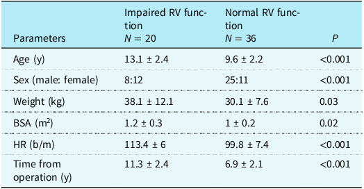
All patients were divided into two groups according to cardiac magnetic resonance-derived right ventricular ejection fraction%; impaired right ventricular systolic function group (n = 20) and normal right ventricular systolic function group (n = 36).The baseline demographic and clinical characteristics of patients stratified by right ventricular ejection fraction% using cardiac magnetic resonance are described in Table 1. Compared with patients with normal right ventricular systolic function, those with impaired right ventricular systolic function were significantly older in age, and predominantly female. Furthermore, patients with impaired right ventricular systolic function had significantly higher body weight, body surface area, HR, and the time from surgical repair compared with patients with normal right ventricular systolic function.
Demographic and clinical characteristics in the studied patients

RV = right ventricle, BSA = body surface area, HR = heart rate.
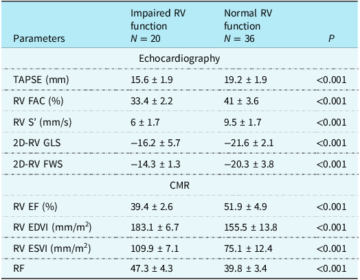
Regarding echocardiographic findings, patients with impaired right ventricular systolic function had significantly lower value of tricuspid annular plane systolic excursion, right ventricular fractional area change%, right ventricular S’, right ventricular global longitudinal strain%, and right ventricular free wall strain% compared with patients with normal right ventricular systolic function (P < 0.001 each). Regarding cardiac magnetic resonance findings, patients with impaired right ventricular systolic function exhibited significantly higher right ventricular end-diastolic volume indexed and right ventricular end-systolic volume compared with normal right ventricular systolic function patients (P < 0.001). Moreover, patients with impaired right ventricular systolic function had significantly more severe pulmonary regurgitation as assessed by pulmonary regurgitant fraction (P < 0.001) (Table 2).
Echocardiographic and CMR variables in the studied groups

TAPSE = tricuspid annular plane systolic excursion, RV FAC = right ventricular fractional area change, RV S’ = tricuspid annular peak systolic velocity, 2D-RV GLS = two-dimensional right ventricular global longitudinal strain, 2D-RV FWS = two-dimensional right ventricular free wall strain, RV EF = right ventricular ejection fraction, RV EDVI = right ventricular end-diastolic volume indexed, RV ESVI = right ventricular end systolic volume indexed, RF = regurgitant fraction.
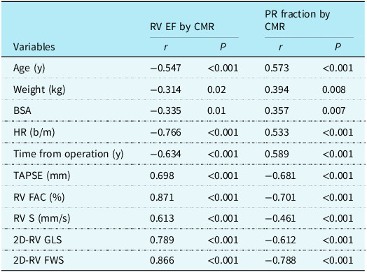
Right ventricular ejection fraction by cardiac magnetic resonance had a significant negative correlation with age, weight, BSA, HR, and the time from operation. Conversely, right ventricular ejection fraction had a significant positive correlation with tricuspid annular plane systolic excursion, right ventricular fractional area change, right ventricular S’, right ventricular global longitudinal strain, and right ventricular free wall strain. In contrast, cardiac magnetic resonance-derived pulmonary regurgitation fraction had a significant positive correlation with age, weight, BSA, HR, and the time from operation but a significant negative correlation with tricuspid annular plane systolic excursion, right ventricular fractional area change, right ventricular S’, right ventricular global longitudinal strain, and right ventricular free wall strain (Table 3).
Correlations between RV EF and PR fraction by CMR and different clinical and echocardiographic variables in the studied patients

PR = pulmonary regurgitation, BSA = body surface area; HR = heart rate; TAPSE = tricuspid annular plane systolic excursion; RV FAC = right ventricular fractional area change; RVEF = right ventricular ejection fraction; RVEDVI = right ventricular end-diastolic volume index; RVESVI = right ventricular end systolic volume index; RV S’ = right ventricular peak systolic velocity; 2D-RV GLS = two-dimensional right ventricular global longitudinal strain; 2D-RV FWD = two-dimensional right ventricular free wall strain.
ROC analysis revealed that 2D-RV free wall strain at a cutoff value of –20 had a sensitivity of 70%, a specificity of 94.4%, diagnostic accuracy of 85.7%, positive predictive value of 87.5%, negative predictive value of 85%, and AUC of 0.946 to predict right ventricular systolic dysfunction in the included patients (Figure 1 ( A )).
( A ) ROC analysis revealed that RV FWS at a cutoff value of −20 had a sensitivity of 70%, a specificity of 94.4%, and diagnostic accuracy of 85.7% to predict RV systolic dysfunction in children with repaired TOF. ( B ) ROC analysis revealed that RV S’ at a cutoff value of 10 cm/s had a sensitivity of 100%, a specificity of 30.6%, and diagnostic accuracy of 56.9% to predict RV systolic dysfunction in children with repaired TOF. ( C ) ROC analysis revealed that RV 2D-GLS at a cutoff value of –18 had a sensitivity of 70%, a specificity of 94.4%, and diagnostic accuracy of 85.7% to predict RV systolic dysfunction in children with repaired TOF. ( D ) ROC analysis revealed that RV FAC at a cutoff value of 35% had a sensitivity of 100%, a specificity of 52.8%, and diagnostic accuracy of 69.6% to predict RV systolic dysfunction in children with repaired TOF.

ROC analysis revealed that right ventricular S’ at a cutoff value of 10 cm/s had a sensitivity of 100%, a specificity of 30.6%, diagnostic accuracy of 56.9%, positive predictive value of 46.8%, negative predictive value of 100%, and AUC of 0.953 to predict right ventricular systolic dysfunction in the included patients (Figure 1 B ).
ROC analysis revealed that 2D-RV global longitudinal strain at a cutoff value of –18 had a sensitivity of 70%, a specificity of 94.4%, diagnostic accuracy of 85.7%, positive predictive value of 87.5%, negative predictive value of 85.7%, and AUC of 0.922 to predict right ventricular systolic dysfunction in the included patients (Figure 1 C ).
ROC analysis revealed that right ventricular fractional area change at a cutoff value of 35% had a sensitivity of 100%, a specificity of 52.8%, diagnostic accuracy of 69.6%, positive predictive value of 54.1%, negative predictive value of 100%, and AUC of 0.953 to predict right ventricular systolic dysfunction in the included patients (Figure 1 D ).
The intra-observer reliability was excellent for different cardiac echocardiographic parameters with intraclass correlation coefficient ranging from 87%–94%.
Discussion
Early identification of right ventricular systolic dysfunction is crucial for patients with repaired tetralogy of Fallot at high risk of poor prognosis. Our study showed that echocardiographic indices, such as right ventricular global longitudinal strain and right ventricular free wall strain, provide strong predictive value for the detection of right ventricular systolic dysfunction in children with repaired tetralogy of Fallot and may be used as a substitution for the expensive cardiac magnetic resonance in the follow-up of these children, especially in the developing countries.
In the current study, 35.7% of paediatric patients following repaired tetralogy of Fallot had right ventricular systolic dysfunction as evidenced by cardiac magnetic resonance. cardiac magnetic resonance-derived right ventricular ejection fraction% was strongly positively correlated to almost all echocardiographic parameters used for right ventricular function assessment, including right ventricular global longitudinal strain, right ventricular free wall strain, right ventricular S’, tricuspid annular plane systolic excursion, and right ventricular fractional area change.
Similarly, Promphan et al. Reference Promphan, Wonglikhitpanya, Katanyuwong and Siripornpitak16 who studied 20 children with repaired tetralogy of Fallot for at least 5 years, reported a strong significant positive correlation between right ventricular ejection fraction by cardiac magnetic resonance and each of right ventricular fractional area change, tricuspid annular plane systolic excursion, and the severity of pulmonary regurgitation as determined by echocardiography. Furthermore, Oliveira et al. Reference Oliveira, de Oliveira and Guimarães17 performed a systematic review that included nine studies to compare cardiac magnetic resonance and global longitudinal strain in patients with repaired tetralogy of Fallot and found that there was a strong positive correlation between right ventricular ejection fraction by cardiac magnetic resonance and right ventricular global longitudinal strain.
CMR findings
Regarding cardiac magnetic resonance findings, patients with impaired right ventricular systolic function exhibited significantly higher right ventricular end-diastolic volume and right ventricular end-systolic volume compared with those with normal right ventricular systolic function. Our results are consistent with the results of previous studies that found that increased preload is implicated as the main contributor to adverse right ventricular remodelling after initial tetralogy of Fallot repair. Reference Thompson, Dong and Kim18–Reference Fernandes, Manlhiot and Roche20 Early, the heart is able to increase stroke volume to maintain cardiac output, but in the long run, progressive ventricular dilation occurs with significant increases in right ventricular end-diastolic volume and right ventricular end-systolic volume.
Interestingly, cardiac magnetic resonance-derived pulmonary regurgitation fraction showed inverse significant correlation with almost all right ventricular systolic functional parameters using echocardiography. Similarly, Eyskens et al. Reference Eyskens, Brown and Claus21 and Li et al. Reference Fernandes, Manlhiot and Roche20 showed that severe pulmonary regurgitation was associated with reduced right ventricular strain. Furthermore, Kidwai et al. Reference Kidwai, Azad, Radhakrishnan, Garg, Yadav and Kumar23 reported that the presence of severe pulmonary regurgitation has a negative impact on right ventricular systolic function, which was better appreciated by right ventricular-fractional area change and right ventricular-global longitudinal strain.
Conventional echocardiographic parameters
Conventional echocardiographic measurements of right ventricular systolic function have limitations in evaluating myocardial function and detecting early stages of decreasing systolic function. They are highly operator-dependent and heavily dependent on the image quality, geometrical variables, image orientation, and the complex shape of the right ventricle. Thus, the reliable quantitative assessment of right ventricular size and function has remained an unresolved issue. Speckle tracking echocardiography overcomes most of the limitations inherent in conventional echocardiography, given that it is independent of cardiac translation, angle- and load-independent, thus, allowing accurate quantification of regional and global right ventricular myocardial function. Reference Meris, Faletra and Conca24–Reference Leibundgut, Rohner and Grize25 In patients with repaired tetralogy of Fallot, systolic dysfunction of the right ventricular remains an important issue. Adverse right ventricular remodelling has been related to right ventricular dilation secondary to PR, electromechanical dyssynchrony, and myocardial fibrosis.
RV global longitudinal strain
The findings in the current study showed that right ventricular 2D-global longitudinal strain was significantly lower than normal values in children with impaired right ventricular systolic function detected by cardiac magnetic resonance. Our findings are consistent with previous studies that have reported significant decrease in longitudinal strain. Reference Meris, Faletra and Conca24–Reference Leibundgut, Rohner and Grize25 The decrease in longitudinal strain values may be due to the selective hypo-perfusion of longitudinal fibres, resulting in severe ischaemia. Reference Scherptong, Mollema and Blom26 Moreover, Kavurt et al. Reference Kavurt, Paç, Koca, Mıhçıoğlu and Yiğit27 found that right ventricular global longitudinal strain values were significantly lower in repaired tetralogy of Fallot patients compared to controls, and right ventricular global longitudinal strain had the best correlation with right ventricular ejection fraction derived from cardiac magnetic resonance.
In the current study, both right ventricular 2D-free wall strain and right ventricular 2D global longitudinal strain were equally predictive for right ventricular systolic dysfunction with a sensitivity of 70%, a specificity of 94%, and an accuracy of 85%. Both right ventricular S’ and right ventricular fractional area change had a high sensitivity of 100% but low specificity of 30% and 50% respectively.
Some authors suggested that right ventricular global longitudinal strain might be more sensitive than cardiac magnetic resonance for early detection of right ventricular systolic dysfunction. This hypothesis is supported by a study done by Scherptong et al., Reference Scherptong, Mollema and Blom26 which showed that, although right ventricular ejection fraction on cardiac magnetic resonance remained unchanged, echo-derived right ventricular-global longitudinal strain already decreased on follow-up. They also reported that right ventricular global longitudinal strain continued to deteriorate in serial assessments despite preserved right ventricular ejection fraction, suggesting that regional wall-motion evaluations may detect early findings of subtle right ventricular systolic dysfunction. They concluded that right ventricular global longitudinal strain is more sensitive to detect changes in right ventricular performance before right ventricularejection fraction starts to decline, and it should be used for serial monitoring of right ventricular systolic function. Moreover, Kavurt et al. Reference Kavurt, Paç, Koca, Mıhçıoğlu and Yiğit27 found that right ventricular global longitudinal strain at a cutoff value of −17.4% had 75% sensitivity and 68.4% specificity in identifying right ventricular systolic dysfunction.
RV free wall strain
Regarding right ventricular free wall strain, we found that it is a good predictive for right ventricular systolic dysfunction with a sensitivity of 70%, a specificity of 94.4%, and a diagnostic accuracy of 85.7% similar to that of right ventricular 2D global longitudinal strain. One previous study showed that right ventricular free wall strain provides superior prognostic value than right ventricular global longitudinal strain in patients with repaired tetralogy of Fallot. Reference Gao, Li and He14 Moreover, Fujiwara et al. Reference Fujiwara, Tatebe and Nochioka28 found that the repaired tetralogy of Fallot group showed a significant decrease in right ventricular free wall strain than the other group, while right ventricular circumferential strain wasn’t affected. This may reflect an abnormal right ventricular contraction pattern where longitudinal contraction is more vulnerable than circumferential contraction in patients with repaired tetralogy of Fallot and right ventricular dilatation.
According to the current American society of Echocardiography, stable patients (over 10 years old) with repaired tetralogy of Fallot should have their right ventricular functions evaluated with cardiac magnetic resonance every 36 months and every 12 months if there is progressive right ventricular dilatation or dysfunction. Reference Valente, Cook and Festa29 Our results suggested that right ventricular global longitudinal strain and right ventricular free wall strain could assist physicians in predicting right ventricular systolic function to determine when cardiac magnetic resonance is most appropriate.
Limitations of the study
Relatively small sample size, cross-sectional design of the study with lack of follow-up, and the complex tripartite morphology of the right ventricular are not entirely represented by the basic 2D evaluation. So, three-dimensional speckle tracking echocardiography is recommended to be the topic of future research in comparison to cardiac magnetic resonance in these children.
Conclusion
Right ventricular free wall strain and right ventricular global longitudinal strain provide stronger predictive value over right ventricular S’ and right ventricular fractional area change, comparable to cardiac magnetic resonance data, in detection of right ventricular systolic dysfunction in children with repaired tetralogy of Fallot. Therefore, we recommend the use of right ventricular global longitudinal strain and right ventricular free wall strain (FWL) for routine follow-up in children with repaired tetralogy of Fallot. Further studies are needed with larger number of patients to estimate right ventricular systolic function by speckle tracking echocardiography that will help to determine the optimal time of cardiac magnetic resonance follow-up.
Financial support
This research received no specific grant from any funding agency, commercial, or not-for-profit sectors.
Ethical standards
The authors assert that all procedures contributing to this work comply with the ethical standards of the relevant national guidelines on human experimentation and with the Helsinki Declaration of 1975, as revised in 2008, and has been approved by the institutional committees of Faculty of Medicine, Tanta University.
Competing interest
None.