INTRODUCTION
Aspidosiphon muelleri Diesing, 1851 is the most widespread species of the family Aspidosiphonidae and presents a cosmopolitan distribution in temperate to subtropical waters. Aspidosiphon muelleri is also the most eurytopic member of the genus and lives in a wide variety of temperatures and depths, being more abundant in littoral waters (Murina, Reference Murina1975; Cutler, Reference Cutler1994). In the Mediterranean Sea, it is the predominant littoral sipunculan species in the western basin, and it is common in both soft and hard substrata (Saiz-Salinas, Reference Saiz-Salinas and Ramos1993; Papazacharias et al., Reference Papazacharias, Koukouras, Kevrekidis and Voultsiadou1998; Pancucci-Papadopoulou et al., Reference Pancucci-Papadopoulou, Murina and Zenetos1999; Açik et al., Reference Açik, Murina, Çinar and Ergen2005; Açik, Reference Açik2008, Reference Açik2011; Ferrero-Vicente et al., Reference Ferrero-Vicente, Loya-Fernández, Marco-Méndez, Martínez–García and Sánchez-Lizaso2011, Reference Ferrero-Vicente, Del-Pilar-Ruso, De-La-Ossa-Carretero and Sánchez-Lizaso2013a, Reference Ferrero-Vicente, Marco-Méndez, Loya-Fernández and Sánchez-Lizasob).
Aspidosiphon muelleri has been reported from many different habitats and associations within the Mediterranean Sea; associated with the algae Padina sp. and Cystoseira crinite Duby, 1830, associated with sponges (Koukouras et al., Reference Koukouras, voultsiadou-koukoura, Chintiroglou and Dounas1985; Açik et al., Reference Açik, Murina, Çinar and Ergen2005), as associated infauna of the echiuran Bonellia viridis Rolando, 1821 (Schembri & Jaccarini, Reference Schembri and Jaccarini1978), in bare rock, inside the rhizome of the seagrass Posidonia oceanica and on coralligenous substrata (Cladocora caespitosa (Linnaeus, 1767) and Pinna nobilis Linnaeus, 1758) (Açik et al., Reference Açik, Murina, Çinar and Ergen2005; Açik, Reference Açik2008). Drilling activity has been observed in individuals inhabiting chalk rock from the coast of Israel, even if the drilling mechanism is unknown (Por, Reference Por, Rice and Todorović1975). Aspidosiphon muelleri has also been reported in soft substrata, from sandy to muddy bottoms along the Mediterranean Sea, often inhabiting gastropod shells or polychaete tubes (Saiz-Salinas, Reference Saiz-Salinas1986, Reference Saiz-Salinas and Ramos1993; Saiz-Salinas & Villafranca-Urchegui, Reference Saiz-Salinas and Villafranca-Urchegui1990; Papazacharias et al., Reference Papazacharias, Koukouras, Kevrekidis and Voultsiadou1998; Somaschini et al., Reference Somaschini, Martini, Gravina, Belluscio, Corsi and Ardizzone1998; Murina et al., Reference Murina, Pancucci-Papadopoulou and Zenetos1999; Klaoudatos et al., Reference Klaoudatos, Klaoudatos, Smith, Bogdanos and Papageorgiou2006; Açik, Reference Açik2008; Ferrero-Vicente et al., Reference Ferrero-Vicente, Loya-Fernández, Marco-Méndez, Martínez–García and Sánchez-Lizaso2011, Reference Ferrero-Vicente, Del-Pilar-Ruso, De-La-Ossa-Carretero and Sánchez-Lizaso2013a, Reference Ferrero-Vicente, Marco-Méndez, Loya-Fernández and Sánchez-Lizasob). It is known that A. muelleri prefers to use empty tubes of the serpulid polychaete Ditrupa arietina (Müller, 1776) as a shelter in soft sediment, and its abundance could be limited by the availability of this particular resource (Ferrero-Vicente et al., Reference Ferrero-Vicente, Marco-Méndez, Loya-Fernández and Sánchez-Lizaso2013b). Ditrupa arietina is a widespread endobenthic species, common in the Mediterranean where it achieves high densities. Although D. arietina is considered temporal fauna in altered sediment (Picard, Reference Picard1965), there is evidence that its abundance has increased in some areas of the western Mediterranean (Grémare et al., Reference Grémare, Amouroux and Vétion1998a, Reference Grémare, Sardá, Medernach, Jordana, Pinedo, Amouroux, Martin, Nozais and Charlesb; Bolam & Fernandes, Reference Bolam and Fernandes2002; Labrune et al., Reference Labrune, Grémare, Guizien and Amouroux2007; Morton & Harper, Reference Morton and Harper2009), showing seasonal peaks of abundance in May–June (Sardá et al., Reference Sardá, Pinedo and Martin1999). Ditrupa arietina lives for approximately 2 years, and after its death (presumably predated by a prosobranch naticid), its tube becomes available for secondary colonization by A. muelleri (Morton & Harper, Reference Morton and Harper2009; Morton & Salvador, Reference Morton and Salvador2009).
Although external signs of sexual dimorphism are lacking, sipunculans are typically dioecious, but cases of hermaphroditic species have been reported (Gibbs, Reference Gibbs1975; Cutler & Cutler, Reference Cutler and Cutler1986). Gametes first develop in the coelomic fluid, before being taken up into the nephridia. They are stored for a short time in the nephridia, and finally released by way of nephridiopores after a short period of storage. Fertilization is external in all cases (Rice, Reference Rice, Giese and Pearse1975a, Reference Rice, Ryl and Tyler1989; Cutler, Reference Cutler1994; Maiorova & Adrianov, Reference Maiorova and Adrianov2005, Reference Maiorova and Adrianov2007). Asexual reproduction is known to occur within the genus Aspidosiphon (Rice, Reference Rice1970), in Aspidosiphon elegans (Chamisso & Eysenhardt, 1821), which is considered to be an alien species in the Mediterranean Sea (Açik, Reference Açik2008). Nonetheless, this reproductive ability has not been reported in A. muelleri, and little is known about the reproductive cycle of this species. Most of sipunculan species with a well-known life cycle show abbreviated development, omitting some stage of the Sipuncula development (trochophore stage, pelagosphera stage or having a lecithotrophic pelagosphera) (Rice, Reference Rice1976). Species with a known life cycle are Apionsoma misakianum (Ikeda, 1904) (Rice, Reference Rice1981), Nephasoma pellucidum (Keferstein, 1865) (Schulze & Rice, Reference Schulze and Rice2009), Phascolion strombus strombus (Montagu, 1804) (Åkesson, Reference Åkesson1958; Wanninger et al., Reference Wanninger, Koop, Bromham, Noonan and Degnan2005), Phascolion cryptum Hendrix, 1975 (Rice, Reference Rice, Rice and Todorović1975b), Phascolosoma turnerae Rice 1985 (Rice et al., Reference Rice, Reichardt, Piraino and Young2012), Siphonosoma cumanense (Keferstein, 1867) (Rice, Reference Rice1988), Themiste pyroides (Chamberlin, 1919) (Rice, Reference Rice1967; Adrianov et al., Reference Adrianov, Maiorova and Malakhov2011), Themiste lageniformis Baird, 1868 (Pilger, Reference Pilger1987), Themiste alutacea (Grube, 1858) (Rice, Reference Rice, Rice and Todorović1975b) and Thysanocardia nigra (Ikeda, 1904) (Rice, Reference Rice1967; Adrianov & Maiorova, Reference Adrianov and Maiorova2010).
However, to our knowledge, the genus Aspidosiphon presents a larval development with two pelagic stages: trochophore and planktotrophic pelagosphera larvae. This pelagosphera larva increases notably in size during its long period in the water (1–6 months) before its metamorphosis to a vermiform juvenile, which could make possible an interoceanic communication between different populations. (Scheltema & Hall, Reference Scheltema, Hall, Rice and Todorović1975; Cutler, Reference Cutler1994). Although planktotrophic pelagosphera larvae of many Aspidosiphonidae have been collected in plankton tows, their complete life cycles are still unknown (Hall & Scheltema, Reference Hall, Scheltema, Rice and Todorović1975; Rice, Reference Rice1981).
The aim of the present study is to describe some aspects related to the reproductive biology and ecology of Aspidosiphon muelleri in temperate waters.
MATERIALS AND METHODS
Study area and sampling design
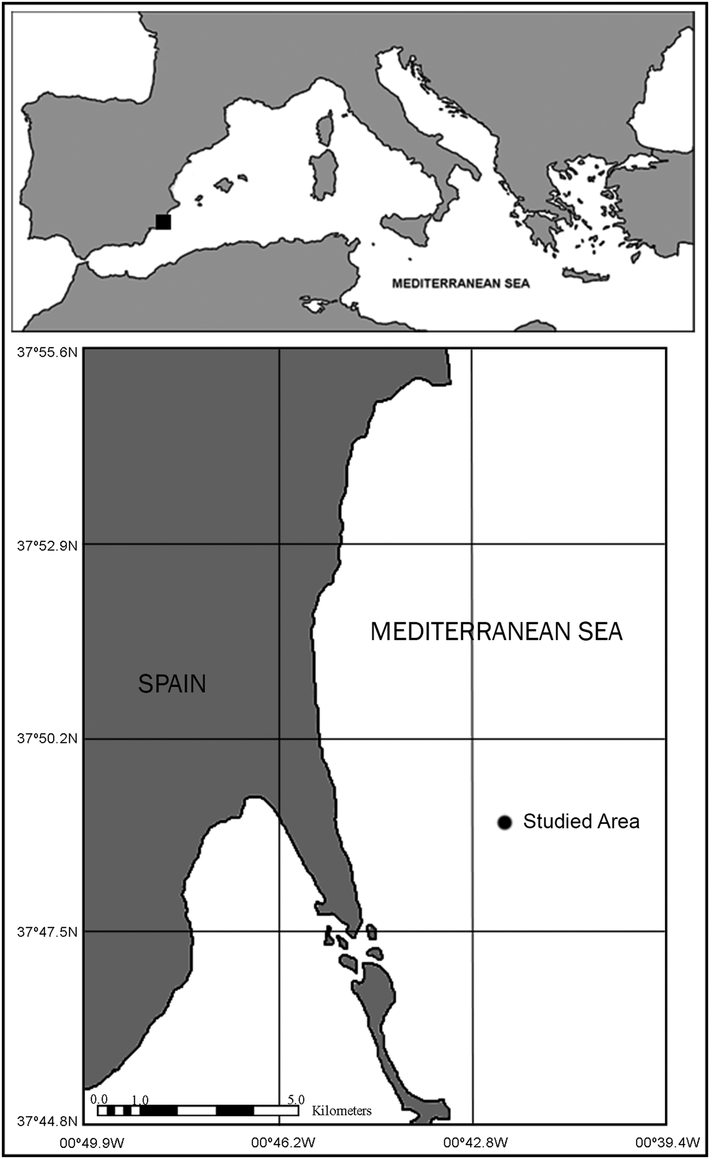
The study area is located near the shoreline of San Pedro del Pinatar (south-eastern Spain) (Figure 1). Samples were randomly taken around the coordinates 37°48.85′N 0°42.37′W (always keeping less than 50 m away from this point). The area of study was selected due to its suitable features for the study of Aspidosiphon muelleri. On one hand, the sipunculan assemblages at this bottom are well characterized, being A. muelleri an abundant species in this area (Ferrero-Vicente et al., Reference Ferrero-Vicente, Loya-Fernández, Marco-Méndez, Martínez–García and Sánchez-Lizaso2011, Reference Ferrero-Vicente, Del-Pilar-Ruso, De-La-Ossa-Carretero and Sánchez-Lizaso2013a), thus ensuring the presence of individuals which is irregular in many areas of the western Mediterranean Sea (Ferrero-Vicente et al., Reference Ferrero-Vicente, Del-Pilar-Ruso, De-La-Ossa-Carretero and Sánchez-Lizaso2013a). On the other hand, there is a high density of empty tubes and shells available to be used as shelter by A. muelleri, which allows the assessment of some aspects of the ecology of A. muelleri related to these resources (Cutler & Cutler, Reference Cutler and Cutler1989; Saiz-Salinas, Reference Saiz-Salinas and Ramos1993; Ferrero-Vicente et al., Reference Ferrero-Vicente, Del-Pilar-Ruso, De-La-Ossa-Carretero and Sánchez-Lizaso2013a, Reference Ferrero-Vicente, Marco-Méndez, Loya-Fernández and Sánchez-Lizasob).
Map showing the location of the studied population of Aspidosiphon muelleri.

Eleven monthly samples were taken from June 2011 to May 2012 using a Van Veen grab (0.028 m2) for a total of 132 samples. One of these samples was assigned to sediment analysis (granulometry, organic matter, pH and Eh). The ten remaining samples were sieved (500 µm) and used to study the ecology and reproductive biology of A. muelleri. Depth, temperature and bottom salinity were also measured using a CTD sensor (RBR-XR-420/620).
Laboratory work and data process
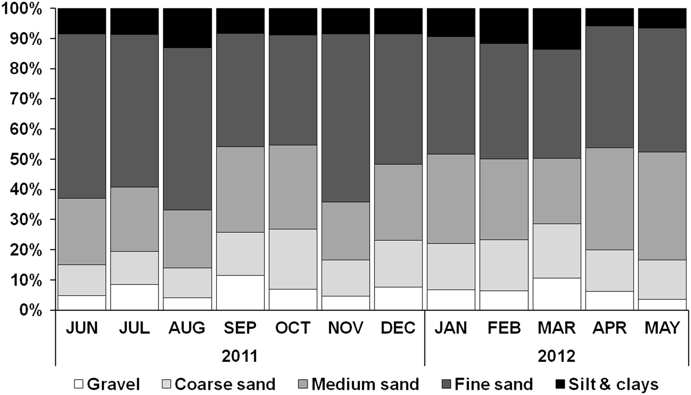
Redox potential (Eh) and pH were measured in the samples designated to the sediment analysis immediately after collection. Organic matter content was measured from a 30 g subsample, taken from the shallow layer of the sediment (0–5 cm), through weight loss on ignition after 4 h at 500°C inside a muffle furnace. The granulometric analysis was carried out following the methodology described by Buchanan (Reference Buchanan, Holme and McIntyre1984), sorting the samples in five categories: gravel; coarse sand; medium sand; fine sand; and silt and clays.
Ten replicates from every month were used for the fauna analysis (120 samples). Samples were sieved through a 500 µm mesh screen to separate the macrofauna and then fixed in 10% buffered formalin. Afterward, the fauna was preserved in 70% ethanol and sorted into different taxa. Abundances of other ecologically related species were also monitored and grouped into shelter suppliers (gastropods and Ditrupa arietina) or shelter dwellers (hermit crabs, polychaetes and tanaids). Sipunculans were identified to the species level (Cutler, Reference Cutler1994) through analysis of their internal and external anatomy using a binocular scope and identifying characteristics of taxonomically-relevant structures. Length and trunk thickness of each specimen and shelter were logged. Specimens of A. muelleri were dissected; gender was determined when it was possible, and oocytes present in the coelom were photographed and their size measured by image analysis techniques. We used 50 oocytes of each female specimen to calculate average size in every month.
Shelter availability (shelters m−2) was calculated for every one of the samples used for the fauna analysis. Shelters usually habited by sipunculans (according to previous observations since 2005) were sorted into empty tubes of D. arietina and empty shells of gastropods.
Univariate techniques and descriptive statistics were used to analyse the population dynamics of A. muelleri and its reproductive behaviour, moreover to define its possible relation with the abiotic factors of the environment. Relation between specimen size and shelter size was evaluated (Pearson's r correlation) as well as relation between reproductive variables (% females with presence of free oocytes in the coelom and oocyte size) and possible trigger factors for reproductive activity (surface/bottom temperature and irradiance).
RESULTS
Abiotic factors
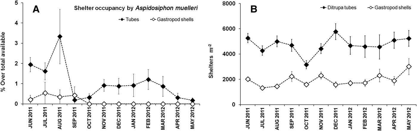
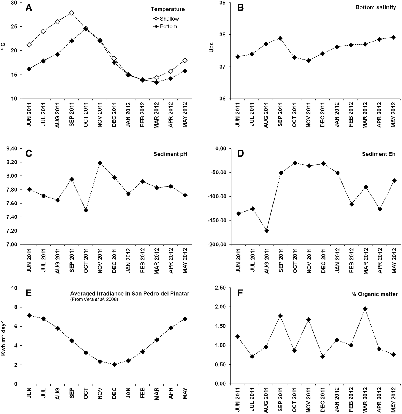
Granulometry of the sediment was homogeneous throughout the studied period (Figure 2). Sediment in the area is dominated by fine sand (43.8 ± 2.17%) and medium sands (25.9 ± 1.6%), with a presence of bioclasts, including gastropod shells and serpulid polychaete tubes. Depth ranged from 34.2 to 36.5 m. Presence of shelters in the sediment was high and constant for the whole study period, with a greater density of tubes than gastropod shells (Figure 3B). Average shelter densities were 4725 ± 174 tubes m−2 and 1934 ± 99 gastropod shells m−2, respectively. Tubes represented 70.9% of the total shelters available, while gastropod shells represented the remaining 29.1%. Organic matter content ranged between 0.76–1.95% (Figure 4F), and the redox potential (Eh) was also variable, but lower values were recorded during the warmer months (Figure 4D). Eh values were negative throughout the year, which shows the reductive environment of this sediment.
Granulometry in the studied area for each studied month.
(A) Ratio of shelter occupancy by Aspidosiphon muelleri for each month; (B) shelters density for each month in the studied area.
Charts from the different parameters measured at the studied area for each month.
Water temperature varied seasonally, following the previous months' irradiance levels (Figure 4A, E). Seabed temperature fluctuated annually, reaching the interannual peak (24.5°C) in October 2011 and the interannual low in March 2012 (13.46°C).There is a clear stratification in the water column that synchronizes with the months of maximum irradiance and weather stability, and there may be as much as 6.8°C difference detectable between surface temperature and bottom temperature in August 2011. The bottom salinity ranged from 37.2 to 37.9 psu, with a slight drop in October 2011, at the beginning of autumn (Figure 4B).
Biology of Aspidosiphon muelleri
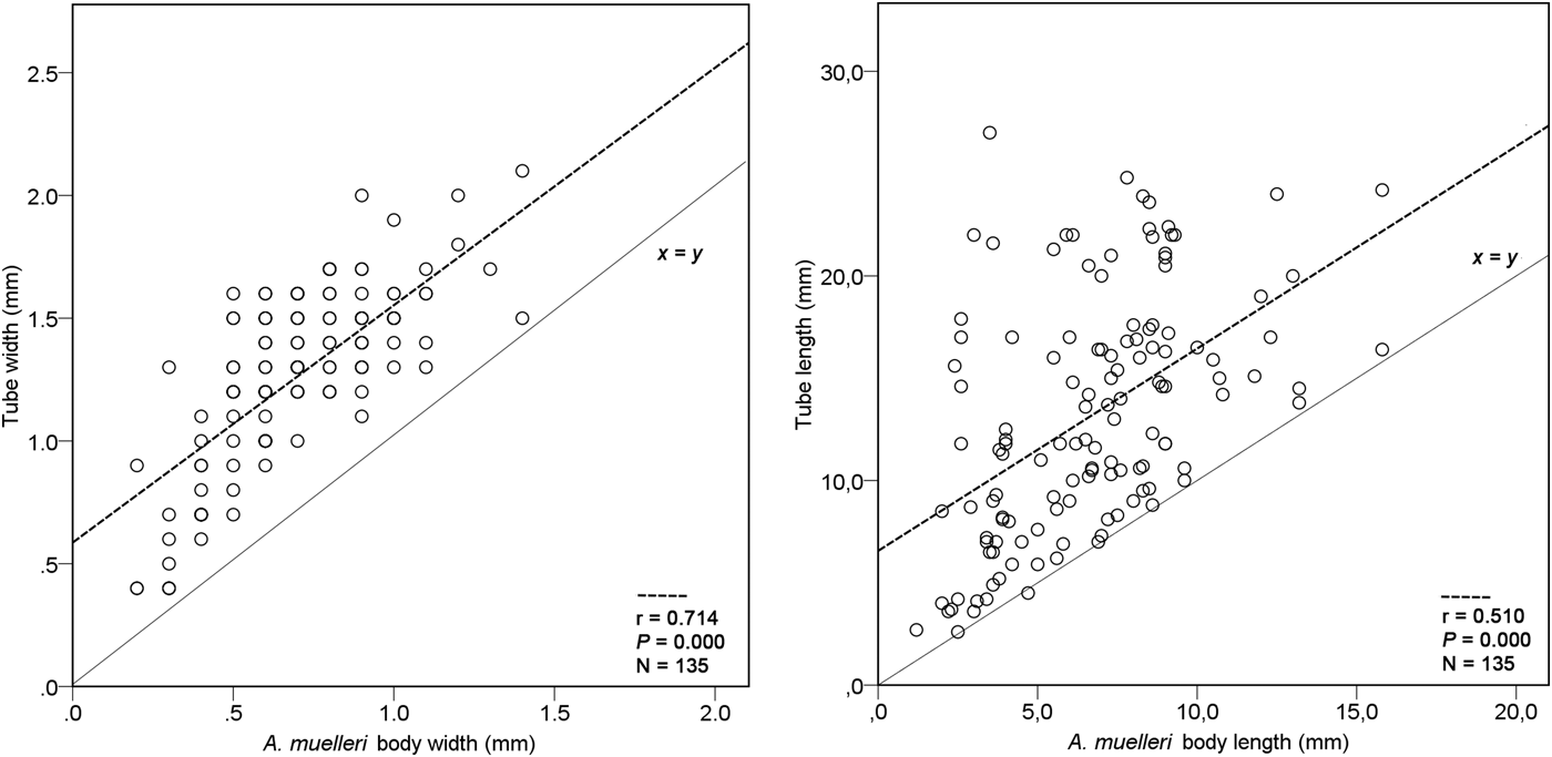
A total of 156 sipunculans were collected, with Aspidosiphon muelleri being the dominant species (146 specimens, 93.6% of the total). All specimens were dissected to analyse their external and internal anatomy. The remaining individuals corresponded to Thysanocardia procera (Möbius, 1875) (two specimens, 1.3%), Sipunculus nudus Linnaeus, 1766 (one specimen, 0.6%), Phascolion strombus strombus (one specimen, 0.6%) and six unidentified specimens (3.9%). Aspidosiphon muelleri sexual identification was possible in 72 of the cases, of which 54.2% were females whereas 45.8% were males. Sexual dimorphism was not detected and sexual identification was only possible through the observation of gametocytes in the coelom. In some cases, gametocytes were present in small specimens (3.7 mm and greater). Sipunculans were found inhabiting Ditrupa arietina tubes in 95% of the cases, in gastropod shells in 3.6% of the cases, and into bare substratum in the remaining 1.4% of cases. A significant correlation was detected between the sipunculan body width and tube width (Pearson correlation; r = 0.707; P = 0.000) and between sipunculan body length and tube length (Pearson correlation; r = 0.478; P = 0.000) (Figure 5). In all cases, tube length was longer than the body length of the animal, which means that the animal was always totally covered by the tube. Abundance of A. muelleri was not significantly related to shelter abundance (Pearson correlation; P = 0.684) and yielded an average density of 46.5 ± 6.0 ind m−2 for the whole study period, with an average body length of 6.6 ± 0.2 mm (mean ± standard error (SE)), and average body width of 0.7 ± 0.0 mm.
Dispersion charts showing the correlations between sipunculan body size and shelter size.
Skin colour was highly variable, but generally dull white, turning orange in some specimens. Anal and caudal shields displayed a darker tonality, usually dark orange. Juvenile specimens usually exhibited a translucent skin. Both types of hooks (uni and bidentate) were present in the introvert (Figure 6D). Individuals presented two elongate nephridia, as long as 20–60% of the trunk length (36.7 ± 2.0%). The number of intestine coils was significantly correlated with the length of the body (Pearson correlation; r = 0.582; P = 0.011) and ranged between 12 and 21 coils.
(A) Specimen of Aspidosiphon muelleri inside of a Ditrupa arietina tube; (B) specimen of Epilepton clarkiae collected inside a D. arietina tube inhabited by A. muelleri; (C) foraminifer species attached to the anal shield of A. muelleri; (D) unidentate pyramidal-shaped hooks from the introvert of A. muelleri.
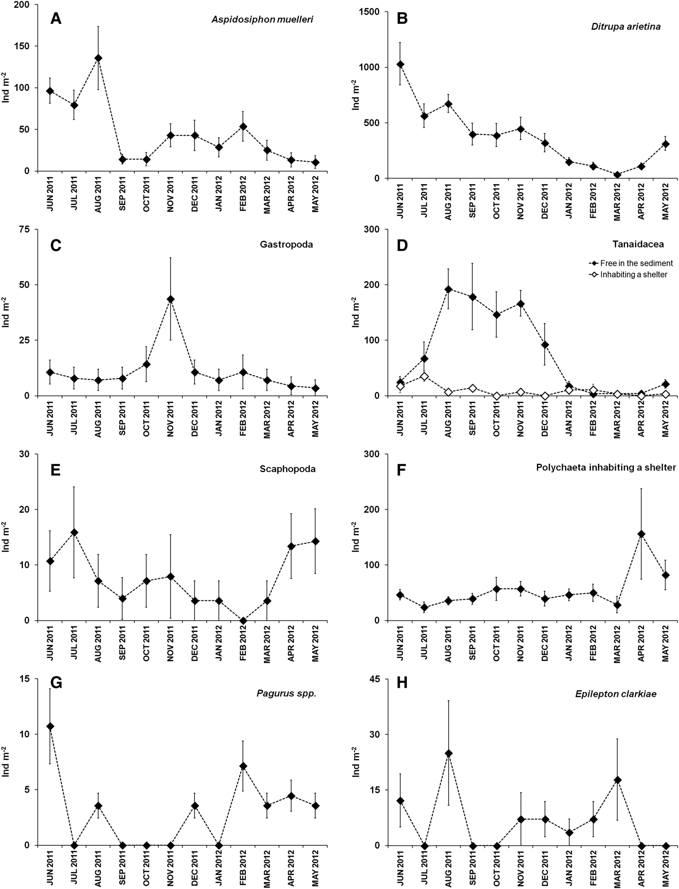
Maximum abundances of A. muelleri occurred in the summer period, with a peak in August (135.7 ± 37.9 ind m−2; mean ± SE). After this month the population decreased drastically, with a posterior slight recovery during the winter months (Figure 7A). Ditrupa tubes occupancy rate by Aspidosiphon muelleri was also higher in the summer months, with a peak in August (3.33 ± 1.34%) matching with the maximum abundance of sipunculans (Figure 3A). Gastropod shells were only used as a shelter in the summer period, when the maximum demand occurred. The rest of the months the occupancy rate for gastropod shells decreased to 0%.
Abundances of the different species and taxonomic groups recorded for each month (mean ± standard error).
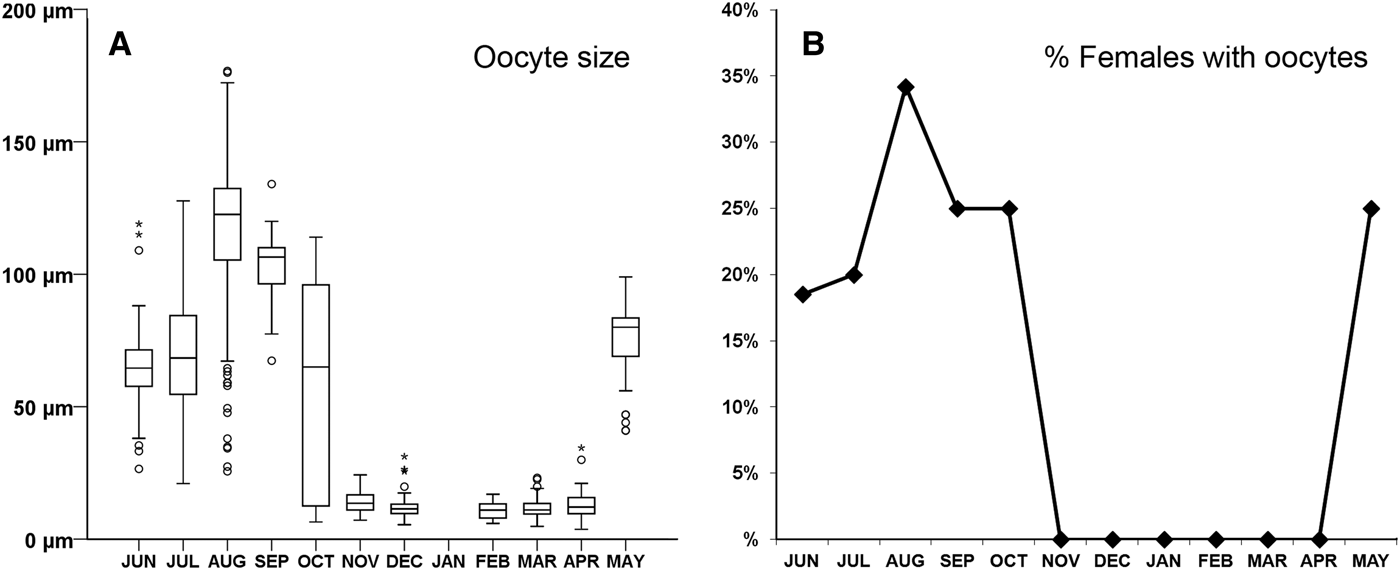
Gametocytes development takes place on the retractor muscle base and is eventually released into the coelomic fluid. Reproductive activity occurred mainly during the summer, beginning in May and extending through October, with a maximum peak in August (Figure 8). Both, the oocyte size and the percentage of females with free oocytes in the coelom, reached their maximum during that month. Average oocyte size was significantly related to shallow water temperature (Pearson correlation; r = 0.808; P = 0.001) as well as to irradiance (Pearson correlation; r = 0.589; P = 0.044). Percentage of females with free oocytes was also related to shallow water temperature (Pearson correlation; r = 0.776; P = 0.003) and showed a non-significant relation to irradiance (Pearson correlation; r = 0.562; P = 0.057).
(A) Averaged oocytes size in each month; (B) percentage of females with presence of free oocytes in the coelom.
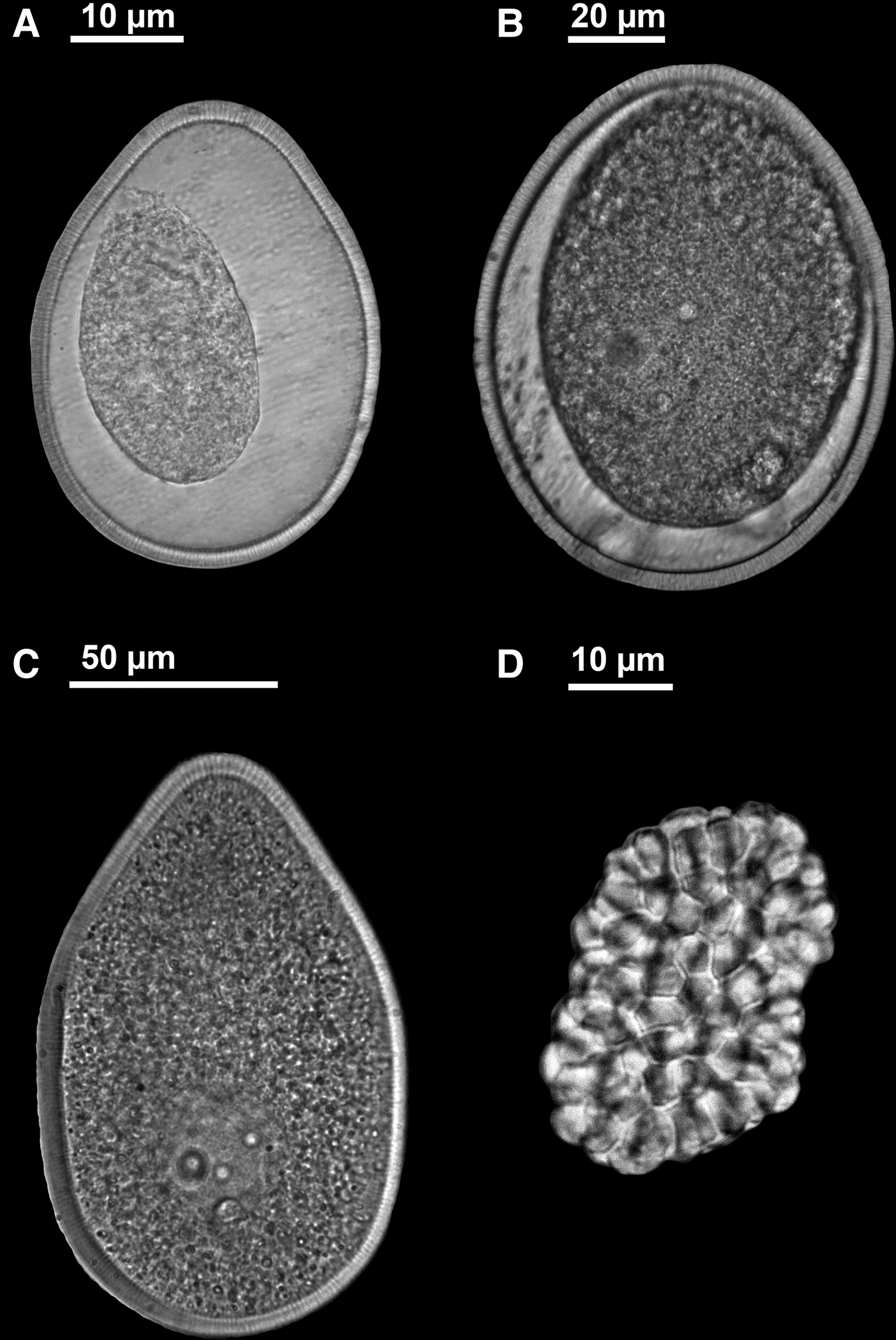
Oocytes presented an asymmetric-ovoid shape, with a pointed pole in most of the cases (Figure 9). Oocytes started to grow in May and August—just prior to spawning—and they had an average size of 118 ± 23 µm along its longer axis.
(A–C) Ovoid-shaped coelomic oocytes of different size; (D) coelomic sperm plates. The abnormal appearance of the cytoplasm, pulled away from the overlaying egg envelope (A–B), could be an artefact due to fixation process in formalin.
Although density of coelomocytes was not measured, a remarkable increase in abundance was observed in October, matching the final stage of the spawning event.
Sampling occurred once a month, and gametes into the nephridia (presumably where they would be hours before being released to the seawater) were not observed.
In regard to shelter supplier organisms (Ditrupa, Gastropoda and Scaphopoda), population density of D. arietina reached its maximum in the month of June, then slowly descended until March when it began to increase again. Abundance of D. arietina was much higher than abundances of Gastropoda or Scaphopoda (Figure 7B, C, E). Gastropoda tripled its population during the month of November (43.6 ± 18.5 ind m−2), and the rest of the year remained a stable population (~7–10 ind m−2); scaphopod abundance was low and generally higher in the summer months, reaching a maximum of 14.3 ± 5.8 ind m−2 in May 2012.
Regarding other shell-dwelling organisms (not Sipuncula), tanaid abundance increased notably, with its population reaching 200.0 ± 40.5 ind m−2 in August 2011. Tanaid abundance was high during the summer and autumn months, although the number of individual tube-dwellers did not increase in relation to total individuals during this period (Figure 7D). Hermit crabs did not yield a high density of individuals and abundance was variable, but more stable from February to June (Figure 7G). Its maximum abundance was 10.7 ± 3.4 ind m−2 in the month of June 2011. Polychaete presence (mainly Syllidae) inside the tubes was high throughout the year, with a peak in April 2012, reaching a maximum density of 156.2 ± 81.5 ind m−2 (Figure 7F). The presence of the commensal bivalve Epilepton clarkiae (Clark, 1852) living together with Aspidosiphon muelleri and sharing the Ditrupa arietina tube was constant throughout the year (Figure 7H). Bivalves were found in the tubes with a prevalence of 11.64%; one bivalve (7.53% of the cases), two bivalves (3.42%) and even three bivalves (0.69%) were found in the same tube attached to the body of the sipunculans. Bivalves were not found in any gastropod shell inhabited by A. muelleri. Foraminifers were also encountered inside some of the tubes attached to the body of the sipunculans (Figure 6C).
DISCUSSION
Aspidosiphon muelleri is a common species within the macrobenthic fauna of soft bottoms from the western Mediterranean, being often a highly abundant species (Saiz-Salinas, Reference Saiz-Salinas1986; Ferrero-Vicente et al., Reference Ferrero-Vicente, Loya-Fernández, Marco-Méndez, Martínez–García and Sánchez-Lizaso2011, Reference Ferrero-Vicente, Del-Pilar-Ruso, De-La-Ossa-Carretero and Sánchez-Lizaso2013a, Reference Ferrero-Vicente, Marco-Méndez, Loya-Fernández and Sánchez-Lizasob). Densities of 1675 ind m−2, the maximum known for this species (Ferrero-Vicente et al., Reference Ferrero-Vicente, Del-Pilar-Ruso, De-La-Ossa-Carretero and Sánchez-Lizaso2013a) have been recorded in this area. The area of study was selected due to its known high abundance of A. muelleri. The averaged abundance of individuals (46.5 ± 6.0 ind m−2) was higher than recorded in other nearby areas of the western Mediterranean (13.78 ± 3.93 ind m−2 in Ferrero-Vicente et al., Reference Ferrero-Vicente, Del-Pilar-Ruso, De-La-Ossa-Carretero and Sánchez-Lizaso2013a), whereas the relative abundance—93.6% of the total sipunculans collected were A. muelleri—was similar to other studies from the area: 89.06 and 74.07% (Ferrero-Vicente et al., Reference Ferrero-Vicente, Loya-Fernández, Marco-Méndez, Martínez–García and Sánchez-Lizaso2011, Reference Ferrero-Vicente, Del-Pilar-Ruso, De-La-Ossa-Carretero and Sánchez-Lizaso2013a).
Sex-ratio was close to 1:1 (54.2% females vs 45.8% males), which fits with a dioecious reproduction typical in Sipuncula (Rice, Reference Rice, Ryl and Tyler1989). Nonetheless not all sipunculans present this pattern, and biased sex-ratios are not uncommon in some species; Rice et al. (Reference Rice, Reichardt, Piraino and Young2012) recorded a 74.0% of females for Phascolosoma turnerae (a deep-sea wood-dwelling sipunculan), and Pilger (Reference Pilger1987) recorded 96% females and 4% males of the parthenogenic species Themiste lageniformis. Many other species present sex-ratios in which females outnumber the males (Pilger, Reference Pilger1987). Even asexual reproduction has also been described for one species of the genus, Aspidosiphon elegans (Rice, Reference Rice1970).
Gametocytes were observed in specimens from 3.7 mm, and 4–5 mm appears to be the approximate size for the gametocytes development in this population of A. muelleri. This size could be reached by A. muelleri in a few months, since growth from larval fixation to adult size (even more than 10 mm) could take less than six months (Ferrero-Vicente et al., Reference Ferrero-Vicente, Marco-Méndez, Loya-Fernández and Sánchez-Lizaso2013b).
According to Ferrero-Vicente et al. (Reference Ferrero-Vicente, Marco-Méndez, Loya-Fernández and Sánchez-Lizaso2013b), approximately 82% of the available shelters in this area correspond to Ditrupa tubes whereas 18% are gastropod shells. This work also suggests a shelter limitation for this species (getting abundances 10 times higher by adding shelters in the sediment) and a preference of A. muelleri to use tubes as shelter instead of snails. Shelter partitioning in the present work was similar; approximately 71% of available shelters were tubes and 29% were gastropod shells. Shelter preference of tubes over gastropod shells fit also well with the present results, since 95% of individuals inhabited a tube and only 3.6% a snail. The remaining 1.4% of individuals was encountered bare into the sediment, but they presumably left the shelter in the fixation process. A linear relation between shelter abundance and sipunculan abundance was not detected due to high shelter availability throughout the whole study period (Figure 3B), thus indicating that shelter availability is not driving the maximum density of A. muelleri in this area. However, there may be some limitation during the summer, matching the maximum population density. At this point, sipunculans start to use gastropod shells as resource in spite of their preference for using tubes, and it might be that Ditrupa tubes are not so easy to find at that point of the year, probably due to the higher demand of this resource by tanaids and A. muelleri. After reproduction, when population density decreased drastically, sipunculans used preferably tubes as shelter. Correlation between body and shelter size was strong, and it is known that A. muelleri left its shelter just when all available space of the shelter—in a contracted state of the animal—is occupied for its body, ensuring that the animal can always be covered by the tube (Ferrero-Vicente et al., Reference Ferrero-Vicente, Marco-Méndez, Loya-Fernández and Sánchez-Lizaso2013b). This preference for Ditrupa tubes was not found for other shell/tube-dwelling species of similar size from the western Mediterranean (Phascolion caupo Hendrix, 1975 and Phascolion strombus strombus) (Ferrero-Vicente et al., Reference Ferrero-Vicente, Loya-Fernández, Marco-Méndez, Martínez–García, Saiz-Salinas and Sánchez-Lizaso2012, Reference Ferrero-Vicente, Marco-Méndez, Loya-Fernández and Sánchez-Lizaso2013b).
Internal anatomy did not yield differences with regard to other specimens from other areas (Saiz-Salinas, Reference Saiz-Salinas and Ramos1993; Cutler, Reference Cutler1994; Pancucci-Papadopoulou et al., Reference Pancucci-Papadopoulou, Murina and Zenetos1999; Açik et al., Reference Açik, Murina, Çinar and Ergen2005; Açik, Reference Açik2011), but specimens showed plasticity in some characters (e.g. skin took different tonalities from translucent skin in juvenile specimens to white or dark orange skin in adult specimens). Presence of unidentate and bidentate hooks, with pyramidal shape in the proximal part of the introvert was also noted. Some specimens presented pointed spines in the anal shield, in the grooves close to the introvert. This characteristic was not discriminative since different degrees of spine length and edge shape were detected, from almost flat to very pointed spines.
The number of intestine coils was significantly correlated with the length of the body (Pearson correlation; r = 0.582; P = 0.011), which indicates that intestine grows in length as long as the animal keeps growing.
Sediment in the area has a negative redox potential, ranging from −30.2 to −170.8 mV, which indicates a reducing environment. This condition does not seem to be relevant for this species, likely due to the fact that A. muelleri lives in the shallowest and most oxygenated layer of the sediment, protected from predation by its shelter.
In temperate waters, marine invertebrates usually exhibit a two- or three-month peak in reproductive activity during the summer or early autumn (depending on latitude) (Cutler, Reference Cutler1994; Ying et al., Reference Ying, Dahms, Liu, Wu, Zhang, Chen and Yang2009). In the case of A. muelleri, the highest density of individuals occurred during July and August, and then decreased drastically in September. This increase in population density coincides with the development of oocytes, which increase its size along these months. Female oocytes average size in its longer axis (118 ± 23 µm) was similar to that logged by Açik (Reference Açik2008) for this species (117.5–145 µm).
Trigger factors for reproduction in sipunculans are poorly understood, but in our case the development of the gametocytes and spawning event might be stimulated by an increase in irradiation and water temperature, as usual in many marine invertebrates from temperate waters. The first females with presence of oocytes appeared in the month of May (following the increase in water temperature). We can indirectly estimate spawning took place between August and September (just before the annual maximum temperature) because the percentage of females with oocytes and oocyte size both reached their maximum in August. This spawning event may last a few weeks, but it has been observed in other species that in colder waters at high latitude as well as in equatorial waters, spawning may be extended for several months (Rice, Reference Rice, Rice and Todorović1975b; Amor, Reference Amor1993; Cutler, Reference Cutler1994; Adrianov & Maiorova, Reference Adrianov and Maiorova2010). Moreover a higher water temperature induces a higher larval survival, but on the other hand, this might mean a shorter larval stage, which implies a lesser dispersion (O'Connor et al., Reference O'Connor, Bruno, Gaines, Halpern, Lester, Kinlan and Weiss2007).
It is known that macroinvertebrates present seasonal life cycles in order to take advantage of optimal environmental conditions or avoid unfavourable conditions (Johnson et al., Reference Johnson, Carreiro, Jin and JacK2012). Fluctuations in sipunculan spawning period are also known to occur due to differences in their conditions of habitation, showing an extended spawning period in a more stable situation (Rice, Reference Rice1967). This fact led us to think that different A. muelleri populations can exhibit different reproductive strategies for different areas, and they could have different spawning timing in areas with a less pronounced seasonality, like high latitude or equatorial areas.
It is known that lifespan can be long for some species (e.g >20 years in Phascolosoma turnerae (Rice et al., Reference Rice, Reichardt, Piraino and Young2012)). In our case, the dramatic decrease in abundance of individuals from August to September suggests that many adult individuals might die after the spawning, maybe as a consequence of this event. Although the abundance in September was low, the percentage of females with oocytes was still high. In October, the oocyte size was smaller and highly variable; a high presence of coelomocytes was observed during this period, and likely their function is the phagocytosis of the oocytes, which were not expelled to the seawater after the spawning (Amor, Reference Amor1993; Adrianov & Maiorova, Reference Adrianov and Maiorova2010).
As with many other sedentary groups, sipunculans attract a variety of associated fauna. Some small bivalves with a known association to sipunculans are Kurtiella bidentata (Montagu, 1803), reported together with Golfingia vulgaris (de Blainville, 1827) (Gibbs, Reference Gibbs1977), Montacuta phascolionis Dautzenberg & Fischer, 1925, and Pythinella cuneata (Verrill & Bush, 1898) together with Phascolion strombus (Gage, Reference Gage1968, Reference Gage1979; Troncoso & Urgorri, Reference Troncoso and Urgorri1992). To our knowledge, previous data about commensalism between bivalves and Aspidosiphon muelleri are limited to the relation with the bivalve Jousseaumiella concharum Knudsen, 1944 (Saiz-Salinas, Reference Saiz-Salinas and Ramos1993). This is the first report of Epilepton clarkiae associated with A. muelleri, although it is not the first record of this bivalve in association with a sipunculan, since it was reported together with Golfingia vulgaris inside the burrows, which this sipunculan digs in the sediment (Gibbs, Reference Gibbs1977), but other associations have been found with Phascolion strombus (Troncoso & Urgorri, Reference Troncoso and Urgorri1992) and Golfingia elongata (McMillan, 1968). The collected specimens of E. clarkiae were juveniles, and an adult specimen might not be able to inhabit the tube since its regular size is bigger than the tube diameter. The fact that E. clarkiae was not found in the gastropod shells inhabited by A. muelleri is probably due to the low number of sipunculans recorded inside gastropod shells. This commensal relationship might play an important role in the distribution of E. clarkiae since any individuals living bare into the sediment, or even inside a shelter not occupied by A. muelleri, were not found.
ACKNOWLEDGEMENTS
We are grateful to Dr Serge Gofas (University of Málaga) for his help in identifying the bivalve Epilepton clarkiae and to Aaron Macy (Dauphin Island Sea Laboratory) for help with language corrections and comments on the manuscript. We also thank the anonymous referees for their comments and suggestions to improve the manuscript.
FINANCIAL SUPPORT
This research was funded by ‘Mancomunidad Canales del Taibilla’.