Implications
The nutritional environment in which the fetus and newborn grow and develop has the potential to have long-term effects on body composition as well as metabolic health of the offspring. These outcomes are highly dependent on the timing of the nutritional intervention and thus reflect the specific organ system that is most rapidly developing at each time point. Adipose tissue development is particularly sensitive throughout this period and there is clearly the potential to promote both brown and white adiposity that may either benefit or adversely affect the offspring.
Introduction
Diet during pregnancy is one important modifiable factor that can have a substantial influence on the viability and body composition of the newborn. This is usually considered to have the largest impact in late gestation when the absolute rate of growth of the fetus is greatest (Symonds et al., Reference Symonds, Stephenson, Gardner and Budge2007). As such, total nutrient requirements through the final 10 weeks of pregnancy were carefully calculated 30 years ago and provide a widely used standard by which to define the feeding requirements for sheep production (Agricultural Research Council 1980). A major factor that, therefore, determines the total metabolizable energy requirements is fetal number (Agricultural and Food Research Council, 1993). The need for this type of information was highlighted by the potential losses in the neonatal period as a result of starvation and hypothermia being greatest in small sized offspring (Symonds and Lomax, Reference Symonds and Lomax1992). These individuals are likely to be characterized as having depleted fat stores in conjunction with an increased surface area to volume ratio, thus subjecting them to much greater thermal demands. Interestingly, in some breeds of sheep, these potential risks have been reduced over the past 20 years as global temperatures have risen (Hellmann et al., Reference Hellmann, Byers, Bierwagen and Dukes2008). Consequently, more small sized offspring have survived into adulthood resulting in mean adult body weight actually decreasing over the same period (Ozgul et al., Reference Ozgul, Tuljapurkar, Benton, Pemberton, Clutton-Brock and Coulson2009).
This review will, therefore, consider the relative impact of changes in maternal dietary intake at different stages of the reproductive cycle to highlight how nutrition can have variable effects on the offspring which are primarily dependent on the developmental process targeted. It will focus on studies in sheep, as these have been the most widely investigated largely because of their extensive use for investigating the fetal or developmental origins of adult health and disease. Indeed, investigations of this type are now the main type of ruminant research funded, emphasizing the current scarcity of funding for large animal research (Roberts et al., Reference Roberts, Smith, Bazer, Cibelli, Seidel, Bauman, Reynolds and Ireland2009).
Critical windows in development and the impact of changes in maternal diet
The main emphasis of recent research into the impact of maternal diet on reproduction in ruminants has focused upon the impact of global reductions in food intake (Symonds and Budge, Reference Symonds and Budge2009). These types of studies have addressed the more widespread concerns relating to inadequate maternal nutrition, rather than excess food intake which may now be more of a problem within the developed world. Although, its relevance to ruminant production is not always direct, it does emphasize the potential long-term outcomes that may have particular relevance to selecting animals for later sheep production particularly ‘replacement ewes’.
Primary factors determining newborn birth weight are fetal number in conjunction with maternal parity (Gardner et al., Reference Gardner, Buttery, Daniel and Symonds2007). Consequently, first time mothers will produce smaller offspring and usually have single rather than multiple fetuses. Then, as the number of pregnancies increases, both fetal number and weight rises. These adaptations are mediated, in part, by the changes in maternal physiology and uterine function after completion of a first pregnancy, in conjunction with more subtle changes in body composition and endocrine sensitivity (Hyatt et al., Reference Hyatt, Keisler, Budge and Symonds2010).
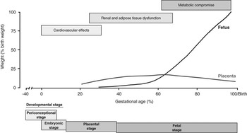
The critical stages of pregnancy after conception are summarized in Figure 1 and cover the peri-conceptional period up to the time of rapid embryo development, followed by the establishment of the placenta as morphogenesis of the fetus occurs enabling the formation of the fetal circulation and all essential organ systems. Subsequently, during late gestation, important adaptations in placental function occur as its total mass declines (Heasman et al., Reference Heasman, Clarke, Firth, Stephenson and Symonds1998). These enable the substantial increase in nutrient requirements by the rapidly growing fetus to be met in conjunction with an appreciable mobilization of maternal fat stores, which is, itself, dependent on the prevailing nutritional and thermal environment (Symonds and Clarke, Reference Symonds and Clarke1996). For individuals that have been subjected to a previous period of more extreme nutritional deprivation, this adaptation may be severely compromised leading to the termination of pregnancy (Bloomfield et al., Reference Bloomfield, Oliver, Hawkins, Campbell, Phillips, Gluckman, Challis and Harding2003) as discussed further below. A final important component of the global nutritional requirements at this stage include mammary gland development, which is obviously necessary to ensure sufficient milk is supplied to the newborn (Agricultural Research Council, 1980).
Summary of the different long-term effects in the offspring after maternal nutrient restriction at defined stages of the reproductive cycle in sheep.

Maternal diet prior to pregnancy and its potential contribution to preterm labor
The extent to which a reduction in maternal food intake before conception has a direct effect on the reproductive cycle, as opposed to an indirect influence as a result of the pronounced changes in the maternal metabolic and hormonal environment, remains an area of debate. This controversy is emphasized by the very variable reproductive effects of reducing maternal food intake from at least 60 days before mating (McMillen and Robinson, Reference McMillen and Robinson2005). In a comparatively small subgroup of sheep, in which a reduction in body weight was greatest before conception, this was accompanied by preterm labor (Bloomfield et al., Reference Bloomfield, Oliver, Hawkins, Campbell, Phillips, Gluckman, Challis and Harding2003). Interestingly, that this finding has not been confirmed by any other groups (Edwards and McMillen, Reference Edwards and McMillen2002; Budge et al., Reference Budge, Edwards, McMillen, Bryce, Warnes, Pearce, Stephenson and Symonds2004) to date, may be related to differences in breed of sheep, body composition or even in the time of year in which such experiments are undertaken. Irrespective of the very different reproductive outcomes, a reduction in maternal food intake either before, or after, pregnancy results in a pronounced reduction in plasma concentration of counter-metabolic hormones particularly cortisol (Bispham et al., Reference Bispham, Gopalakrishnan, Dandrea, Wilson, Budge, Keisler, Broughton Pipkin, Stephenson and Symonds2003; Jaquiery et al., Reference Jaquiery, Oliver, Bloomfield, Connor, Challis and Harding2006). As such the lack of a persistent increase in maternal plasma cortisol suggests that a reduction in food availability of this magnitude is within that normally experienced by the animal. It also reinforces the point that maternal nutrient restriction is not equivalent to a biological and/or psychological stress (Budge et al., Reference Budge, Stephenson and Symonds2007), which may be due in part to the fact that food (i.e. roughage) is actually available throughout the majority of the day in these studies. The net effect is to promote the mobilization of maternal fat stores and maintenance of glucose production that would be important for supporting either the rapidly dividing embryo and/or the growing placenta (Symonds and Clarke, Reference Symonds and Clarke1996).
It is not normal agricultural practice to suddenly restrict maternal food intake before conception, as this will reduce both ovulation and rates of conception, thereby having the net effect of reducing the mother’s fertility. This type of adaptation would be particularly detrimental in lowland breeds of sheep for which twin or triplet bearing pregnancies are the norm. The long-term impact of improving the overall plane of nutrition of the mother before conception is emphasized when comparing fetal number in highland breeds of sheep in which simply bringing them down to a lowland pasture for one season before mating can result in a majority bearing twin, as opposed to singleton, pregnancies (M. E. Symonds unpublished).
The impact of maternal diet on growth and metabolism of the placenta
After uterine attachment that in the sheep occurs between approximately 22 and 28 days gestation, a reduction in maternal diet can restrict overall growth of the placenta, although the magnitude of this response is strongly influenced by the timing of any nutritional intervention (Yiallourides et al., Reference Yiallourides, Sebert, Wilson, Sharkey, Rhind, Symonds and Budge2009). Interestingly, in sheep, reducing maternal food intake from the time of mating up to the period when placental growth has commenced in both the maternal caruncle and adjoining fetal cotyledon (i.e. 0 to 30 days gestation), or continuing up to the time at which fetal growth becomes exponential (i.e. 0 to 110 days gestation) (Reynolds et al., Reference Reynolds, Borowicz, Vonnahme, Johnson, Grazul-Bilska, Wallace, Caton and Redmer2005), has no effect on either placental or fetal weight in later gestation (Yiallourides et al., Reference Yiallourides, Sebert, Wilson, Sharkey, Rhind, Symonds and Budge2009). A decrease in maternal food intake from ∼28 days gestation continuing up to the end of the period of maximal placental growth that is 80 days gestation, does restrict placental mass primarily as a consequence of reducing growth of the placental component (Heasman et al., Reference Heasman, Clarke, Firth, Stephenson and Symonds1998). At the same time, this results in a reduction in epithelial cell proliferation, which is most likely mediated by the increased glucocorticoid sensitivity exhibited by the placenta, which persists up to term (Gnanalingham et al., Reference Gnanalingham, Williams, Wilson, Bispham, Hyatt, Pellicano, Budge, Stephenson and Symonds2007). This, in turn, appears to be primarily a consequence of the reduction in maternal plasma cortisol which persists throughout the period of nutrient restriction (Bispham et al., Reference Bispham, Gopalakrishnan, Dandrea, Wilson, Budge, Keisler, Broughton Pipkin, Stephenson and Symonds2003) and which, in conjunction with a reduction in both gene expression and activity of the enzyme 11β-hydroxysteriod dehydrogenase type 2, would be expected to enhance glucocorticoid sensitivity within the placenta (Whorwood et al., Reference Whorwood, Firth, Budge and Symonds2001). However, although placental growth is initially restricted, the normal decrease in placental mass that occurs between mid and late gestation in the sheep fails to occur which means that previously nutrient restricted mothers have larger placenta at term and there are no negative effects on fetal growth in the second half of gestation (Heasman et al., Reference Heasman, Clarke, Firth, Stephenson and Symonds1998). It is thus possible that placental efficiency is subsequently enhanced in mothers that were previously nutrient restricted (Fowden et al., Reference Fowden, Sferruzzi-Perri, Coan, Constancia and Burton2009). Indeed, the growth of some fetal organs, including the kidney and adipose tissue, can be enhanced (Whorwood et al., Reference Whorwood, Firth, Budge and Symonds2001; Bispham et al., Reference Bispham, Gopalakrishnan, Dandrea, Wilson, Budge, Keisler, Broughton Pipkin, Stephenson and Symonds2003; Brennan et al., Reference Brennan, Gopalakrishnan, Kurlak, Rhind, Brooks, Rae, Olson, Stephenson and Symonds2005).
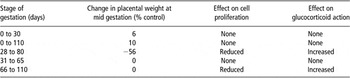
The magnitude of endocrine and related adaptations within the placenta after shorter term reductions in maternal food intake (<30 days) is strongly dependent on the stage of gestation in which it is introduced as summarized in Table 1. Under these conditions, adaptations within the mother and/or within the placenta appear to minimize any structural changes in the placenta or alterations in glucocorticoid action (Yiallourides et al., Reference Yiallourides, Sebert, Wilson, Sharkey, Rhind, Symonds and Budge2009). The period of maternal nutrient restriction that has the greatest endocrine effect within the placenta after maternal nutrient restriction is, therefore, seen when this is targeted to between 66 and 110 days that is commencing at the time of maximal growth. In addition to increased glucocorticoid sensitivity, gene expression for the insulin-like growth factor II receptor is raised together with markers of lipid metabolism (Yiallourides et al., Reference Yiallourides, Sebert, Wilson, Sharkey, Rhind, Symonds and Budge2009) of which peroxisome-activated receptor (PPAR)γ has a primary role (Nunn et al., Reference Nunn, Bell and Barter2007). It is, therefore, possible that, as in the placenta of other species in which PPARγ modulates lipid uptake and metabolism (Schaiff et al., Reference Schaiff, Barak and Sadovsky2006), a comparable effect is seen in the sheep. Increased action of uncoupling protein (UCP)2 in the placenta during maternal nutrient restriction would promote mitochondrial fatty acid oxidation while limiting glucose metabolism. UCPs are able to uncouple the oxidation of fatty acids by affecting the electron transport chain from ATP synthesis (Echtay et al., Reference Echtay, Winkler and Klingenberg2000). Fatty acids also affect the expression of UCPs by acting as ligands for PPARs, with the main products of fatty acid metabolism that is acetate and β-hydroxybutyrate being readily transferred across the placenta (Hull et al., Reference Hull, Elphick and Broughton1979). These could, therefore, be used as an alternative fuels to glucose for fetal metabolism and lipogenesis (Herrera, Reference Herrera2002). Clearly, further studies are required to establish the role of lipid metabolism in the sheep placenta after changes in the maternal diet and the extent to which this impacts on nutrient supply to the fetus.
Summary of the influence of stage of gestation on the effect of maternal nutrient restriction on placental weight, cell proliferation and glucocorticoid action in sheep.

Based on Gnanalingham et al., Reference Gnanalingham, Williams, Wilson, Bispham, Hyatt, Pellicano, Budge, Stephenson and Symonds2007; Yiallourides et al., Reference Yiallourides, Sebert, Wilson, Sharkey, Rhind, Symonds and Budge2009
Effect of maternal nutrient restriction between early-to-mid gestation on fetal growth and organ development
One of the primary effects of maternal nutrient restriction either before, or during, pregnancy is a reduction in plasma concentration of catabolic hormones including cortisol, thyroid hormones and insulin. These adaptations ensure that the maternal plasma glucose concentration is maintained and, thus, fetal growth is not compromised. The endocrine sensitivities of a large number of fetal organs are, however, reset particularly with regard to glucocorticoid action. As in the placenta (see above), this response is mediated by an increase in both gene expression for the glucocorticoid receptor and reduced action of the enzyme 11β-hydroxysteriod dehydrogenase type 2 which occurs in a range of newborn tissues including heart, kidney, liver and lungs (Whorwood et al., Reference Whorwood, Firth, Budge and Symonds2001). These changes would be predicted to enhance glucocorticoid action even in the absence of any change in plasma cortisol (Gardner et al., Reference Gardner, Van Bon, Dandrea, Goddard, May, Wilson, Stephenson and Symonds2006). These have no immediate adverse effect on the offspring and persist in some tissues into juvenile life when offspring are raised in an outdoor-free living environment. Under these conditions, their body composition remains the same as those born to control fed mothers and they show no signs of metabolic or cardiovascular compromise (Gopalakrishnan et al., Reference Gopalakrishnan, Gardner, Dandrea, Langley-Evans, Pearce, Kurlak, Walker, Seetho, Keisler, Ramsay, Stephenson and Symonds2005).
When these animals are raised under an obesogenic environment with comparable thermal and photoperiod conditions that is inside within a barn in which their activity is reduced (by 75% compared with free-living animals maintained within an adjacent field), they show a majority of the adverse symptoms of the metabolic syndrome as summarized in Table 2. These include an accelerated increase in resting blood pressure (Symonds et al., Reference Symonds, Stephenson and Budge2009b), greatly enhanced ectopic lipid accumulation (Chan et al., Reference Chan, Sebert, Hyatt, Stephenson, Budge, Symonds and Gardner2009) and insulin resistance (Sebert et al., Reference Sebert, Hyatt, Chan, Patel, Bell, Keisler, Stephenson, Budge, Symonds and Gardner2009). Although, such disease symptoms are not obviously relevant to agricultural practice, they illustrate the added value of using sheep as a model for human metabolic disease.
Summary of the effect of maternal nutrient restriction between early-to-mid gestations on the primary symptoms of the metabolic syndrome after adolescent onset obesity in the sheep

Increased maternal food intake from mid gestation and its effect on adipose tissue development and function
Increasing the amount of food provided to pregnant sheep from the time at which placental mass has peaked can have positive effects in promoting fetal growth and repartitioning of nutrients between skeletal muscle and adipose tissue growth (Budge et al., Reference Budge, Bispham, Dandrea, Evans, Heasman, Ingleton, Sullivan, Wilson, Stephenson and Symonds2000). The magnitude of this response is dependent, in part, on the amount of feed consumed in the earlier stages of gestation (Bispham et al., Reference Bispham, Gardner, Gnanalingham, Stephenson, Symonds and Budge2005). One of the major adaptations is to increase the abundance of brown adipose tissue, clearly benefiting the newborn’s ability to effectively adapt to the cold exposure of the extra-uterine environment (Budge et al., Reference Budge, Bispham, Dandrea, Evans, Heasman, Ingleton, Sullivan, Wilson, Stephenson and Symonds2000). Brown adipose tissue is uniquely characterized as possessing UCP1 which enables the rapid generation of heat through the uncoupling of electron transport from oxidative phosphorylation (Cannon and Nedergaard, Reference Cannon and Nedergaard2004). One hormone that can act to promote brown adipose tissue function in the newborn is prolactin (Pearce et al., Reference Pearce, Budge, Mostyn, Genever, Webb, Ingleton, Walker, Symonds and Stephenson2005). Plasma prolactin concentrations are positively correlated with gestational age in infants (Lucas et al., Reference Lucas, Baker and Cole1990), and in the sheep fetus there is an increase in both prolactin gene expression and circulating prolactin concentrations during the last 10 to 15 days of gestation to peak at birth (term, 147 ± 3 days gestation) (Gluckman et al., Reference Gluckman, Marti-Henneberg, Kaplan and Grumbach1983). Prolactin acts through its receptor of which there are at least two forms, the long and short form (Bignon et al., Reference Bignon, Binart, Ormandy, Schuler, Kelly and Djiane1997). In the fetal sheep, changes in gene expression and protein abundance of the prolactin receptor, both with gestational age and the maternal nutritional environment, are key factors determining both the initial appearance in UCP1, as well as changes in its activity around the time of birth (Symonds et al., Reference Symonds, Phillips, Anthony, Owens and McMillen1998 and Reference Symonds, Mostyn and Stephenson2001).
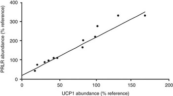
The extent to which an increase in prolactin receptor abundance may determine the rate of loss of UCP1 after birth has yet to be established. Given the close relationship between the rate of decline in both UCP1 and prolactin receptor (Figure 2), it is likely to delay this process. This may have practical implications because, after birth when UCP1 is rapidly lost, there is a period of pronounced hyperplasia within the same perirenal-abdominal fat depot, which adopts primarily white adipose tissue characteristics (Clarke et al., Reference Clarke, Buss, Juniper, Lomax and Symonds1997b). This type of transformation within one fat depot, may be very different to that seen in small mammals in which it, has been suggested that brown and white fat have very different origins (Timmons et al., Reference Timmons, Wennmalm, Larsson, Walden, Lassmann, Petrovic, Hamilton, Gimeno, Wahlestedt, Baar, Nedergaard and Cannon2007). In these species, however, the main site of brown fat is within the intrascapular region which does not undergo the same type of transition to white fat after birth but is instead retained as brown fat throughout the life cycle (Cannon and Nedergaard, Reference Cannon and Nedergaard2004).
Linear correlation between abundance of the brown adipose tissue specific uncoupling protein (UCP)1 and the abundance of the prolactin receptor (PRLR) over the first month of postnatal life in sheep. (Based upon Pearce et al., Reference Pearce, Budge, Mostyn, Genever, Webb, Ingleton, Walker, Symonds and Stephenson2005).

In utero determinants of fat distribution and function: potential role of photoperiod
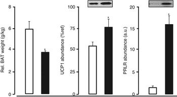
Recent studies have highlighted the importance of early life events in determining further aspects of adipocyte function and distribution (Budge et al., Reference Budge, Sebert, Sharkey and Symonds2009). This includes the finding that white adipocyte progenitor cells become committed to the adipose lineage during the late fetal/early postnatal period, and that there is also a marked expansion of this cellular pool because of the proliferation during postnatal life (Tang et al., Reference Tang, Zeve, Suh, Bosnakovski, Kyba, Hammer, Tallquist and Graff2008). Furthermore, it has also been suggested that brown adipocytes have the same lineage as skeletal myoblasts, a process that may be regulated by bone morphogenetic protein acting through PRD1-BF1-RIZ1 homologous domain containing 16 gene (Seale et al., Reference Seale, Bjork, Yang, Kajimura, Chin, Kuang, Scime, Devarakonda, Conroe, Erdjument-Bromage, Tempst, Rudnicki, Beier and Spiegelman2008; Tseng et al., Reference Tseng, Kokkotou, Schulz, Huang, Winnay, Taniguchi, Tran, Suzuki, Espinoza, Yamamoto, Ahrens, Dudley, Norris, Kulkarni and Kahn2008). Alternatively, there may be two types of brown adipose tissue cells that are either positive or negative for Myf-5 (Enerback, Reference Enerback2009) and, thus, are either ‘normal’ or ‘recruitable’ brown adipose tissue and whose origin may be set very early in development. Ultimately, this would provide a common mechanism relating brown adipocyte and muscle development, differing significantly from white adipocytes, and is in accord with the distinct differences in myogenic gene expression found between brown and white cells. This is also in accord with some phenotypic similarities observed between brown adipocytes and skeletal muscle (Timmons et al., Reference Timmons, Wennmalm, Larsson, Walden, Lassmann, Petrovic, Hamilton, Gimeno, Wahlestedt, Baar, Nedergaard and Cannon2007). The main regulators of this process remain to be established but it is notable that prolactin receptor knockout mice not only show reduced UCP1 abundance at birth (Viengchareun et al., Reference Viengchareun, Servel, Feve, Freemark, Lombes and Binart2008) but also less fat as adults (Freemark et al., Reference Freemark, Fleenor, Driscoll, Binart and Kelly2001). It has yet to be established whether the same plethora of factors that have been implicated in the regulation of brown fat development in rodents have the same role in large mammals including ruminants. Given the strong link between maternal diet, prolactin receptor abundance and UCP1 (Figure 3) may provide a mechanism by which to promote brown fat abundance both at birth and in the longer term.
Effect of increased maternal food intake over the final half of gestation on relative fat mass, the brown adipose tissue specific uncoupling protein (UCP)1 and the abundance of the prolactin receptor (PRLR) in the newborn sheep. Mothers were either fed to 100% of total metabolisable energy requirements (open boxes) or ad libitum (i.e. 150% of metabolisble energy requirements; closed boxes). (Based upon Budge et al., Reference Budge, Bispham, Dandrea, Evans, Heasman, Ingleton, Sullivan, Wilson, Stephenson and Symonds2000).

The potential importance of the established relationship between the prolactin receptor and brown fat function to both fetal growth and development, but also to longer term fat development, is highlighted by the recent finding of brown fat in adult humans (Virtanen et al., Reference Virtanen, Lidell, Orava, Heglind, Westergren, Niemi, Taittonen, Laine, Savisto, Enerback and Nuutila2009) and for which a strong seasonal influence has been demonstrated (Au-Yong et al., Reference Au-Yong, Thorn, Ganatra, Perkins and Symonds2009). It is established that one of the major hormones that responds to photoperiod is prolactin (Bassett et al., Reference Bassett, Bomford and Mott1988) (Goldman et al., Reference Goldman, Matt, Roychoudhury and Stetson1981) which also increases with day length (Steger et al., Reference Steger, Bartke, Goldman, Soares and Talamantes1983). Studies in adult hamsters have also indicated an important role for melatonin in mediating the effect of day length on brown adipose tissue function (Heldmaier et al., Reference Heldmaier, Steinlechner, Rafael and Vsiansky1981). The melatonin receptor is present on fetal adipose tissue, however, melatonin seems to inhibit the lipolytic actions of noradrenaline on fetal adipose tissue, at least when studied in vitro at ∼130 days of gestation under hypothermic conditions (Torres-Farfan et al., Reference Torres-Farfan, Valenzuela, Mondaca, Valenzuela, Krause, Herrera, Riquelme, Llanos and Seron-Ferre2008) (i.e. 37°C compared with the normal fetal body temperature of ∼40°C (Power, Reference Power1989)).
The extent to which photoperiod, as opposed to ambient temperature, is a primary regulator of brown adipose tissue function remains uncertain as, although photoperiod can determine brown adipose tissue activity irrespective of ambient temperature, its effect is enhanced in the cold (Klingenspor et al., Reference Klingenspor, Klaus, Wiesinger and Heldmaier1989; Wiesinger et al., Reference Wiesinger, Heldmaier and Buchberger1989). In mammals that naturally show a seasonal change in body composition according to day length, exposure to short days for only 5 weeks promotes brown adipose tissue function and is accompanied by marked fat mobilization (Demas et al., Reference Demas, Bowers, Bartness and Gettys2002). However, in this model the effect declines with the duration of exposure that may be related to compensatory changes in food intake.
Prolactin administration also promotes the loss of UCP1 in lactating rats (Chan and Swaminathan, Reference Chan and Swaminathan1990) and prolactin secretion can be temperature sensitive (Vigas et al., Reference Vigas, Celko and Koska2000). The prolactin receptor is essential for brown adipose tissue function in the newborn (Viengchareun et al., Reference Viengchareun, Servel, Feve, Freemark, Lombes and Binart2008), in which its direct stimulation promotes brown adipose tissue thermogenesis (Pearce et al., Reference Pearce, Budge, Mostyn, Genever, Webb, Ingleton, Walker, Symonds and Stephenson2005). Indeed, during normal development, an increase in prolactin receptor abundance could be a key stage in fetal brown adipose tissue development (Symonds et al., Reference Symonds, Phillips, Anthony, Owens and McMillen1998). In this regard, photoperiod is the primary determinant of plasma prolactin in maternal and thus fetal circulation with values more than a 100 fold different between pregnant sheep (and their fetuses) maintained under long daylight (>16 h) compared with short daylight (<9.5 h) (Bassett et al., Reference Bassett, Bomford and Mott1988). Clearly, future studies are necessary to establish the role of photoperiod on fetal development and how this may interact with maternal diet.
The influence of temperature on fetal development
One of the best examples of the potential influence of the thermal environment on fetal development comes from studies that have examined the impact of chronic maternal cold exposure induced by winter shearing (Symonds et al., Reference Symonds, Bird, Clarke, Gate and Lomax1995). This procedure is often utilized by lowland farmers to increase stocking rates close to the time of lambing when all sheep are barn-housed to aid animal husbandry during lambing. At the same time, the beneficial effects on reproductive performance are only seen in twin or triplet bearing sheep (Symonds and Lomax, Reference Symonds and Lomax1992). The primary effect of this challenge to both the mother and the fetus is to induce chronic maternal adaptations to the cold that result in enhanced secretion and/or action of thyroid hormones, noradrenaline and growth hormone, whereas the action of insulin is reduced (Symonds et al., Reference Symonds, Bryant and Lomax1988 and Reference Symonds, Bryant and Lomax1986). The magnitude of these adaptations is dependent, in part, on the prevailing ambient temperature. Overall, the net effect of these long-term changes in energy metabolism within the mother is that her ability to mobilize and then utilize her own fat stores is increased, thereby preventing the late gestational decline in plasma glucose that often occurs in unshorn mothers (Symonds et al., Reference Symonds, Bryant and Lomax1988).
The beneficial effects on the fetus and newborn include not only an increase in brown fat but also a larger liver with greater glycogen stores (Symonds et al., Reference Symonds, Bryant, Clarke, Darby and Lomax1992; Clarke et al., Reference Clarke, Bryant, Lomax and Symonds1997a). This means that newborn is better adapted not only to meet the thermal challenge of the extra-uterine environment but also to improve the thyroid and respiratory function (Symonds et al., Reference Symonds, Lomax, Kenward, Andrews and Johnson1993; Clarke et al., Reference Clarke, Bryant, Lomax and Symonds1997a). These effects then persist through at least the first month of life, so that even when the offspring are artificially reared on a comparatively low plane of nutrition, they still deposit more fat in which brown adipose tissue characteristics are retained (Symonds et al., Reference Symonds, Bryant, Clarke, Darby and Lomax1992). In those offspring that are reared by their mothers, growth rate is increased through lactation primarily as a result of increased milk production (Symonds et al., Reference Symonds, Bryant and Lomax1990).
Future studies into epigenetic control mechanisms
Now it appears that those individuals exposed to an inadequate diet during very early development may exhibit a range of adaptations that not only relate to up or down-regulation of gene expression but also this could include epigenetic modifications (Symonds et al., Reference Symonds, Sebert, Hyatt and Budge2009a). To date, all of the experimental evidence relating to this process has been largely confined to studies using rodents with extreme dietary modifications such as consumption of a hypermethylating diet through pregnancy and lactation (Waterland and Jirtle, Reference Waterland and Jirtle2003) that contains a ∼9-fold increase in choline and folic acid, ∼3 times or more increase in methionine and ∼60 times or more increase in vitamin B12 compared with control diets (Reeves, Reference Reeves1997). The one exception involved sheep that were conceived in elderly (i.e. cull-ewes) fed with a diet that was lacking in sulphur and cobalt with result that the recipients were depleted in sulphur containing amino acids, specific B group vitamins (vitamin B12 and folate), as well as having reduced plasma glucose (Sinclair et al., Reference Sinclair, Allegrucci, Singh, Gardner, Sebastian, Bispham, Thurston, Huntley, Rees, Maloney, Lea, Craigon, McEvoy and Young2007). Subsequently, only after 6 days of exposure to this embryonic environment, all blastocysts were transferred to sheep fed with a standard diet. As such, it is not possible to fully dissociate the suggested adverse outcomes from either maternal age, diet, embryo transfer or accelerated postnatal growth. In addition, the high embryo loss rate of ∼45% (Sinclair et al., Reference Sinclair, Allegrucci, Singh, Gardner, Sebastian, Bispham, Thurston, Huntley, Rees, Maloney, Lea, Craigon, McEvoy and Young2007) may exaggerate the reported outcomes as this is much greater than is seen in normal agricultural practice, especially when using a breed of sheep that normally produces singleton fetuses.
An alternative experimental manipulation that has been used to compromise fetal growth in the rodent is that of uterine artery ligation in late gestation which reduces uterine artery blood flow by ∼50% (Simmons et al., Reference Simmons, Flozak and Ogata1993) and results in substantial intra-uterine growth retardation (Wigglesworth, Reference Wigglesworth1974). When these offspring are cross-fostered onto control dams they show rapid catch-up growth, an adaptation that is likely to be as important in determining the subsequent adverse outcomes as the preceding growth restriction in utero (Symonds, Reference Symonds2007). As adults, these growth manipulated offspring exhibit type 2 diabetes that is associated with progressive epigenetic silencing of the homeobox 1 transcription factor Pdx1 which is critical for pancreatic β-cell function and development (Park et al., Reference Park, Stoffers, Nicholls and Simmons2008). Nevertheless, during the neonatal period, the reduction in Pdx1 expression could be reversed in vitro by inhibition of histone deacetylase action. Potential epigenetic effects may well be extended to a number of other tissues and cells including the regulation of intracellular energy locus, as recently shown in vitro (by using human neonatal skin fibroblasts), at least under conditions of 0 or very high (i.e. 1000 mg/l) glucose concentration (Murayama et al., Reference Murayama, Ohmori, Fujimura, Minami, Yasuzawa-Tanaka, Kuroda, Oie, Daitoku, Okuwaki, Nagata, Fukamizu, Kimura, Shimizu and Yanagisawa2008).
The extent to which such mechanisms operate under the range of normal circulating plasma glucose, particularly during early development, is important as it has also been shown that glucose metabolism is closely linked to chromatin modification and global transcription control (Wellen et al., Reference Wellen, Hatzivassiliou, Sachdeva, Bui, Cross and Thompson2009). This involves the production of acetyl-coenzyme A from glucose and ATP-citrate lyase, which is a key intermediate in regulating energy metabolism within mitochondria, the cytoplasm and the nucleus. It is now necessary to translate these in vitro findings of a novel regulatory mechanism that produces a substrate for chromatin modification to the in vivo situation (Rathmell and Newgard, Reference Rathmell and Newgard2009). This is especially the case for the fetus in which changes in plasma glucose are much more subtle, but can have much greater and long-term effects (Symonds et al., Reference Symonds, Stephenson, Gardner and Budge2007). For example, it has been shown that exposure to the Dutch Famine during the end of World War II can result in subtle changes in the methylation status of the insulin-like growth factor II receptor as determined in genomic DNA extracted from blood samples of the survivors aged ∼60 years (Heijmans et al., Reference Heijmans, Tobi, Stein, Putter, Blauw, Susser, Slagboom and Lumey2008). Whether this is a direct nutritional effect remains to be established as, interestingly, such an adaptation was only seen in those offspring that were actually conceived during the famine and not in those that were exposed to the famine during the final trimester and who were smaller at birth. It remains to be seen whether the types of adaptations to targeted reductions, or increases, in the maternal diet are accompanied by comparable epigenetic adaptations that ultimately underpin the long-term outcomes.
In conclusion, changes in the amount or composition of feed consumed by the mother from the time of ovulation through to lactation have the potential to significantly reset the growth trajectory of a majority of fetal organs and tissues. Ultimately, this will determine size at birth which in conjunction to other external stimuli, such as temperature and/or photoperiod, will not only determine size at birth but long term health and survival. An increased understanding of these processes may be particularly important over the next 10 years or so as the gradual changes in the planet’s temperature and the effects of climate change start to have further impact on agricultural production.
Acknowledgments
The authors would like to acknowledge the support of the European Union Sixth Framework Program for Research and Technical Development of the European Community – The Early Nutrition Programming Project (FOOD-CT-2005-007036) and the Nottingham Respiratory Biomedical Research Unit in their research.