INTRODUCTION
Plant-induced resistance is the process whereby biotic or abiotic elicitors activate the innate immunity present in plants against diseases (Strobel et al. Reference Strobel, Ji, Gopalan, Kuc and He1996; Sato et al. Reference Sato, Kubo and Watanabe2003; Kawahara et al. Reference Kawahara, Namba, Toyoda, Kasai, Sugimoto, Inagaki, Ichinose and Shiraishi2006). The biotic elicitors, which include different chemical compounds such as proteins, glycoproteins, lipids and oligosaccharides, have been extracted from various sources such as fungi, bacteria, viruses and herbivores (Veit et al. Reference Veit, Wörle, Nürnberger, Koch and Seitz2001; Paré et al. Reference Paré, Farag, Krishnamachari, Zhang, Ryu and Kloepper2005; Zhao et al. Reference Zhao, Davis and Verpoorte2005; Sharathchandra et al. Reference Sharathchandra, Geetha, Amruthesh, Kini, Sarosh, Shetty and Shetty2006). Over the past few decades, various types of protein elicitors have been found in bacterial and fungal pathogens (Wei et al. Reference Wei, Laby, Zumoff, Bauer, He, Collmer and Beer1992; He et al. Reference He, Huang and Collmer1993; Arlat et al. Reference Arlat, Van Gijsegem, Huet, Pernollet and Boucher1994; Baillieul et al. Reference Baillieul, Genetet, Kopp, Saindrenan, Fritig and Kauffmann1995; Veit et al. Reference Veit, Wörle, Nürnberger, Koch and Seitz2001; Mao et al. Reference Mao, Liu, Yang, Long, Zhao, Zeng, Liu, Yuan and Qiu2010; Zhang et al. Reference Zhang, Yang, Liu, Qiu, Zhang, Zeng, Yuan and Mao2010). Protein elicitors from plant pathogens induce systemic acquired resistance (SAR) in plants by activating the plant's natural defence system (Peng et al. Reference Peng, Qiu, Ruan, Zhou and Sun2011). Recently, plant pathologists have been interested in detecting effective protein elicitors by manipulating the inducible defence responses of plants so that these protein elicitors may be used for crop disease protection.
In the process of extracting protein elicitors from plant fungal pathogens, it was discovered that the protein lysis buffer demonstrated the ability to induce defence responses in cucumber plants (Cucumis sativus L.) and reduced the disease severity of anthracnose caused by Colletotrichum orbiculare (Berk. & Mont.) Arx. The present study attempted to ascertain the effect of both the protein lysis buffer and its individual components on disease development of cucumber anthracnose in cucumber plants and on the spore germination of this pathogen. Furthermore, expression levels of certain disease resistance-related genes were also examined to explain the possible underlying mechanism of the disease control.
MATERIALS AND METHODS
Fungal and plant materials
The cucumber anthracnose pathogen, C. orbiculare (Berk. & Mont.) Arx.CO-51 was isolated from samples collected from a field in Dali, Taichung, Taiwan (24°5′N, 120°39′E, 40 m a.s.l.). In order to obtain a stock culture and to prevent mutation, a single spore of C. orbiculare CO-51 was transferred each month to a slant of a new potato dextrose agar (PDA, Difco Laboratories, Detroit, USA) and maintained at room temperature (22–25 °C) with a 12 h light period. Eight replicates of the cultures were usually prepared, and those that mutated or showed no sporulation were discarded. The working culture, to be used for preparation of conidial suspensions, and an increase in sporulation of the C. orbiculare CO-51 was obtained by sub-culturing single-spore cultures of the pathogen weekly to a potato sucrose yeast extract agar medium (PSYA; potato extract 4 g, yeast extract 5 g, sucrose 10 g, agar 15 g, distilled water 1 litre), using the conditions described above. Again, eight replicates were prepared and any cultures that mutated or failed to sporulate were discarded. The four-leaf stage (c. 4-week-old) of Cucumber (C. sativus L.) cultivar Swallow (Known-You Seed Co., Ltd, Kaohsiung, Taiwan) was used for all of the experiments in the current study.
Preparation of the conidial suspensions
The cultures of C. orbiculare CO-51 were grown on PSYA medium for 5–7 days before being flooded with distilled water to wash the spores; this was followed by filtration through a single layer of Miracloth (Calbiochem, San Diego, USA) to remove any mycelial residue from the conidial suspension. The final concentration of the suspension was adjusted to 2×10⁴ spores/ml and used for both inoculation and spore germination experiments.
Preparation of the protein lysis buffer
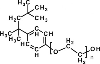
The buffer solution consisted of 57·06 g urea (Bio-Rad, Hercules, USA) dissolved in 50 ml of deionized water; 4 g CHAPS (Bio Basic Inc., Toronto, Canada), 0·48 g Tris-base (Sigma-Aldrich Co., St Louis, USA), 4 ml Triton X-100 (Bio Basic Inc.) and 4 ml Nonidet P-40 (Bio Basic Inc.) were added and the final volume of the solution was adjusted to 100 ml with deionized water. The buffer solution was then filtered through a 0·22 μ m membrane, and stored at −20 °C until it was needed for the experiments. Nonidet P-40 (Fig. 1) is no longer commercially available from Sigma-Aldrich. An equivalent detergent, IGEPAL CA-630, is sold by Sigma-Aldrich, but was not tested in this study.
The chemical structure of Nonidet P-40.

Effect of protein lysis buffer and its individual components on the suppression of anthracnose disease of cucumber seedlings
The protein lysis buffer was diluted with water to make ×100, ×250, ×500, ×750, ×1000 and ×1500 dilutions. Leaves of cucumber plants (4-leaf stage) were sprayed with different concentrations of the protein lysis buffer, or with distilled water (DW) as a control. After drying (c. 30 min), entire cucumber plants were spray-inoculated with the conidial suspension (2×10⁴ spores/ml) of C. orbiculare CO-51. The plants were incubated in a moist chamber (dark, 24 °C) for 24 h, and then transferred to a plant growth chamber (28 °C) with a 12 h light period until a disease assessment was performed (6 days after inoculation). In addition, the leaves of cucumber plants were sprayed either with a ×1000 dilution of the protein lysis buffer, or one of the individual components of the buffer, or with DW as a control. The methods used for both the inoculation and the disease assessment were as described above. Each treatment contained four replicates and the experiment was carried out three times.
Effect of protein lysis buffer and its individual components on the germination of anthracnose fungal spores
Aliquots (5 μl) of the ×500 dilution of the protein lysis buffer or one of the individual components of the buffer were dropped onto glass slides and mixed with an equal volume of a conidial suspension (4×10⁴ spores/ml), to make the final dilution ×1000, with the final concentration of the conidial suspension at 2×10⁴ spores/ml. The mixtures were covered with cover slips and each slide was placed on an L-shaped glass rod in a Petri dish (9 cm diameter) containing moistened filter paper. The Petri dishes were incubated at 26±2 °C; DW was used as a control. Germination of the conidia (c. 100 spores) was observed and recorded 16 h after incubation. Each treatment contained three replicates and the experiment was carried out three times.
Effect of Nonidet P-40 on the suppression of anthracnose disease of cucumber seedlings
Nonidet P-40 was dissolved in DW to obtain concentrations of 50, 100, 200, 400, 800 and 1000 μl/l, which were then sprayed onto the leaves of cucumber plants (4-leaf stage); DW was used as a control. The methods used for inoculation and disease assessment were as described above. Each treatment contained four replicates and the experiment was carried out three times.
Effect of Nonidet P-40 on the germination of anthracnose fungal spores
Aliquots (5 μl) of Nonidet P-40 solutions were dropped onto glass slides and mixed with an equal volume of conidial suspensions (4×10⁴ spores/ml), to make final concentrations of Nonidet P-40 of 0, 50, 100, 200, 400, 800 and 1000 μl/l, with a final concentration of the conidial suspension at 2×10⁴ spores/ml. The incubation conditions used were as described above, with DW as a control. Germination of conidia (c. 100 spores) was observed and recorded 16 h after incubation. Each treatment contained three replicates and the experiment was carried out three times.
Effect of application time point of Nonidet P-40 on the suppression of anthracnose disease of cucumber seedlings
The leaves of cucumber plants (4-leaf stage) were sprayed with a Nonidet P-40 solution (50 μl/l), and whole plants were spray-inoculated with a conidial suspension (2×10⁴ spores/ml) of C. orbiculare CO-51 at 0, 1, 2, 3, 4, 5, 6 and 7 days after Nonidet P-40 treatment. The leaves of cucumber plants sprayed with DW were used as a control. The incubation conditions and disease assessments utilized were as described above. Each treatment contained four replicates and the experiment was carried out three times.
Effect of Nonidet P-40 on expression levels of genes related to plant resistance
The leaves of cucumber plants (4-leaf stage) were sprayed with either a Nonidet P-40 solution (50 μl/l) or DW. After drying, whole plants were spray-inoculated with or without a conidial suspension (2×10⁴ spores/ml) of C. orbiculare CO-51; the incubation conditions utilized were as described above. Samples from the third leaves of cucumber plants were collected using a cork borer at 0, 6 and 12 h after inoculation. Leaf samples were frozen with liquid nitrogen and stored at −80 °C until used for experiments. The experiments were replicated three times. The disease assessment was performed 6 days after inoculation.
The leaf samples (c. 0·1 g) and five steel beads were placed into micro-centrifuge tubes (1·5 ml), which were then frozen with liquid nitrogen and homogenized using an electric homogenizer (model MM 400, Retsch GmbH, Haan, Germany) (1800 rpm, 20 s). The total RNA was extracted using a TRIzol® reagent (Invitrogen, Carlsbad, USA), according to the manufacturer's instructions. The RNA samples were treated with DNase (DNA-free kit, Ambion, Austin, USA), and the concentration of RNA for each sample was estimated by measuring its absorbance at 260 nm (GeneQuant II spectrophotometer, Pharmacia Biotech, Piscataway, USA). The total RNA (1 μg) was then reverse-transcribed using Moloney Murine Leukemia Virus (MMLV) reverse transcriptase (Promega, Madison, USA) with oligo-d (T) primers to obtain first-strand cDNA, by following the PCR reaction conditions recommended by the manufacturer.
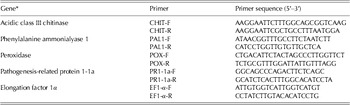
The gene expression experiments were conducted using a quantitative polymerase chain reaction analyser (Real time PCR Prism 7000, Applied Biosystems Inc., Foster City, USA). The PCR reaction mixtures contained: 1 μl of cDNA, 4 μl of 5× HOT FIREPol® EvaGreen® qPCR Mix Plus (Solis BioDyne, Tartu, Estonia) and 250 nm of each gene-specific primer (Table 1) (Narusaka et al. Reference Narusaka, Narusaka, Horio and Ishii1999; Cools & Ishii Reference Cools and Ishii2002; Lin & Ishii Reference Lin and Ishii2009) in a final volume of 20 μl. Controls for each primer pair consisted of PCR tubes with no template. The PCR reaction conditions were: 95 °C for 15 min, followed by 40 cycles of: 95 °C for 15 s, 60 °C for 20 s, and 72 °C for 20 s. Each treatment contained three replicates of duplicated real-time PCR reactions and the experiments were carried out three times. The ratio of gene expression between the target gene and a housekeeping gene, elongation factor 1-α (EF1-α) (Lin & Ishii Reference Lin and Ishii2009), was determined and plotted as fold-increases above values measured at 0 h for the uninoculated, distilled water (DWU) treatment.
PCR primers used in the current study

* Gene fragments of acidic class III chitinase (CHIT), phenylalanine ammonialyase 1 (PAL1), peroxidase (POX), pathogenesis-related protein 1-1a (PR1-1a) and elongation factor 1α (EF1-α) were amplified using specific primers designed by Narusaka et al. (Reference Narusaka, Narusaka, Horio and Ishii1999), Cools & Ishii (Reference Cools and Ishii2002), and Lin & Ishii (Reference Lin and Ishii2009).
Statistical analysis
The data were analysed with one-way ANOVA and Fisher's least significant difference (LSD) test using statistical analysis software (SAS/STAT, V 9.1, SAS institute Inc., Cary, USA) to assess significant differences between treatments.
RESULTS
Effect of protein lysis buffer and its individual components on the suppression of anthracnose disease of cucumber seedlings and on the germination of anthracnose fungal spores
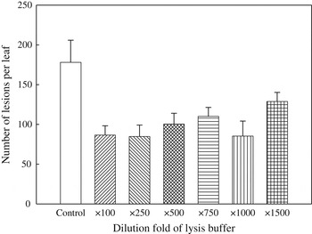
Different concentrations of protein lysis buffer were applied to cucumber leaves and the whole plants were then spray-inoculated with a conidial suspension of C. orbiculare CO-51. The results indicated that the application of protein lysis buffer was effective in reducing disease severity of cucumber anthracnose by 27·7–52·4% (Fig. 2). The ×1000 dilution of the protein lysis buffer was equally as effective as both the ×250 and the ×100 dilutions; therefore, this dilution was used for all of the remaining experiments.
Effect of various concentrations of protein lysis buffer on the suppression of anthracnose disease of cucumber seedlings caused by Colletotrichum orbiculare CO-51. Error bars represent s.e.m., d.f.=6.

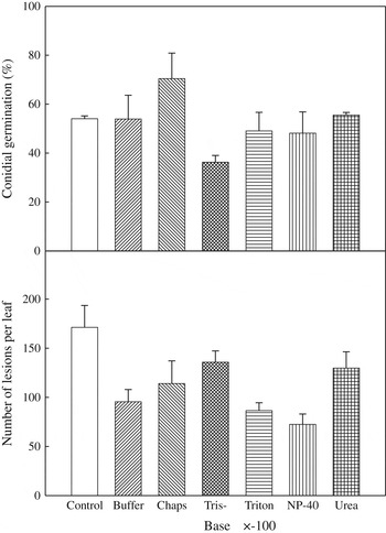
Protein lysis buffer (×1000) and all the individual components of the buffer were applied to cucumber leaves. Protein lysis buffer (×1000) and all the individual components showed different levels of effectiveness in reducing the number of lesions caused by C. orbiculare CO-51 on cucumber leaves. Among these treatments, Nonidet P-40 showed a significantly (P<0·01) higher level of disease development suppression (number of lesions was reduced by 57·7%) as compared with the DW treatment (Fig. 3). The protein lysis buffer (×1000) and all the individual components were also examined for their effects on conidial germination of C. orbiculare CO-51. The results indicated that only Tris-base had a significant effect (P<0·05) on suppressing conidial germination, while CHAPS actually promoted it. None of the other lysis buffer ingredients, including Nonidet P-40, were effective in lowering the spore germination of the pathogen (Fig. 3).
Effect of protein lysis buffer and its individual components on both the conidial germination of Colletotrichum orbiculare CO-51, and the suppression of cucumber anthracnose. Error bars represent s.e.m., d.f.=6.

Effect of Nonidet P-40 on the suppression of anthracnose disease of cucumber seedlings and on the germination of anthracnose fungal spores
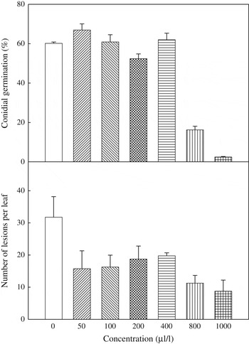
Cucumber leaves were sprayed individually with various concentrations of Nonidet P-40 solutions and air dried before inoculation with cucumber anthracnose fungus. Treatment of leaves with Nonidet P-40 significantly (P<0·01) reduced anthracnose disease on the inoculated cucumber seedlings (Fig. 4). Concentrations of Nonidet P-40>50 μl/l provided effective control against cucumber anthracnose, reducing disease severity by 37·8–72·4% compared with the DW treatment (Fig. 4). Among the concentrations tested, 50 μl/l of Nonidet P-40 showed a significantly (P<0·01) higher suppressive effect, reducing disease severity by 50·4% compared with the DW treatment and with no interruption in the conidial germination of this pathogen (Fig. 4). Furthermore, the conidial germination of anthracnose fungus was significantly (P<0·01) suppressed only when the concentration of Nonidet P-40 solution was >800 μl/l (Fig. 4).
Effect of various concentrations of Nonidet P-40 on both the conidial germination of Colletotrichum orbiculare CO-51, and the suppression of cucumber anthracnose. Error bars represent s.e.m., d.f.=6.

Effect of application time on the suppression of anthracnose disease of cucumber seedlings
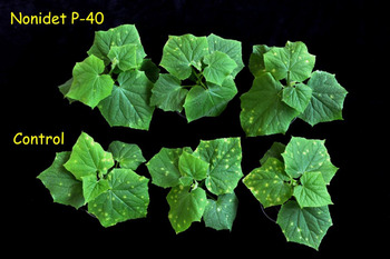
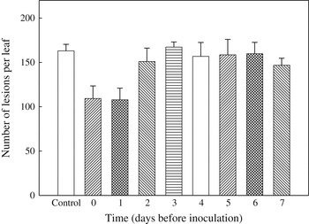
The leaves of cucumber plants were sprayed with a Nonidet P-40 solution (50 μl/l) at different time intervals prior to the inoculation of the plants with anthracnose fungus. The results indicated that Nonidet P-40 significantly (P<0·01) reduced the number of lesions caused by cucumber anthracnose fungus when Nonidet P-40 solution (50 μl/l) was applied either 1 or 0 days before plants were challenged with this pathogen (Figs 5 and 6).
The effect of Nonidet P-40 (50 μl/l) on the control of cucumber anthracnose. (Colour version available online)

Effect of the timing of the Nonidet P-40 application (50 μl/l) on the control of cucumber anthracnose. Error bars represent s.e.m., d.f.=8.

Effect of Nonidet P-40 on expression levels of genes related to plant resistance
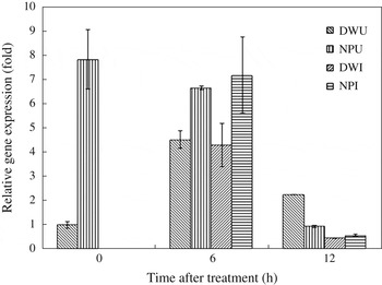
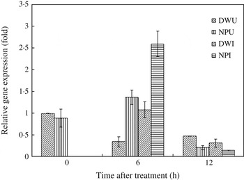
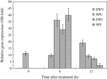
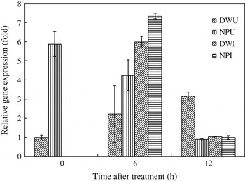
The gene expression of peroxidase (POX) increased significantly (P<0·01) under treatment with Nonidet-40 at 0 and 6 h after treatment; this expression was further increased by inoculation with anthracnose fungus 6 h after treatment (Fig. 7). Inoculation of anthracnose fungus in plants treated with DW slightly increased expression of the phenylalanine ammonialyase 1 (PAL1) gene 6 h after treatment. Pre-treatment of cucumber leaves with Nonidet P-40 also slightly increased PAL1 gene expression 6 h after treatment and this expression was further enhanced upon challenge by the pathogen at this time (P<0·01) (Fig. 8). Inoculation of anthracnose fungus increased the expression of the acidic class III chitinase (CHIT) gene 6 h after treatment in the DW treatment (P<0·01) (Fig. 9). Nonidet P-40 treatments showed an increase in CHIT transcripts at 0 (P<0·05) and 6 h (P<0·01); furthermore, an increase of gene expression was evident in cucumber plants pre-treated with Nonidet P-40 and inoculated with anthracnose fungus 6 h after treatment (P<0·01) (Fig. 9). In addition, the pathogenesis-related protein 1-1a (PR1-1a) transcripts were higher in Nonidet P-40 pre-treated cucumber plants 0 (P<0·01) and 6 h (P<0·01) after treatment (Fig. 10). Inoculation of DW-treated plants with the anthracnose fungus also increased expression of the PR1-1a gene 6 h after treatment (P<0·01). The expression of this gene was further enhanced in Nonidet P-40 pre-treated cucumber plants upon inoculation with the pathogen 6 h after treatment (P<0·01) (Fig. 10).
The real-time RT-PCR analysis of peroxidase gene (POX) expression in cucumber plants. The abbreviations for the experimental plots are: DWU=DW treated and uninoculated; DWI=DW treated and inoculated; NPU=Nonidet P-40 treated and uninoculated; NPI=Nonidet P-40 treated and inoculated. Error bars represent s.e.m., d.f.=9.

The real-time RT-PCR analysis of phenylalanine ammonialyase 1 gene (PAL 1) expression in cucumber plants. DWU=treated with distilled water (DW) and uninoculated; DWI=treated with DW and inoculated; NPU=treated with Nonidet P-40 and uninoculated; NPI=treated with Nonidet P-40 and inoculated. Error bars represent s.e.m., d.f.=9.

The real-time RT-PCR analysis of acidic class III chitinase gene (CHIT) expression in cucumber plants. DWU=treated with DW and uninoculated; DWI=treated with DW and inoculated; NPU=treated with Nonidet P-40 and uninoculated; NPI=treated with Nonidet P-40 and inoculated. Error bars represent s.e.m., d.f.=9.

The real-time RT-PCR analysis of pathogenesis-related protein 1-1a gene (PR1-1a) expression in cucumber plants. DWU=treated with DW and uninoculated; DWI=treated with DW and inoculated; NPU=treated with Nonidet P-40 and uninoculated; NPI=treated with Nonidet P-40 and inoculated. Error bars represent s.e.m., d.f.=9.

DISCUSSION
The chemical structure of Triton X-100 is very similar to that of Nonidet P-40 and the names are sometimes reported as synonyms (Sigma Product Information Sheet). The results of the current study showed that both Triton X-100 and Nonidet P-40 were effective in suppressing the disease development of cucumber anthracnose; however, Nonidet P-40 did show a better effect on control of this disease (Fig. 3). This may be because Nonidet P-40 is slightly more hydrophobic than Triton X-100, and is therefore able to pass through the wax layer of cucumber leaves more easily than Triton X-100.
Nonidet P-40, octylphenoxypolyethoxyethanol, is a nonionic, non-denaturing detergent commonly used in the isolation of membrane complexes. Nonidet P-40 (5 ml/l) has been shown to solubilize the majority of membrane proteins and lipids (0·78 and 0·81, respectively) from BRI 8-cell plasma membranes as assessed by radioactivity, where the samples of BRI 8-cell plasma membrane were labelled with either [35S]methionine as a marker for protein or [32P]Pi as a marker primarily of lipid fractions (Davies et al. Reference Davies, Wigglesworth, Allan, Owens and Crumpton1984). Moskvina et al. (Reference Moskvina, Imre and Ruis1999) found that Nonidet P-40 (100 μl/l) treated Saccharomyces cerevisiae caused relocalization of the transcription factor Msn2p to the cell nucleus, a process important for stress response element-mediated gene expression. In addition, the sterol biogenesis of S. cerevisiae was disrupted by a low concentration of Nonidet P-40, indicating that changes in the plasma membrane structure caused by the presence of detergents may trigger a stress response in yeast (Hronská et al. Reference Hronská, Mrózová, Valachovič and Hapala2004).
Like animals, plants are able to perceive the conserved microbial structures named microbe-associated molecular patterns (MAMPs) and endogenous elicitors, which are termed damage-associated molecular patterns (DAMPs), both of which are able to trigger defence signalling followed by the expression of innate immunity (Boller & Felix Reference Boller and Felix2009). In the current study, cucumber plants treated with Nonidet P-40 showed a reduction in the number of leaf lesions caused by anthracnose fungus, accompanied by an increase in the expression levels of several disease resistance-related genes such as POX, PAL1, PR1-1a and CHIT (Narusaka et al. Reference Narusaka, Narusaka, Horio and Ishii1999; Cools & Ishii Reference Cools and Ishii2002; Lin & Ishii Reference Lin and Ishii2009). There are several processes potentially affected by the presence of Nonidet P-40 that might explain the underlying changes in the inducible responses of the cucumber plants observed in the current study. Furthermore, the severity of the disease on cucumber leaves treated with Nonidet P-40 and rinsed with DW was even higher than for the DW treatment alone (unpublished data). This indicated that the integrity of cucumber leaves was compromised by Nonidet-40 and that the endogenous elicitors (DAMPs) of cucumber leaves released by Nonidet P-40 were removed by DW, thus making the disease more severe. Treatment with Nonidet P-40 may either release DAMPs from cucumber leaf cells or MAMPs from anthracnose fungal cells, or the treatment may simultaneously release both. Consequently, cucumber plants perceive these molecular patterns and induce a stereotypic defence response before penetration of the anthracnose fungus into the plant cuticle. Further studies are needed to test these hypotheses. In addition, the polyether detergents such as Tween, Triton and Brij series are known to have a tendency to accumulate peroxide (Jaeger et al. Reference Jaeger, Sorensen and Wolff1994). Triton X-100 (from different suppliers) and Brij 35 have been found to contain variable amounts of powerful oxidizing impurities representing equivalents of 0·4–2·2‰ H2O2 (Ashani & Catrovas Reference Ashani and Catrovas1980). Owing to the similar chemical structures of Nonidet P-40 and Triton X-100, the involvement of impurities such as H2O2 in Nonidet P-40 that might induce disease resistance should be studied in the future. The current paper is the first report of the involvement of Nonidet P-40 in controlling the anthracnose disease of cucumber plants and provides a new insight into specific detergents such as Nonidet P-40 that may be used in agricultural systems for plant protection.
This research was funded by Grant numbers NSC97-2313-B-005-035-MY3, NSC99-2811-B-005-012, NSC101-2313-B-055-001-MY2 and NSC101-2911-I-005-301 (PFBC) from the National Science Council, Taiwan.