Implications
Modern commercial chickens have a high capacity for fatty acid biosynthesis and fat accumulation. Supplementation with L-arginine (L-Arg) is effective for reducing unfavorable fat levels in pigs, meat ducks and chickens. However, the dietary L-Arg levels that may reduce fat deposition in broilers were unknown. It was also not known whether supplementation with L-Arg would improve fatty acid oxidation as a possible mechanism for reducing fat deposition. This study answered both questions. The findings of this study help to understand the fat-reducing effects of dietary L-Arg supplementation.
Introduction
L-arginine (L-Arg) is a critical amino acid for chickens because, like other birds, they are unable to obtain Arg from endogenous sources because of the absence of most of the enzymes involved in the urea cycle. It has also been shown that chickens have the highest Arg requirement of all animals studied (Ball et al., Reference Ball, Urschel and Pencharz2007). L-Arg is a precursor required for the biosynthesis of many molecules, including proteins, nitric oxide, creatine, ornithine, glutamate, polyamines, proline, glutamine, agmatine and dimethylarginine, hence it has multiple physiological functions in poultry (Khajali and Wideman, Reference Khajali and Wideman2010). Experimental studies have shown that L-Arg supplementation alleviated oxidative stress and improved the antioxidant capacity (Atakisi et al., Reference Atakisi, Atakisi and Kart2009), attenuated pulmonary hypertension syndrome, reduced ascites mortality (Tan et al., Reference Tan, Sun, Li, Pan and Wang2006), and enhanced innate, cellular and humoral immune responses in broiler chickens (Tayade et al., Reference Tayade, Koti and Mishra2006; Munir et al., Reference Munir, Muneer, Masaoud, Tiwari, Mahmud, Chaudhry and Rashid2009).
Modern commercial broilers have a high capacity for lipid biosynthesis (Cui et al., Reference Cui, Zheng, Liu, Zhao, Chen and Wen2012) because selection strategies have focused on improved BW gain, breast yield and feed efficiency, whereas they have ignored fat deposition in the chicken abdomen (Havenstein et al., Reference Havenstein, Ferket and Qureshi2003). Choct et al. (Reference Choct, Naylor, Hutton and Nolan2000) demonstrated that modern strains of broilers contain 15% to 20% fat and >85% of this fat is not physiologically required for body function. Therefore, the excessive accumulation of fat in the abdomens of modern broiler chickens is one of the major problems facing the broiler industry (Le Mignon et al., Reference Le Mignon, Pitel, Gilbert, Le Bihan-Duval, Vignoles, Demeure, Lagarrigue, Simon, Cogburn, Aggrey, Douaire and Le Roy2009) because it is considered to be wasted dietary energy and a waste product with low economic value, which also reduces the carcass yield and affects consumer acceptance (Emmerson, Reference Emmerson1997).
Studies with mice (Clemmensen et al., Reference Clemmensen, Madsen, Smajilovic, Holst and Brauner-Osborne2012), obese rats (Jobgen et al., Reference Jobgen, Meininger, Jobgen, Li, Lee, Smith, Spencer, Fried and Wu2009), growing-finishing pigs (Tan et al., Reference Tan, Yin, Liu, Li, Xu, Kong, Huang, Tang, Shinzato, Smith and Wu2009), White Pekin ducks (Wu et al., Reference Wu, Fang and Guo2011) and chickens (Corzo et al., Reference Corzo, Moran and and Hoehler2003) suggest that dietary L-Arg supplementation is effective for reducing body fat deposition. L-Arg supplementation at a level of 1.00% reduced body fat deposition in meat-type ducks by suppressing the activities of malate dehydrogenase, glucose-6-phosphate dehydrogenase and fatty acid synthase (FAS; Wu et al., Reference Wu, Fang and Guo2011). Similarly, 1.00% L-Arg reduced the mRNA levels of the acetyl-coenzyme A carboxylase-α gene in the abdominal adipose tissue of finishing pigs (Tan et al., Reference Tan, Yin, Liu, Tang, Xu, Kong, Li, Yao, Gu, Smith and Wu2011). Therefore, dietary L-Arg supplementation reduces fat accumulation by modulating de novo lipogenesis, although theoretically it might also decrease the expression of lipogenesis genes and/or increase the expression of lipolysis genes to reduce fat accumulation. However, the effects of L-Arg supplementation on lipolysis, blood lipid profiles and the expression of genes related to lipid metabolism have not been investigated, and only one study reported a reduction in the abdominal fat pad at 42 to 56 days after increasing the L-Arg concentration in broiler chicken diets (Corzo et al., Reference Corzo, Moran and and Hoehler2003). Thus, we investigated the effects of supplementation with different levels of L-Arg in the diets of broiler chickens on fat deposition, plasma lipid profiles, FAS gene expression in the liver (a key indicator used to monitor lipogenesis), and the expression levels of the carnitine palmitoyl transferase1 (CPT1) and 3-hydroxyacyl-CoA dehydrogenase (3HADH) genes in the heart (the main indicators used to evaluate lipolysis levels), as well as the growth performance.
Material and methods
Birds, diets and management
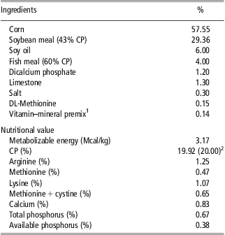
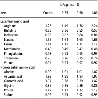
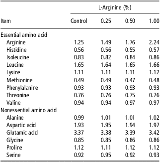
A total of 256 1-day-old Cobb 500 male broiler chicks were obtained from a commercial hatchery (Hua Dacheng, Xianyan, China). The chicks were placed in cages, exposed to continuous light and fed a commercial starter diet (22% CP and 3000 kcal ME/kg; Hua Qing, Yanling, China). The temperature was maintained at 35°C during the first 3 days and then reduced weekly by 3°C until it reached 22°C. Before the experiment, all chickens were weighed individually after withdrawal of feed overnight (12 h) and some of the chickens with the lowest or highest weights were eliminated. A total of 192 21-day-old broiler chickens with similar average BWs (920 ± 15 g) were selected and assigned randomly to four dietary treatments, that is, eight replicate cages (length 100 cm × width 50 cm × height 45 cm) with six birds per replicate. Broiler chickens were fed ad libitum with the same basal diets (in mash form), which were supplemented with 0 (control), 0.25%, 0.50% or 1.00% Arg in the form of L-arginine monohydrochloride (99.3% L-Arg, Binhai Hanhong Bio-Chemical Co. Ltd, Shanghai, China) from 21 to 42 days of age. The control diet (Table 1) was prepared to meet or exceed the National Research Council (NRC, 1994) requirements for broiler chickens and the dietary treatments were analyzed (Table 2) in duplicate to determine the amino acid contents (Beijing Zhongxu Yangguang Animal Husbandry Technology Development Co. Ltd, China).
Ingredients and chemical composition of the basal diet
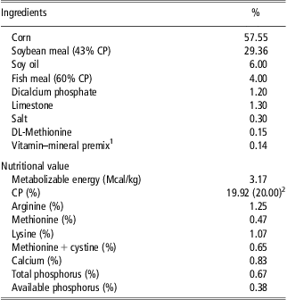
1Supplied per kilogram of diet: vitamin A, 12 000 IU; vitamin D3, 4000 IU; vitamin E, 30 IU; vitamin K3, 3 mg; thiamine, 2.2 mg; riboflavin, 10 mg; D-pantothenic acid, 7 mg; folic acid, 0.55 mg; pyridoxine, 4 mg; niacin, 37 mg; cobalamin, 0.02 mg; biotin, 0.20 mg; choline chloride, 400 mg; iron, 85 mg; copper, 8 mg; manganese, 75 mg; zinc, 69 mg; iodine, 0.4 mg; and selenium, 0.3 mg.
2Analyzed.
Concentrations of amino acids determined in the experimental diets
Performance parameters
BWs were recorded and the feed intake was observed daily and weighed on a replicate basis at the start and the end of the 3-week test period to determine the average daily gain (ADG), average daily feed intake (ADFI) and feed conversion rate (FCR). Mortalities were recorded daily throughout the experimental period (3 weeks). All experimental procedures were reviewed and approved by the Animal Care Committee of the Northwest Agriculture & Forestry University (Shaanxi, China).
Sample collection
At 42 days of age, the live BWs were recorded after an overnight fast (12 h) and 16 chickens per treatment (two chickens per replicate) with a BW close to the replicate average BW were selected and slaughtered. First, eight chickens per treatment (one chicken per replicate) were slaughtered to collect blood samples for lipid analysis, liver and heart samples for RNA isolation, the abdominal fat pad to calculate the abdominal fat pad percentage, and breast muscles to determine the intramuscular fat content (IMF). Second, eight chickens per treatment were slaughtered to collect only the abdominal fat pad and breast muscles. After bleeding, the abdominal fat pad was removed from the proventriculus surrounding the gizzard down to the cloaca and weighed. The right breast muscles were removed from the sternum, minced after removing any obvious fat, and stored at −20°C. The IMF of the breast muscle was determined in triplicate, according to the method described by Zhao et al. (Reference Zhao, Chen, Zheng, Wen and Zhang2007), and expressed as the percentage total fat (dry weight). Blood samples were collected using a 5-ml anticoagulant syringe (Shanghai K & G International Co. Ltd, Shanghai, China) via the wing vein, centrifuged at 4°C and 5900 × g for 10 min to obtain the plasma, and stored in 1.5-ml Eppendorf tubes at −70°C until analysis. Liver and heart samples were collected rapidly in 1.5-ml RNase-free centrifuge tubes, frozen immediately in liquid nitrogen, and stored at −80°C until RNA isolation.
Assay of lipid parameters
The levels of plasma triglyceride (TG), total cholesterol (TC), high-density lipoprotein cholesterol (HDL-C), low-density lipoprotein cholesterol (LDL-C) and nonesterified fatty acids (NEFA) were estimated spectrophotometrically using commercial diagnostic kits (Jiancheng Bioengineering Institute, Nanjing, China; Chen et al., Reference Chen, Wang, Kong, Ma and Zou2011). Each sample was analyzed in triplicate.
RNA isolation and real-time PCR
Total RNA was extracted from the liver and heart samples using RNAiso Plus (TaKaRa, Biotechnology Co. Ltd, Dalian, China), according to the manufacturer's protocol. The total RNA was dissolved in 0.1% DEPC-treated water and the integrity of the total RNA was estimated using agarose gel electrophoresis, whereas the concentration and purity of the RNA were evaluated spectrophotometrically at 260/280 nm. The RNA concentrations of all samples were 1.9 to 2.0, according to estimates made by measuring the A260. The total RNA was reverse transcribed to cDNA using a PrimeScript RT Reagent Kit (Perfect Real Time; TaKaRa, Biotechnology Co. Ltd). Briefly, 2 μg of RNA was reacted with 4 μl of 5× PrimeScript, 1 μl of PrimeScript RT Enzyme Mix, 1 μl of oligo dT primer (50 μM), 1 μl of random 6-mers (100 μM) and RNase-free water (TaKaRa, Biotechnology Co. Ltd) to produce a final volume of 20 μl. Reverse transcription was performed at 37°C for 15 min, followed by heat inactivation for 5 s at 85°C. The chicken 3HADH primer used for real-time PCR was prepared as reported by Mujahid and Furuse (Reference Mujahid and Furuse2008) and synthesized by TaKaRa Biotechnology Co. Ltd. The other primers were designed based on chicken mRNA gene sequences in GenBank and were also synthesized by TaKaRa Biotechnology Co. Ltd (Supplementary Table S1).
The gene expression levels in all samples were measured in triplicate using three individual preparations of reverse-transcribed cDNA by real-time PCR with the Bio-Rad MiniOpticon quantitive system (Bio-Rad, Hercules, CA, USA) and SYBR® Premix Ex TaqTM (Perfect Real Time; TaKaRa Biotechnology Co. Ltd), according to the manufacturer's instructions. Briefly, 2 μl of cDNA was mixed with 12.5 μl of SYBR® Premix Ex Taq®, 1 μl of each primer (10 μM) and sterile distilled water to produce a final volume of 25 μl. The thermal cycle was as follows: 30 s at 95°C for one cycle, 5 s at 95°C and 34 s at 60°C for 40 cycles. The mRNA expression of β-actin was used as an internal control (Chen et al., Reference Chen, Wang, Kong, Ma and Zou2011), which was not significantly different between the two experimental groups. The values were normalized against the mRNA expression of β-actin and calculated as the percentage of the control value.
Statistical analysis
The experimental data were analyzed by one-way ANOVA using the GLM procedure in SAS (version 8.02; SAS Institute Inc., Cary, NC, USA) where the cage was the experimental unit for growth performance and the individual bird served as the experimental unit for plasma lipids and gene expression. The linear and quadratic effects of L-Arg among treatments were analyzed using a contrast statement. Significant differences among treatments were tested using Duncan's multiple-range test at a significance level of P < 0.05.
Results and discussion
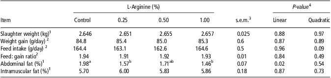
All chickens were healthy and no mortality occurred throughout the entire experimental period (data not shown). The slaughter weight and growth performance (ADG, ADFI, and FCR) were not significantly affected (P > 0.05) by L-Arg supplementation in the diet, as shown in Table 3. Similarly, Fernandes et al. (Reference Fernandes, Murakami, Martins, Sakamoto and Garcia2009) reported that the average live BW of broiler chickens at 42 days was not affected by L-Arg supplementation. Tan et al. (Reference Tan, Sun, Li, Pan and Wang2006) also found that supplementing broiler chicken diets with 1.00% L-Arg did not affect the BW gain, whereas Lee et al. (Reference Lee, Austic, Naqi, Golemboski and Dietert2002) showed that increasing the L-Arg level from 100% to 300% NRC (1994) did not affect the BW gain in White Leghorn chickens. D'Amato and Humphrey (Reference D'Amato and Humphrey2010) reported that the addition of L-Arg at 2 g/kg did not affect the BW, feed intake or FCR compared with chickens fed a basal diet. A study using White Pekin ducks found that diet supplementation with 1.00% L-Arg did not affect the ADFI or feed efficiency (Wu et al., Reference Wu, Fang and Guo2011). By contrast, Emadi et al. (Reference Emadi, Jahanshiri, Kaveh, Hair-Bejo, Ideris and Alimon2011) reported that feeding chicks from 1 to 49 days with diets supplemented with 2.5% L-Arg above their requirements according to NRC (1994) recommendations improved the ADG, ADFI and FCR compared with those fed the control diet. These differences may have been due to the period and/or levels of supplementation.
Growth performance, abdominal fat content and fat deposition in the breast muscle of broiler chickens fed diets with or without L-arginine supplementation
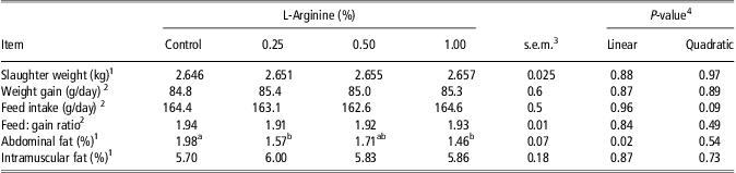
1Values are means with n = 16 chickens per treatment (two chickens/replicate).
2Values are means with n = 8 cages per treatment (six chickens/cage).
3Pooled s.e.m.
4Linear and quadratic effects of L-arginine levels.
a,bMeans within a row that do not share a common superscript are significantly different (P < 0.05).
The results showed that IMF of breast muscle was not significantly (P > 0.05) affected by L-Arg supplementation (Table 3). We also found that the abdominal fat content expressed as the percentage BW was significantly (P < 0.02; Table 3) lower with the 0.25% and 1.00% L-Arg treatments compared with the control, whereas they did not differ significantly from each other. IMF is one of the major factors that affects meat quality because of its effects on other traits such as the flavor, juiciness and tenderness of meat (Zhao et al., Reference Zhao, Chen, Zheng, Wen and Zhang2007). The abdominal fat tissue grows faster compared with other fat tissues (Butterwith, Reference Butterwith1989) and it reaches its maximum rate of growth at 42 days of age (Havenstein et al., Reference Havenstein, Ferket and Qureshi2003). It is the key indicator used to judge total body fat (Chen et al., Reference Chen, Wang, Kong, Ma and Zou2011) because it is linked directly to the total body fat content in broiler chickens (Sonaiya, Reference Sonaiya1985). Our results agreed with previous studies of broiler chickens, pigs and White Pekin ducks (Corzo et al., Reference Corzo, Moran and and Hoehler2003; Tan et al., Reference Tan, Yin, Liu, Li, Xu, Kong, Huang, Tang, Shinzato, Smith and Wu2009; Wu et al., Reference Wu, Fang and Guo2011). Corzo et al. (Reference Corzo, Moran and and Hoehler2003) detected a significant reduction in the abdominal fat content after increasing the L-Arg concentration in the diet of broiler chickens between 42 and 56 days of age. Tan et al. (Reference Tan, Yin, Liu, Li, Xu, Kong, Huang, Tang, Shinzato, Smith and Wu2009) also showed that dietary L-Arg supplementation led to a significant reduction in total body fat deposition in pigs. Wu et al. (Reference Wu, Fang and Guo2011) reported that supplementing the diet of meat-type ducks with 1.00% L-Arg significantly decreased the abdominal fat pad percentage by reducing the activities of malate dehydrogenase, glucose-6-phosphate dehydrogenase and FAS (lipogenic enzymes) in the liver.
However, Fernandes et al. (Reference Fernandes, Murakami, Martins, Sakamoto and Garcia2009) reported that increasing the dietary L-Arg concentration from 1.49% to 1.79% during the starter phase had no effect on the abdominal fat content of broiler chickens on day 42. However, Fernandes et al. (Reference Fernandes, Murakami, Martins, Sakamoto and Garcia2009) supplemented the diet with L-Arg from 1 to 21 days of age, whereas all chickens consumed the same diet without Arg supplementation from 22 to 42 days of age. Therefore, the difference between these two results may be attributed to the timing of the supplementation because the starter phase is associated with a rapid growth rate, but it is not linked to excessive fat deposition in broiler chickens (Tzeng and Becker, Reference Tzeng and Becker1981).
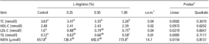
Table 4 shows the TC, HDL-C, LDL-C, TG and NEFA concentrations in the plasma of broilers fed diets containing different levels of L-Arg. The plasma concentrations of TC with the L-Arg treatments were significantly (P < 0.0002) lower than those of the control, whereas they did not differ significantly from each other. The concentration of HDL-C was not affected by the dietary treatments, whereas the concentration of LDL-C was significantly (P = 0.0219) lower with 1.00% L-Arg at compared with the control group. Our results agree with Emadi et al. (Reference Emadi, Jahanshiri, Kaveh, Hair-Bejo, Ideris and Alimon2011), who demonstrated that supplementation of L-Arg in the basal diet significantly reduced the blood TC concentration in broiler chickens. Similar findings were also reported in rats by El-Kirsh et al. (Reference El-Kirsh, Abd El-Wahab and Abd-Ellah Sayed2011), who found that an oral dose of L-Arg three times per week did not significantly affect the plasma HDL-C, whereas the plasma TC and LDL-C were lower. Our results and previous studies suggest that L-Arg supplementation could reduce the blood TC concentration. To the best of our knowledge, no information is available about the mechanism underlying the cholesterol-lowering effect of dietary L-Arg supplementation. In chickens, the TC content of muscle is associated with the blood cholesterol level and both are controlled by hepatic hydroxyl-3-methylglutaryl-CoA reductase (HMGR) gene expression (Cui et al., Reference Cui, Yang, Wang, Zhao, Jiang, Zhao, Chen, Zheng, Li and Wen2010). Thus, we analyzed HMGR expression in the liver of chickens as a possible mechanism for explaining the plasma TC reduction. HMGR is a crucial enzyme during cholesterol biosynthesis (Cui et al., Reference Cui, Yang, Wang, Zhao, Jiang, Zhao, Chen, Zheng, Li and Wen2010). In the present study, L-Arg inclusion in the diets of broilers significantly (P < 0.0001; Table 5) reduced HMGR mRNA expression, which agreed with Jobgen et al. (Reference Jobgen, Fried, Fu, Meininger and Wu2006) who reported that L-Arg was involved in the regulation of cholesterol metabolism via nitric oxide. However, it is still unknown whether L-Arg inhibited the expression of HMGR directly or indirectly, therefore further studies are required to elucidate how L-Arg affects hepatic HMGR mRNA expression.
Plasma lipid concentrations of broiler chickens fed diets with or without L-arginine supplementation
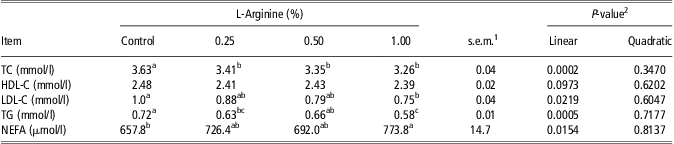
TC = total cholesterol; HDLC = high-density lipoprotein cholesterol; LDL-C = low-density lipoprotein cholesterol; TG = triglyceride; NEFA = nonesterified fatty acid.
1Pooled s.e.m., n = 8 (eight chickens/treatment).
2Linear and quadratic effects of L-arginine levels.
a,b,cMeans within a row that do not share a common superscript are significantly different (P < 0.05).
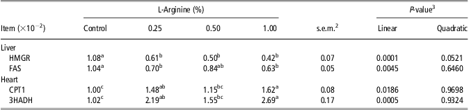
mRNA expressionFootnote 1 levels of genes related to lipid metabolism in the tissues of broiler chickens fed diets with or without L-arginine supplementation
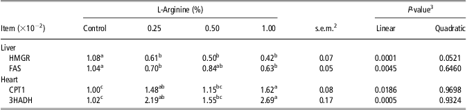
HMGR = 3-hydroxyl-3-methylglutaryl-CoA reductase; FAS = fatty acid synthase; CPT1 = carnitine palmitoyl transferase1; 3HADH = 3-hydroxyacyl-CoA dehydrogenase.
1 Values are expressed as percentages of the control value.
2 Pooled s.e.m., n = 8 (eight chickens/treatment).
3 The linear and quadratic effects of L-arginine levels.
a,b,c Means within a row that do not share a common superscript are significantly different (P < 0.05).
The level of TG was significantly lower (P < 0.0005; Table 4) in broiler chickens fed diets containing 0.25% or 1.00% L-Arg compared with those fed the control diet, whereas the NEFA level was significantly (P = 0.0154; Table 4) increased by 1.00% L-Arg supplementation compared with the control group. These findings suggest that L-Arg promotes the conversion of TG to glycerol and free fatty acids. Previous studies also showed that L-Arg dietary supplementation reduced the blood TG concentration in obese rats (Jobgen et al., Reference Jobgen, Meininger, Jobgen, Li, Lee, Smith, Spencer, Fried and Wu2009), growing-finishing pigs (Tan et al., Reference Tan, Yin, Liu, Li, Xu, Kong, Huang, Tang, Shinzato, Smith and Wu2009), laying quail (Atakisi et al., Reference Atakisi, Atakisi and Kart2009) and broiler chickens (Emadi et al., Reference Emadi, Jahanshiri, Kaveh, Hair-Bejo, Ideris and Alimon2011).
To acquire a comprehensive understanding of the molecular mechanisms underlying the fat-reducing effect of dietary L-Arg supplementation, we quantified the gene expression of FAS in the liver, and CPT1 and 3HADH in the heart of broiler chickens (Table 5). Our results showed that FAS gene expression in the liver was significantly (P = 0.0045) lower, whereas the expression levels of CPT1 and 3HADH in the heart were increased significantly (P < 0.05) with 0.25% or 1.00% L-Arg supplementation compared with the control. However, there were no significant differences between L-Arg supplementation at levels of 0.25% and 1.00%. FAS is an essential enzyme in the final step of the lipogenic pathway (Pitel et al., Reference Pitel, Fillon, Heimel, Le Fur, el Khadir-Mounier, Douaire, Gellin and Vignal1998) and the liver is the most important vital organ for fatty acid synthesis in avian species, in which almost 85% of the fat accumulated by growing birds is produced in the liver (Molette et al., Reference Molette, Theron, Marty-Gasset, Fernandez and Remignon2012). Therefore, we estimated the FAS mRNA expression in the liver as a biomarker of fatty acid synthesis. Our results showed that dietary L-Arg supplementation reduced fatty acid synthesis by downregulating hepatic FAS gene expression. The downregulation of hepatic FAS gene expression will reduce the availability of fatty acids for esterification into TGs and storage in adipose tissue. Thus, our results partly explain why dietary L-Arg supplementation reduced the abdominal fat content and lowered the plasma TG level. Santoso et al. (Reference Santoso, Tanaka and Ohtani1995) noted that the blood TG level was correlated with fatty acid synthesis in chickens. Tan et al. (Reference Tan, Yin, Liu, Tang, Xu, Kong, Li, Yao, Gu, Smith and Wu2011) and Wu et al. (Reference Wu, Fang and Guo2011) also reported that dietary L-Arg supplementation inhibited de novo fatty acid synthesis in the adipose tissue of pigs by downregulating the expression of the acetyl-coenzyme A carboxylase-α gene and by reducing the activities of malate dehydrogenase, glucose-6-phosphate dehydrogenase and FAS (lipogenic enzymes) in the livers of meat-type ducks.
Significant increases were also noted in the CPT1 and 3HADH mRNA expression levels of the control and 0.25% and 1.00% L-Arg treatments, which are summarized in Table 5. Our study is not the first experiment to investigate the effects of dietary L-Arg supplementation on fatty acid β-oxidation, but it is the first experiment designed specifically to investigate the fat-reducing effects of amino acids on the β-oxidation of fatty acids. A previous report suggested that dietary L-Arg supplementation improved fatty acid oxidation via nitric oxide due to the elevation of CPT1A (Jobgen et al., Reference Jobgen, Fried, Fu, Meininger and Wu2006). However, it is very difficult to compare our findings with other studies. CPT1 is an integral outer mitochondrial membrane enzyme that catalyzes the first step in the β-oxidation of fatty acids. In broiler chickens, L-CPT1 (liver-type CPT1) and M-CPT1 (muscle-type CPT1) are the main CPT1 isoforms involved with transporting fatty acids into the mitochondria and they control the flux of fatty acids entering the β-oxidation pathway (Skiba-Cassy et al., Reference Skiba-Cassy, Collin, Chartrin, Medale, Simon, Duclos and Tesseraud2007), whereas 3HADH is involved with the oxidation phases of fatty acid β-oxidation (Eaton, Reference Eaton2002). Thus, the mRNA expression levels of CPT1 and 3HADH in the hearts of chickens were used as biomarkers of fatty acid β-oxidation because Mujahid and Furuse (Reference Mujahid and Furuse2008) used the CPT1 and 3HADH mRNA levels in the hearts of chickens as reliable parameters for detecting changes in the mitochondrial fatty acid oxidation level. For the first time, our study suggested that dietary supplementation with 0.25% L-Arg increased the mRNA expression of CPT1 and 3HADH in the hearts of chickens, which could help to explain the lower fat accumulation in chickens at 42 days of age. This is because the balance of the fatty acid uptake, hepatic de novo fatty acid synthesis (anabolism), and fatty acid oxidation via β-oxidation (catabolism) determines the amount of fat that can be deposited in the body.
We detected linear effects of L-Arg levels but the addition of 0.50% L-Arg did not appear to significantly affect the abdominal fat content or the mRNA expression of genes involved with lipid metabolism compared with the control, unlike the other L-Arg treatments. This may have been due to the interaction between L-Arg and other nutrients when the diet was supplemented with 0.50% L-Arg. It is possible that the diets supplemented with 0.50% L-Arg did not modulate the mRNA expression of genes involved with lipid metabolism sufficiently to reduce the abdominal fat content in the same way as the other Arg treatments. Indeed, this confirmed that supplementation with 0.25% L-Arg was sufficient to modulate the lipid metabolism, whereas supplementation with 1.00% L-Arg led to a reduction in the abdominal fat content and modulated the lipid metabolism, but not significantly compared with 0.25% L-Arg supplementation, although the L-Arg concentration was four times higher in the 1.00% L-Arg treatment.
In conclusion, this study showed that 0.25% L-Arg supplementation in the diets of broiler chickens reduced the blood plasma TC and TG concentrations and abdominal fat deposition. The lower abdominal fat deposition may have been attributable to reduced hepatic FAS gene expression and enhanced fatty acid β-oxidation via increased expression of the CPT1 and 3HADH genes. These findings suggest that dietary L-Arg supplementation is effective for reducing unfavorable fat deposition (abdominal fat) and it had no negative effects on the favorable fat (IMF) content. We also proposed a novel biochemical basis to explain the beneficial effects of L-Arg in reducing fat accumulation in avian species. Further studies are required to determine whether dietary L-Arg supplementation in high-energy diets produces the same results.
Acknowledgments
This study was supported by the Science & Technological Project of Shaanxi Province, China (2010ZDGC-02, 2011KTCQ02-02, and 2011KTDZ02-01-01).
Supplementary materials
For supplementary material referred to in this article, please visit http://dx.doi.org/10.1017/S1751731113000347