Introduction
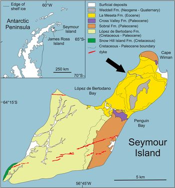
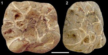
This paper arises following re-evaluation of a unique specimen of fossil cirripede that was collected from Seymour Island, off the Antarctic Peninsula, in the 1980s (Fig. 1) by the senior author, J. Stilwell. The Eocene (ca. 55–45 Ma) Age La Meseta Formation, exposed on the northern part of Seymour Island, hosts a thick succession of richly fossiliferous siliciclastic rocks of with a biodiversity of >150 species of invertebrates preserved within a spectrum of tidal channel and barrier island settings (Stilwell and Zinsmeister, Reference Stilwell and Zinsmeister1992). The late Eocene investigated specimen (NMV P139129; Museums Victoria, Australia) from the La Meseta Formation is semi-circular in transverse section (partially due to weathering), possesses six-fold symmetry, and thus, is superficially akin to cirripedes within the Coronulinae (whale barnacles) (Fig. 2). If it were confirmed as such, it would push the earliest coronulines back ca. 30 Ma, at a time when the mysticetid cetaceans were beginning to evolve (Bianucci and Landini, Reference Bianucci, Landini and Miller2007). As such, this Antarctic fossil barnacle group needs to be evaluated for any potential connection to the evolutionary history of cetaceans and cirripedes phylogeny.
Location of Seymour Island, Antarctic Peninsula, Weddell Sea. The barnacle specimen was collected from the middle units of the La Meseta Formation as float along the coast shown by the black arrow, which is the area shaded in light color surrounding the meseta. The southern part of the island and its represented strata are highlighted in different shades and make up the López de Bertodano and Sobral formations, which have been cut by volcanic dykes.

Macrophotographs of late Eocene Antarctic barnacles group (NMV P139129) from the La Meseta Formation. (1) Dorsal view of specimen, note encrusted serpulid worms; (2) lateral profile of specimen. Scale bars equal 1 cm.

The subfamily Coronulinae are host specific, obligate commensals that favor attachment within the skin of members of the Mysticeti (baleen whales). Their absence from the Odontoceti (toothed whales), except in rare circumstances, is not fully resolved, but is, in part, most likely due to the greater velocity with which the latter move through the water, preventing settlement of larvae and inhibiting feeding. Moreover, toothed whales also have relatively smooth skin relative to baleen whales. Fossils of Coronula are reported worldwide, and there are recorded sites, which have dense accumulations of Coronula fossils, preserved with the fossil mandible of the baleen whale bones (Buckeridge et al., Reference Buckeridge, Chan and Lee2019a).
The relationship between a commensal taxon and its host is biologically complex—and with whale barnacles perhaps more so than most, because the earliest known coronulines possess unique features, such as the complex infolding in the parietes, and are thus highly derived. Transition from a circular shell wall to one that develops the intricacies of Coronula involves exaptation, which is a process wherein features acquire functions for which they were not originally selected. Seilacher (Reference Seilacher2005) assessed the mechanism by which Coronula attached to cetaceans, and concluded that embedding within the whale skin was achieved by a narrow ring of sutural tissue at the base of the barnacle shell during its cypris larval stage that evolved to clean surfaces prior to attachment of sessile cirripedes. The ability of barnacles to excavate (and dissolve) substrate was noted as early as Darwin (Reference Darwin1854); more recent observations of this phenomenon are provided in Santos et al. (Reference Santos, Mayoral and Muñiz2005, p. 184), Buckeridge and Newman (Reference Buckeridge and Newman2017, p. 229), and Buckeridge et al. (Reference Buckeridge, Kočí, Schlögl, Tomašových and Kočová Veselská2019b). In combination with shell growth, this sutural tissue has the ability to penetrate the skin of the whale sufficiently deeply to avoid dislodgment during periods of skin shedding. Although the Cretaceous barnacle Archaeochionelasmus nekvasilovae provides some appreciation of how early sessile barnacles may have colonized the shells of marine turtles and/or ammonites (Kočí et al., Reference Kočí, Kočová Veselská, Newman, Buckeridge and Sklenář2017), Archaeochionelasmus is primarily a smooth cone that offered low resistance to water flow. Knowing how early coronulines were constructed is very informative in unravelling how they became such successful commensals on whales.
The earliest Coronuloidea
Taxa within the superfamily Coronuloidea include Emersonius cybosyrinx Ross and Newman, Reference Ross and Newman1967 from the late Eocene of Florida. Although it is the oldest known coronuloid, Emersonius possessed a low shell, within which are numerous rows of square parietal tubes. Ross and Newman (Reference Ross and Newman1967, p. 16) considered this form to be closely allied to Platylepas, an epizoan taxon on present day sea snakes, turtles, and dugongs, but they concluded that the demise of Emersonius was due to extinction of the host (i.e., that it represented an evolutionary dead-end rather than a transitionary form). Emersonius is not a coronuline because it lacks the complex infolding in the parietes, and is thus not the missing link leading to modern taxa such as Coronula.
The Eocene Epoch marked an interval of morphological “experimentation” for sessile barnacles, and Emersonius was one of these, but apparently was not perpetuated. Emersonius also predates the earliest baleen whale, Toipahautea waitaki Tsai and Fordyce, Reference Tsai and Fordyce2018, from the late Oligocene (early Chattian), by ca. 10 Myr. As noted, cirripedes rarely settle on toothed whales, such as basilosaurids, which are known from the La Meseta Formation (Buono et al., Reference Buono, Férnandez, Reguero, Marenssi, Santillana and Mörs2016), the rationale being that these carnivores moved too swiftly through the water, disrupting both initial adhesion and subsequent feeding. Nonetheless, there were marine vertebrates, such as cetaceans, evolving during the Eocene in Antarctica and elsewhere globally (e.g., Stilwell and Zinsmeister, Reference Stilwell and Zinsmeister1992), and these were potential hosts for any barnacle cyprids seeking a substrate.
Preservation of the barnacles(s)
The specimen (NMV P139129; Museums Victoria, Australia) is enclosed within a dense, well-cemented, calc-arenaceous matrix, the removal of which would almost certainly irreparably damage it and degrade it beyond any hope of attributing the barnacle to any group. Unfortunately, the full nature of the shells, especially in the specimen, is obscured by matrix, making a definitive taxonomic assessment impossible. Importantly, both the matrix and the barnacle shell are calcareous, precluding any chemical dissolution of the matrix.
Thus, we have a potentially very significant, but unique, fossil that is only partially exposed from the matrix. The problem is compounded by the age of the specimen, which, being significantly older (mid-Eocene Age) than any known members of their potential families, would not necessarily have demonstrated the complex intricacies of younger taxa in their respective groups (i.e., they would be expected to possess more plesiomorphic features and few, if any derived characters of modern coronulids).
Resolution of these unique fossils required non-destructive analysis—where subtle differences in rock chemistry could be enhanced to produce a 3-D model of the enclosed fossil. Although X-ray computed tomography has been utilized successfully in paleontology endeavors since the dawn of the new millennium (see Sutton, Reference Sutton2008; Abel et al., Reference Abel, Laurini and Richter2012), as far as we are aware, this is the first attempt on barnacles to more elucidate their complex internal morphologies, which are notoriously challenging to extract through mechanical fossil preparation means. This study highlights the importance of 3-D tomographic imaging techniques in cirriped morphology and taxonomy.
Material and methods
Collection site
The studied specimen was collected as float (i.e., it was not in situ). The barnacle was found during the 1986–87 season by lead author, J.S. The “coronulid-like” specimen is quite worn down, a result of being a beach float. The matrix is a fine sandy grit, with some shell fragments; lithologically, it conforms to the informal stratigraphic unit, Telm 3, of Sadler (Reference Sadler, Feldmann and Woodburne1988). Telm 3, ~92 m thick and sedimentologically complex, comprising cross-bedded sandstones and siltstones with abundant shell beds and lenses, was deposited in in a barrier island setting, and is interpreted as a tidal channel deposit with associated facies (Stilwell and Zinsmeister, Reference Stilwell and Zinsmeister1992).
Non-destructive assessment of fossils
X-ray CT is a radiological imaging technique that is designed to estimate the 3-dimensional spatial distribution of materials (or their electron densities). The attenuation of the transmission X-ray is measured at many points during translation motion of the X-ray source and detector. This is not an option for well-indurated fossils, but may be suited to samples with heterogeneous distribution of materials with different electron densities.
The CT module within the pre-clinical Siemens Inveon PET/CT system at the Australian Nuclear Science and Technology Organisation (ANSTO) was used to discern/evaluate if this imaging modality could determine significant differences between calcite (the composition of the barnacle shell) and the matrix. The sample was scanned using an 80 kV, 500 μA X-ray with a fixed X-ray focal spot of 50 μm, and detector pixel resolution of 38.076 μm. The image was reconstructed using the Feldkamp algorithm with no down-sampling to maintain a voxel (3D pixel) resolution of 38.076 μm. Due to the high density of the sample and limitations of the Siemens Inveon Acquisition Workplace (IAW) reconstruction software, no beam hardness correction was applied. Scans were completed with an acquisition time of 17 minutes. Given the limitations of the X-ray source itself with the current equipment, higher resolution scans will add more noise and make the image volume more difficult to handle from a computer power and visualization point of view.
Repository and institutional abbreviation
The figured specimen (NMV P139129) examined in this study is deposited in the Museums Victoria, Melbourne, Victoria, Australia, paleontology collection.
Results
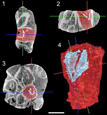
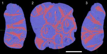
Analysis of the coronulid-like specimen with ANSTO's pre-clinical CT instrument demonstrated that the calcareous compartmental plates were easily distinguished from the rock matrix in the reconstructed image (Figs. 3, 4). Although the possible morphology of an Eocene ‘coronuline’ is speculative, it is unlikely that the earliest epibiots would have possessed porous paries (as this specimen appeared to do). However, CT imaging showed that the specimen was made up of a tight grouping of at least ten separate barnacles (Fig. 3). The compartmental walls in each were solid, such that the specimens conformed to either the Archaeobalanidae or Austrobalanidae, both of which include taxa with six non-porous compartmental plates. The Archaeobalanidae typically possess six solid compartmental plates (i.e., these are the structures that make up the shell wall). The shells commonly have a calcareous base, and compartments may have well-developed, non-porous radii. In comparison, the Austrobalanidae can have either six or four compartmental plates, and, in some genera, these have internal chitinous lamella; the radii, if present, are solid and narrow. Bases are commonly membranous (i.e., not mineralized). If only compartmental plates were available, an accurate allocation of a specimen to either family is rarely possible; opercula are needed for this. Unfortunately, opercula are not present in this material.
X-ray CT image of the fossil barnacles in Figure 2, depicting virtual slices of the 3-D volume (4) employing the CT scanning technique with highlighted single barnacle (shaded) to illustrate its position in the scan: (1–4) revealing stark contrast between the calcium carbonate barnacle plates in various orientations and the surrounding rock matrix, as shown by the shaded lines, which would not be possible to reveal by traditional mechanical preparation methods. Scale bar equals 1 cm.


The CT imaging penetrated the matrix with “virtual slices” at any depth without damaging the specimen. The resulting images showed that grouping of barnacles was roughly circular—thus, having the appearance of a radially symmetrical barnacle, such as Coronula. Arrangements such as this are not uncommon when barnacles grow around tubular substrates, such as a seaweed stipe (or a submerged tree; see Kočí et al., Reference Kočí, Kočová Veselská, Newman, Buckeridge and Sklenář2017). Unfortunately, we were not able to discern any opercula or sufficient fine detail in the specimens, which precluded a definitive taxonomic placement beyond the archaeobalanids or the austrobalanids. Three species are known from the La Meseta Formation (Solidobalanus sp. from the Archaeobalanidae, and Austrobalanus macdonaldensis Buckeridge, Reference Buckeridge1983 plus Hexaminius venerai Buckeridge et al., Reference Buckeridge, Chan and Lee2019a from the Austrobalanidae) (see Kočí, et al., Reference Kočí, Vodrážka, Kočová Veselská and Buckeridge2018 for list of references). Of these three, it is most like either Solidobalanus sp. or H. venerai, both of which were shallow water, and both of which grew around weed or submerged plant material.
Conclusion
The taxonomy of the Antarctic fossil barnacle was shown to be not what was originally thought. There was the possibility that it could have been a remarkable find, and thus have significant bearing on our understanding of the evolution of the Coronulidae. Prior to CT imaging, the only way to determine the true nature of this specimen would have been to break it, or to slice it up with a diamond saw. In both cases, the systematic value of the specimen would have been significantly degraded. Moreover, there would be little hope of reconstructing the specimen in 3-D with sufficient accuracy relative to advanced tomographic imaging, employing the CT technique, which highlights the use of accessible, pre-clinical X-rays. CT imaging reveals an assignment to either the Archaeobalanidae or Austrobalanidae, because each family has six, non-porous compartmental plates. Further refinement of the technique in the future may resolve the identification of the barnacles. This article testifies, as a proof of concept, to the value of non-destructive testing, especially for samples that are comprised of distinct mineral phases.
Acknowledgments
The authors wish to thank the NSTLI Biosciences Platform of ANSTO and S. Morton (Monash University) for his expertise in refining the figures in this paper. This research was supported by an Earth, Atmosphere and Environment Allocation grant (Monash University) to JDS, who originally collected the fossil barnacle as part of a joint National Science Foundation (USA) expedition to Antarctica during the 1986–87 Austral summer, when he was conducting his MSc research at Purdue University, Indiana (USA) under the expedition leadership of W.J. Zinsmeister.
Accessibility of supplemental data
Data available from the Morpho Source Digital Repository: https://n2t.net/ark:/87602/m4/M108964.