Recent advances in three-dimensional imaging techniques mean that it is now feasible to obtain real-time three-dimensional images in patients with congenital heart disease.1–3 Three-dimensional imaging has the potential to provide comprehensive information to guide surgical intervention. Most real-time data has been obtained by a transthoracic approach, using a specially designed “matrix” ultrasonic probe.4 Limitations to the transthoracic approach include the adequacy of echocardiographic windows,5 but this problem can be overcome by using the same probe placed on the epicardial surface on the heart during surgery. We report eight cases where three-dimensional intraoperative epicardial echocardiography was used to provide immediate guidance for surgery on congenital abnormalities of the atrioventricular valves. The aim of the study was to demonstrate the feasibility of this approach, and the achievable quality of the images.
Methods
All our patients had surgical repair of the atrioventricular valves undertaken at our centre between November, 2003, and December, 2004. Intraoperative transoesophageal echocardiography, and/or cross-sectional epicardial echocardiography, are routinely performed at our centre during surgery on the atrioventricular valves. The patients selected for three-dimensional echocardiography were chosen consecutively on the basis of surgery to the atrioventricular valves where it was felt that three-dimensional data would be clinically desirable to delineate the anatomy in those deemed at high risk for mortality or residual lesions. The echocardiographers, JMS and DR, had used the same system to provide transthoracic three-dimensional echocardiographic reconstructions. Transoesophageal and cross-sectional epicardial echocardiography were undertaken in the usual way. The reports entered at the time of surgery were reviewed retrospectively. A Philips 7500 ultrasonic system with the X4 matrix probe (Philips inc., Andover, Mass, United States of America) was used in all cases. The probe was covered using a sterile plastic sheath containing sterile ultrasound gel. During acquisition of the three-dimensional dataset, respiration was suspended for from four to seven cardiac cycles. For each patient, we obtained from 3 to 4 three-dimensional volumetric datasets. These were obtained using a parasternal long-axis view of the heart, with additional short axis views in two patients. In seven of the eight cases, the surgeon held the probe, the surgeon already being familiar with cross-sectional epicardial echocardiography. The three-dimensional images were then immediately analysed in the operating theatre on the ultrasonic system itself without the need for a separate workstation. The time required for analysis was not measured precisely, but it typically took from 3 to 4 minutes before the findings could be discussed with the surgeon. During this time, the surgeon continued with preparation for surgery prior to review of the three-dimensional data.
Results
Diagnostic accuracy
Adequate three-dimensional images were obtained in all cases. Examples of the images obtained are demonstrated in Figures 1 to 3. In seven of the eight cases, the three-dimensional images matched entirely the findings at surgery. In the sixth patient, however, the three-dimensional echocardiogram was interpreted as showing a cleft in the aortic leaflet of the mitral valve. When the heart was opened, two orifices were found in the mitral valve, with the orifice nearest the aortic valve being slit-like, accounting for the three-dimensional echocardiographic findings. Review of the transoesophageal echocardiographic findings showed the double orifice more clearly than did the epicardial three-dimensional study.

Figure 1. Long axis view (a) of the heart of our fourth patient, a 10 week-old infant, demonstrating the accessory valvar tissue causing obstruction of the left ventricular outflow tract obstruction. The echocardiographic reconstruction (b) has been cropped to show the left atrioventricular valve from the left atrial aspect. The bridging leaflets and the mural leaflet of the left atrioventricular valve can be clearly appreciated. The same three-dimensional dataset has now been cropped further (c) to show the left atrioventricular valve as seen from the left ventricle. AVT: accessory valvar tissue; IVS: interventricular septum; AoV: aortic valve; LA: left atrium; SBL: superior bridging leaflet; IBL: inferior bridging leaflet; ML: mural leaflet.

Figure 2. The long axis echocardiographic view (a) from our second patient demonstrates a supravalvar mitral shelf. The long axis view in diastole (b) demonstrates the extent of the shelf at the left atrioventricular junction. The reconstruction (c) shows the view of the shelf from the left atrial aspect. The stars indicate the extent of the obstructive lesion around the left atrioventricular junction. SMM: supravalvar mitral membrane; LA: left atrium; AoV: aortic valve; LV: left ventricle; AMVL: aortic leaflet of the mitral valve; PMVL: mural leaflet of the mitral valve.

Figure 3. Panel (a) shows the long axis three-dimensional echocardiographic view of a 19-year old with mitral valvar stenosis. Addition of colour (b) shows flow through the stenotic mitral valve. Panel (c) shows the view of the stenotic valve from the ventricular aspect. CS: coronary sinus; LA: left atrium; LV: left ventricle; AMVL: aortic leaflet of mitral leaflet; RV: right ventricle; IVS: interventricular septum; MVO: mitral valve orifice; LVPW: left ventricular posterior wall; PMVL: posterior mitral valve leaflet; AoV: aortic valve.
Additional value of three-dimensional echocardiography
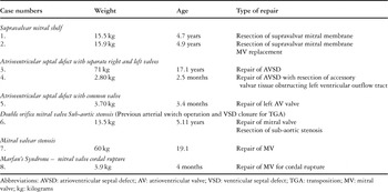
Our use of three-dimensional echocardiography complemented rather than replaced other imaging modalities. The nature of the lesions requiring surgery is shown in the table. For the supramitral shelf found in the first two cases, the three-dimensional data provided a far clearer indication of the extent of the shelf than did conventional techniques, and allowed atrial views to be projected (Figs 2a–c). For the third, fourth, and fifth patients, all having atrioventricular septal defects with common atrioventricular junctions, the morphology of the left atrioventricular valve could be shown in detail (Figs 1a–c), along with delineation of areas of atrioventricular valvar regurgitation which we regarded as superior to that obtained using conventional cross-sectional techniques. The ultrasonic system permits the operator to “cut” the valve sequentially, thus showing precisely the regions of regurgitation. The double orifice in the mitral valve of our sixth patient, and already discussed, was delineated better by transoesophageal echocardiography than three-dimensional echocardiography, but the relationship of the valvar orifice to the aortic valve was better seen on the three-dimensional study. For mitral stenosis, as encountered in the seventh patient, the morphology of the valve could be shown in “en-face” views from the ventricle, while views from the atrium were obtained which are not possible by conventional means. The cordal rupture found in our eighth patient, with Marfan’s syndrome, had already been visualised conventionally, but the three-dimensional technique allowed a more accurate delineation of the part of the leaflets of the mitral valve affected. The flail cords were seen better on epicardial cross-sectional echocardiography, nonetheless, because of the higher frequency and frame rates of the transducer.
Table 1. Diagnoses and procedures evaluated.

Outcome data
Seven of the eight patients survived, and have now been followed over periods ranging from two to sixteen months. We lost one infant with atrioventricular septal defect in the setting of Noonan's syndrome, weighing 2.8 kilograms, who had severe obstruction of the left ventricular outflow tract produced by accessory tissue originating from the left atrioventricular valve. The stitches in the region of apposition of the bridging leaflets gave way, and a second operative procedure was required. This was complicated by renal failure and sepsis, and the child died at the age of four months.
Of the remaining seven patients, two required further surgery within 30 days, including further repair of the left atrioventricular valve in the fifth patient, and additional repair of cordal rupture in the infant with Marfan's syndrome. The child with a double orifice in the mitral valve required additional repair subsequent to one month due to persisting mitral regurgitation. In the second patient, resection of the supravalvar mitral shelf led to severe mitral regurgitation necessitating subsequent replacement of the mitral valve.
Discussion
Three-dimensional echocardiographic imaging has a number of attractions for guiding surgical intervention. The three-dimensional dataset can be presented in a number of different projections. Some of these, such as visualisation of the atrioventricular valve from the atrial aspect, are impossible to achieve using cross-sectional echocardiography. Images are presented which are more akin to the anatomy as seen by the surgeon, rather than a series of two-dimensional slices of the heart. Previous methods to produce a three-dimensional echocardiographic dataset prior to surgery have utilised a transoesophageal approach. This involves rotation of the imaging plane of the probe through 180 degrees. Because of this, the images are closely sequential rather than truly “real-time”.6, 7 This approach requires a dedicated workstation, meaning that the times required for acquisition and analysis are longer, thus hampering its usefulness in the surgical setting.
The data we present demonstrates that high quality three-dimensional information can readily be obtained intraoperatively. The quality of the images obtained using the epicardial approach is superior to that obtained transthoracically, due to the absence of structures intervening between the probe and the heart, and lack of movement by the patient under anaesthesia. This initial experience has been limited to two operators who had already gained experience in acquisition and interpretation of three-dimensional transthoracic images. We cannot prove that surgical results are improved by the use of the three-dimensional approach to imaging, although three-dimensional data is increasingly being demanded by our surgeons prior to surgery on the atrioventricular valves. The rapidity with which images can be displayed and processed on the ultrasound system in the operating theatre is a major advantage over previous techniques.
A number of limitations currently exist. The highest frequency of the three-dimensional ultrasound probe is 4 Megahertz, which is lower than the frequencies from 8 to 12 Megahertz now available for many of the probes used for cross-sectional epicardial echocardiography. This impacts on the resolution of the image obtained but, in our practice, three-dimensional data complements rather than substitutes other techniques. The three-dimensional approach permits a frame rate of 25 frames per second, which is also less than the rates in excess of 100 per second achievable using standard ultrasonic probes. The limitations of frequency and frame rates meant that our subjective impression was that we obtained images of better quality in older and larger patients. Acquisition of a full volumetric dataset would ideally be achieved in a single cardiac cycle, but the currently available system acquires the data in four sections, or seven sections if colour flow mapping is used, so that movement artefact can be a factor. All our patients were under anaesthesia, which meant that movement was not a problem. The matrix probe that we used is larger than the high frequency probes typically used for conventional cross-sectional interrogations, but it was still possible to use the probe in an infant weighing no more that 2.8 kilograms. We anticipate that, in the future, smaller probes of higher frequency will become available, with increased frame rates, which will enhance the quality of the images still further.
Acknowledgements
The post occupied by Debbie Rawlins for training in echocardiography is supported by the British Heart Foundation. The three-dimensional echocardiographic equipment used for the study was purchased using a grant from the Guy's and St Thomas' Hospitals' Charity.