Klippel–Feil syndrome is a rare skeletal disorder primarily characterised by low hairline and a short, usually immobile, neck caused by abnormal fusion of two or more cervical vertebrae.Reference Tracy, Dormans and Kusumi 1 However, congenital heart disease, hearing loss, and skeletal, eye, brainstem, and urinary system abnormalities may accompany this syndrome.Reference Senoglu, Ozbag and Gumusalan 2 According to cervical involvement, Andre Feil subsequently classified this syndrome into three categories: type I is described as a massive cervical fusion of the cervical spine (40% of the cases), type II if there is fusion of 1 or 2 cervical vertebrae (47%), and Type III includes fusion of the cervical vertebrae with concomitant fusion of the thoracic or lumbar vertebrae (13%).Reference Senoglu, Ozbag and Gumusalan 2 Although this disorder is present at birth, mild cases may go undiagnosed until late in life when symptoms worsen or first become apparent.
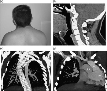
We present the case of a 14-year-old boy who was referred to our adolescent and adult congenital heart disease unit to continue follow-up evaluation of his congenital heart disease. His school performance was normal and there was no family history of heart disease. The New York Heart Association functional class was I/IV and on physical examination he was an intelligent, prepubertal boy with a normal facial appearance, except for the existence of a short neck with restricted upper spine mobility and low occipital hairline (Fig 1a). Written informed consent was obtained from the patient’s family and assent was obtained from the 14-year-old boy.
( a ) Low posterior hairline and short neck characteristic findings of the syndrome. ( b ) Cervical lateral-computed tomography (CT) showing fusion of the vertebral bodies of C2-C3-C4 (arrow head) and C7-D1. ( c ) Angio CT evidencing agenesis of the left upper bronchus and lung parenchyma with the absence of the ipsilateral pulmonary artery. ( d ) Angio CT with levo-looped transposition of the great arteries. Ao=aorta; PA=pulmonary artery; RV=right ventricle.

From a cardiovascular perspective, the patient was asymptomatic. No heart murmurs were heard, although decreased breath sounds were noted in the left lung apex. His hearing assessment and ophthalmological examinations were normal and the patient had a normal 46, XY karyotype.
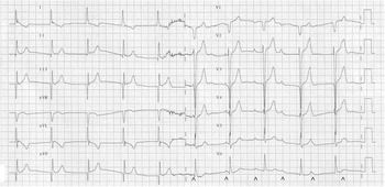
Computed tomography of the cervical spine showed congenital fusion of the vertebral bodies of C2-C3-C4, C7-D1, D1-D2, and D2-D3 with associated scoliosis and fusion of the posterior arch of the second and third ribs of the left side, data consistent with a Klippel–Feil syndrome type III (Fig 1b). Meanwhile, the angiothoracic computed tomography showed agenesis of the left upper bronchus and lung parenchyma with the absence of the ipsilateral pulmonary artery (Fig 1c), a bovine aortic arch, and levo-looped transposition of the great arteries (Fig 1d). Meanwhile, the 12-lead electrocardiogram revealed sinus rhythm with a complete atrioventricular block (Fig 2). Transthoracic echocardiogram confirmed the computed tomography findings and showed moderate dysfunction of the systemic morphologically right ventricle.
12-lead electrocardiogram showing a third-degree atrioventricular block (complete heart block) with complete dissociation of the atrial (arrow head) and ventricular activity. The risk of complete heart block rises over time, in patients with levo-looped transposition of the great arteries, with a 2% per year increase in incidence because of abnormalities of the conduction system. The existence of a narrow QRS complex escape rhythm implies that the pacemarker is above the His bundle. Patients with a junctional pacemaker frequently are haemodynamically stable, and their heart rate increases in response to exercise as occurred in our patient. In addition, in levo-looped transposition of the great arteries, the interventricular septum is depolarised in the opposite direction of normal. This results in the absence of initial Q waves that are typically seen in lateral precordial leads (V5–V6), as well as the abnormal presence of initial Q waves in anterior precordial leads (V1). Moreover, Q waves in the inferior leads (III and aVF) may be misinterpreted as an inferior myocardial infarction.

Klippel–Feil syndrome affects 1:40,000 live births, with a female preponderance (3:1), and is often sporadic. However, familial occurrence and the observation of Klippel–Feil syndrome in association with various chromosomal aberrations support a genetic origin of this syndrome. In fact, several missense variants have been identified in PAX1, GDF6, GDF3, and MEOX1 genes in a small percentage of patients with Klippel–Feil syndrome and other associated anomalies.Reference Mohamed, Faqeih, Alsiddiky, Alshammari, Ibrahim and Alkuraya 3 PAX family of regulatory genes is implicated in sclerotomal resegmentation necessary for proper vertebra development. GDF6 and GDF3 genes are precursors of the bone morphogenetic protein family, involved in the regulation of bone and cartilage differentiation, and MEOX1 encodes homeobox proteins that are localised to many mesodermal structures. In this context, Klippel–Feil syndrome probably represents genetic heterogeneity, with at least two inherited forms: one autosomal dominant and the other autosomal recessive. However, many presumed pathogenic variants are now also recognised as population variants, with a frequency much higher than that of Klippel–Feil syndrome, raising doubts about the genetic proposed link.
Embryologically, Klippel–Feil syndrome arises from a failure of proper segmentation of vertebrae in the cervical region, during the third and fourth week of embryonic development, in relation to faulty segmentation of the mesodermal somites. It is known that paraxial mesoderm is responsible for the formation of the vertebrae as well as the dermis of the skin, striated skeletal muscle, muscles of the head, and connective tissue.Reference Kaplan, Spivak and Bendo 4 Meanwhile, intermediate and lateral mesoderms are involved in the development of the urogenital, pulmonary, and cardiac systems.
The importance of recognising Klippel–Feil syndrome lies in the fact that there is a strong association of this anomaly with significant abnormalities of other systems in the body. These include skeletal system abnormalities such as scoliosis and/or kyphosis (60%), Sprengel deformity of the scapula (30%) and torticollis, urinary system abnormalities (35%), different types of hearing loss (30%), facial asymmetry and flattening of the neck (20%), synkinesis or mirror movements (20%), and congenital heart defects (4.2–14%).Reference Tracy, Dormans and Kusumi 1 Although the ventricular septal defect is the most common heart anomaly, other cardiac malformations, such as cor triatriatum,Reference Abraheem and Potter 5 coarctation of the aorta with dextrocardia and situs inversus,Reference Subramaniam, Babu and Sugnani 6 total anomalous pulmonary venous connection,Reference Masuda, Arikawa, Yuda and Taira 7 levo-looped transposition of the great arteries,Reference Drose 8 and anomalous atrioventricular conduction defects,Reference Bhagat, Pant, Singh, Pant, Gupta and Jaggi 9 have also been described. Similarly, pulmonary developmental anomalies have been reported.Reference Bhagat, Pant, Singh, Pant, Gupta and Jaggi 9
Levo-looped transposition of the great arteries in patients with Klippel–Feil syndrome is a very rare entity. However, this cardiac finding supports the hypothesis that cardiac abnormalities may be related to the development of the spine providing insight into the clinical and embryological data of this syndrome.
Acknowledgements
None.
Financial Support
This research received no specific grant from any funding agency, commercial, or not-for-profit sectors.
Conflicts of Interest
None.