Introduction
The Problem: Antibiotic-resistant Pathogens
Antimicrobial resistance poses a growing threat to public health. Globally, ~700,000 people die annually due to infection from antimicrobial-resistant bacteria and it is projected that by 2050 that figure could rise to 10 million, costing >$100 trillion USD (WHO/IACG, 2019). Development of new antimicrobial strategies is an essential goal in the battle against antibiotic resistance. While infections with antibiotic-resistant bacteria have historically been limited to hospital settings, they are increasingly common in the community. Infections with resistant bacteria are associated with increased lengths of hospital stay, cost, and mortality. The ‘cost’ of antimicrobial resistance has been estimated to be >$105 billion worldwide (Naylor et al., Reference Naylor, Atun, Zhu, Kulasabanathan, Silva, Chatterjee, Knight and Robotham2018). The proliferation of acquired resistance mechanisms in bacteria challenges treatment; currently available antibiotics have limited or, in some cases, no activity against some multidrug-resistant bacteria. Further use of antibiotics selects for further resistance, in target and/or commensal bacteria, accelerating the challenge. On a biological level, genes carry antibiotic resistance mechanisms either on bacterial chromosomes, mobile genetic elements, or chromosomal mutations, which may confer resistance. A parallel challenge is resistance associated with microbial biofilms – communities of microorganisms that form on surfaces, whether artificial (e.g. prosthetic joints) or natural (e.g. wounds). Bacteria in biofilms are resistant to antibiotics through mechanisms other than acquired antibiotic resistance (e.g. Miller & Bassler, Reference Miller and Bassler2001; Naik et al., Reference Naik, Sjolin, Ching, Chi and Herpfer2018), though they may harbor acquired antibiotic resistance.
Wound infections are encountered commonly in today’s clinical practice and are affected by both acquired and biofilm-associated antibiotic resistance. Given that management of wound infections is difficult and that antibiotic-resistant bacteria are oftentimes involved, novel and innovative therapeutic and preventive non-antibiotic approaches are needed. Strategies that limit emergence of conventional antimicrobial resistance are ideal. While clays have been used for medicinal applications since ancient times (Photos-Jones & Hall, Reference Photos-Jones and Hall2011; Gomes et al., Reference Gomes, Rautureau, Niel and Gomes2021), only recently have the technological capabilities existed to examine physical and chemical interactions of clays with microbes and to understand what may make certain clays antibacterial (Williams et al., Reference Williams, Metge, Eberl, Harvey, Turner, Prapaipong and Poret-Peterson2011).
Based on anecdotal observations of the ability of certain natural clays to heal bacterial infections, Williams et al. (Reference Williams, Holland, Eberl, Brunet and Brunet de Courssou2004) initiated research on natural antibacterial clays. More than a decade of testing a variety of clay assemblages collected worldwide, and comparisons made with standard reference materials from the Source Clays Repository of The Clay Minerals Society (www.clays.org), has led to identification of certain clay assemblages with unique geochemical properties capable of in vitro reduction of bacteria in both planktonic and biofilm states, including antibiotic-resistant strains (Williams et al., Reference Williams, Haydel, Giese and Eberl2008; Caflisch et al., Reference Caflisch, Schmidt-Malan, Mandrekar, Karau, Nicklas, Williams and Patel2018; Williams, Reference Williams2019). One clay studied was notably effective at killing bacteria in vitro. It was formed by hydrothermal alteration of a porphyritic andesite-hosted massive sulfide deposit that produced an illite-smectite containing primarily reduced iron clay, quartz, pyrite (coarse crystalline pyrite and spherules <1 μm in diameter), and minor Ca-feldspar. This deposit was mined by Oregon Mineral Technologies (OMT, Inc.) in Douglas County, Oregon (USA). In the present study, the reduced Fe clay is referred to as the OMT Blue clay, because of its distinctive blue color in the field (Morrison et al., Reference Morrison, Williams and Williams2017). The OMT was first used as a soil amendment due to its high sulfur content and low pH (<4); the geologic emplacement of this clay was studied by Morrison et al. (Reference Morrison, Williams and Williams2017) who produced the first antibacterial map showing the antibacterial effectiveness of the clay as a function of the deposit mineralogy and geochemistry. The Morrison et al. (Reference Morrison, Williams and Williams2017) study showed that antibacterial activity is not ubiquitous across the deposit, but rather that the clay assemblage must have specific geochemical properties to exhibit in vitro antibacterial activity (Williams, Reference Williams2019).
Many studies of natural antibacterial clays from a variety of clay depositional environments show common geochemical elements among the clays (Williams, Reference Williams, Finkelman, Seigel and Selinus2021). From the illitic ‘French Green clays’ used for healing Buruli ulcer in the Ivory Coast, Africa (Haydel et al., Reference Haydel, Remenih and Williams2008; Williams et al., Reference Williams, Haydel, Giese and Eberl2008; Williams & Haydel, Reference Williams and Haydel2010), to kaolin-rich clay from the Colombian Amazon (Londoño & Williams, Reference Londoño and Williams2016; Londoño et al., Reference Londoño, Hartnett and Williams2017), and even a glacial deposit from British Columbia – the Kisameet clay (Behroozian et al., Reference Behroozian, Svensson, Li and Davies2020) made of glacial flour from igneous and metamorphic parent materials – all show similar antibacterial properties to the OMT Blue clay when studied in detail (Williams et al., Reference Williams, Metge, Eberl, Harvey, Turner, Prapaipong and Poret-Peterson2011; Morrison et al., Reference Morrison, Underwood, Metge, Eberl and Williams2014, Reference Morrison, Misra and Williams2016, Reference Morrison, Williams and Williams2017; Caflisch et al., Reference Caflisch, Schmidt-Malan, Mandrekar, Karau, Nicklas, Williams and Patel2018). Each natural antibacterial clay contains reduced metals (e.g. Fe(II), Cu(I), and Zn(II)), and the silicate minerals buffer the pH when water is added to conditions that stabilize reduced metals and Al3+ in solution. These are unlike other types of clays used for wound healing (e.g. Garcia-Villén et al., Reference Garcia-Villén, Souza, Barbosa, Borrego-Sánchez, Sámcjez-Espejo, Ojeda-Riascos and Iborra2020) which may be manipulated chemically for their antibacterial action or used for their physical adsorption capacity to reduce edema and stimulate tissue growth (Cervini-Silva et al., Reference Cervini-Silva, Nieto-Camacho, Ramirez-Apan, Gomez-Vidales, Palacios, Montoya and Ronquillo de Jesus2015). Understanding natural antibacterial clay mechanisms and biogeochemical processes involved in reduction of antibiotic-resistant pathogens is necessary for designing new antibacterial agents based on geomimicry and is the primary objective of this research.
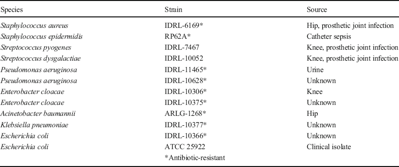
The current study presents a preliminary assessment of the antibacterial action of the OMT Blue clay for topical treatment of wound infections in mice. The clay was previously shown to be active at killing a broad spectrum of pathogenic bacterial biofilms in vitro (Table 1; Fig. 1; Caflisch et al., Reference Caflisch, Schmidt-Malan, Mandrekar, Karau, Nicklas, Williams and Patel2018), therefore, in the present study, experiments were conducted to evaluate its application in vivo, a necessary step before human testing.
Bacterial biofilm species studied (modified from Caflisch et al., Reference Caflisch, Schmidt-Malan, Mandrekar, Karau, Nicklas, Williams and Patel2018)

Results of the OMT Blue clay and aqueous leachate (200 mg clay/mL, shaken 24 h) treatment of established staphylococcal biofilms. Error bars are from triplicate independent experiments, with statistical significance p < 0.05

BACKGROUND
Clay-mineral Antibacterial Mechanisms
Physical clay–microbe interactions
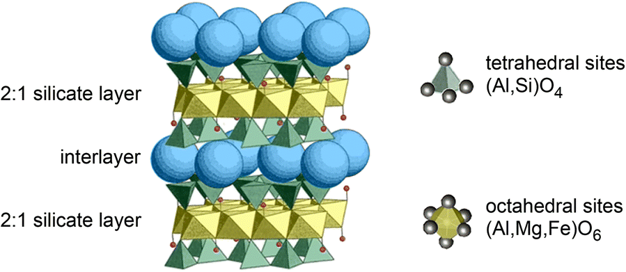
The physical attraction of some clays to bacteria is uncommon as most layered clay minerals have basal surfaces with negative charges and most bacteria also have negatively charged surfaces (Silhavy et al., Reference Silhavy, Kahne and Walker2010). The negative charge on layered clay minerals is derived from cation substitutions in tetrahedral or octahedral sites (e.g. Al3+ for Si4+ in tetrahedral sites or Li+ for Mg2+ in octahedral sites; Moore & Reynolds, Reference Moore and Reynolds1997) that leave a net negative charge distributed across basal planes (Fig. 2). The average surface potential of montmorillonite is −62.8 ± 10.6 mV, and the average surface potential of kaolinite is −40.9 ± 15.5 mV (Yuan & Xiong, Reference Yuan and Xiong2017). Depending on the zeta potential, the potential difference between the particle surface in an aqueous suspension and the fluid, negatively charged surfaces will repel to varying degrees (Londoño & Williams, Reference Londoño and Williams2016). The pH of the fluid in which the particles (clay or bacteria) are suspended determines the potential attraction or repulsion of any particle; the pH at which the particle has a net zero charge is known as the point of zero charge (pzc). Therefore, when evaluating physical mechanisms by which a clay may be antibacterial, the pzc is important for determining the surface charge (energy) of particles under the chemical conditions of the wound fluids.
Structural schematic of smectite showing layered tetrahedral (green) and octahedral (yellow) sheets of the silicate framework. Blue spheres represent interlayer hydrated metal ions attracted to the basal planes (modified from Williams et al., Reference Williams, Haydel and Ferrell2009)

Broken edges of layered silicates may be positively charged (Moore & Reynolds, Reference Moore and Reynolds1997), so it is possible that some negatively charged bacteria may be attracted to clay edges, but the edge charge is highly pH dependent (Zhao et al., Reference Zhao, Bhattacharjee, Chow, Wallace, Maslinyah and Xu2008). Subsequently, edge adsorption is minor compared to the surface area of clay basal planes (Fig. 2). Several studies have shown the importance of clay–microbe interactions in nature and in the laboratory (Müller, Reference Müller2015) and those interactions can be assisted by bacterial production of siderophores or other chelators. While a clear physical association exists between clays and microbes, the scale of interlayer sites in smectites or lumen of kaolins is far too small to host a bacterium; instead, chemical dissolution products from exudates of bacteria may be partially responsible for clay-mineral dissolution (Jaisi et al., Reference Jaisi, Dong, Kim, He and Morton2007) in addition to abiotic dissolution occurring in soils and during mineral weathering (e.g. Li et al., Reference Li, Zhou, Fiore and Yu2019; Fomina & Skorochod, Reference Fomina and Skorochod2020).
One way to increase the force of attraction between a clay and bacterial surface is to exchange interlayer cations with an alkylammonium compound such as cationic hexadecyl-trimethylammonium (HDTMA) or chitosan (Haque et al., Reference Haque, Nasar, Inamuddin, Inamuddin and Ali2018). Both effectively change the basal surface charge with chitosan in particular, changing the clay basal surfaces to a positive charge (Darder et al., Reference Darder, Colilla and Ruiz-Hitzky2003); thus, exterior surfaces of the aluminosilicate may attract negatively charged bacteria. Chitosan was intercalated (Xia et al., Reference Xia, Wang, Zeng, Guo, Zhu, Chen and Dong2020) on a chemically reduced nontronite to make the clay surface charge positive and demonstrated attraction of the iron-bearing clay to bacteria. In addition, reduced iron released by clay dissolution or cation exchange produces hydroxyl radicals through the Fenton reaction series (Morrison et al., Reference Morrison, Misra and Williams2016), causing cell death through hydroxyl radical attack of certain lipids recently identified as cardiolipin in the cell membrane (Wang et al., Reference Wang, Dong, Zeng, Xia, Zhang and Zhou2017; Xia et al., Reference Xia, Wang, Zeng, Guo, Zhu, Chen and Dong2020). It is evident, therefore, that physical interactions between clays and microbes led to chemical interactions, which may be involved in antibacterial processes, and/or contribute to wound healing.
Chemical Clay–microbe Interactions
Natural antibacterial clays generally contain reduced metals (e.g. Fe(II)) and most have an expandable clay component (smectite) containing structural iron (Fe(III) and Fe(II)) in their octahedral sheets and in the interlayer that acts as a reservoir for reduced metal cations (Morrison et al., Reference Morrison, Misra and Williams2016). Oxidation of reduced metals occurs upon hydration of the clay with oxygenated water, which promotes degradation of biomolecules through Fenton reaction series (e.g. Valko et al., Reference Valko, Morris and Cronin2005). The source of reduced iron in solution (Fe2+) may be a variety of minerals in the clay assemblage (e.g. pyrite, magnetite, biotite, and various other clay minerals). The role of smectite in an antibacterial clay is related possibly to its large relative surface area (>100 m2/g) that buffers the water pH in a hydrated suspension (50–200 mg/mL) to conditions where the aluminosilicate releases Fe2+ and Al3+. The dissolution of Fe and Al is primarily an inorganic aqueous reaction in the OMT Blue clay (Morrison et al., Reference Morrison, Misra and Williams2016); certain bacteria may enhance clay dissolution and the release of aqueous Fe and Al, however (Dong, Reference Dong2012; Müller, Reference Müller2015; tsaveLi et al., Reference Li, Zhou, Fiore and Yu2019). OMT Blue clay produces these solutes at concentrations of 3–5 mM at pH < 5, where aluminosilicates become unstable (Morrison et al., Reference Morrison, Misra and Williams2016). Over 24 h, dissolved O2 in the hydration water promotes oxidation of aqueous Fe2+ producing >100 μM H2O2, which crosses bacterial cell membranes and enters the periplasmic space. Although Imlay (Reference Imlay2019) showed that intracellular H2O2 can be used by bacterial cells for respiration and that cells can tolerate significant amounts of this oxidant, Morrison et al. (Reference Morrison, Misra and Williams2016) deduced that the H2O2 reactant along with Fe2+ produces hydroxyl radicals through the reaction: Fe2++H2O2→Fe3++•OH+OH– and that the hydroxyl radical product is responsible for damage to intracellular proteins and DNA in bacteria. Other products of the Fenton reaction series include the superoxide radical, and other reactants (N- and S-species) also produce highly reactive intermediate compounds that may contribute to overall oxidation, but the •OH was confirmed to be a dominant reactant causing bacterial cell-membrane damage (Wang et al., Reference Wang, Dong, Zeng, Xia, Zhang and Zhou2017). The •OH is a highly reactive intermediate radical that exists only for nanoseconds and can diffuse just nanometers before reacting with biomolecules (Winterbourn, Reference Winterbourn2008); •OH would not travel through the cell membrane and, therefore, must be produced intracellularly to decompose DNA and intracellular proteins (Williams, Reference Williams2019). Intracellular opaque precipitates in Escherichia coli killed by treatment with OMT Blue clay were first identified by Williams et al. (Reference Williams, Metge, Eberl, Harvey, Turner, Prapaipong and Poret-Peterson2011), and scanning transmission electron microscopy (STEM) and electron energy loss (EELS) were used by Morrison et al. (Reference Morrison, Underwood, Metge, Eberl and Williams2014) to identify the opaque precipitates as Fe(III) oxides. Later, Wang et al. (Reference Wang, Dong, Zeng, Xia, Zhang and Zhou2017), who identified cardiolipin as the lipid damaged by reduced-iron clays, suggested a pathway for Fe2+ entry through the cell membrane, allowing the intracellular Fenton reactions previously identified. Two additional studies of E. coli treated with different antibacterial clays (Morrison et al., Reference Morrison, Misra and Williams2016; Londoño et al., Reference Londoño, Hartnett and Williams2017) used ion imaging by nano-secondary ion mass spectrometry (NanoSIMS) and documented elevated intracellular Fe compared to untreated bacteria. Furthermore, these studies found Al to be concentrated on the cell exterior (Fig. 3). Evidence for Al binding to phospholipids in the cell membrane, which can cause poration, allowing influx of Fe to the cytoplasm through damaged membranes, was presented by Londoño et al. (Reference Londoño, Hartnett and Williams2017).
SIMS ion images across an Escherichia coli (ATCC 25922) cell. a 12C14N image of a single cell indicating cross section A-A’; b cross-section showing the distribution of Al/CN before (blue) and after (red) treatment with antibacterial clay; c cross-section showing the distribution of Fe/CN (modified from Londoño et al., Reference Londoño, Hartnett and Williams2017)

Materials and Methods
Mineral Preparation
The OMT Blue clay was collected from an open-pit mine, the Oregon Mineral Technologies Pit #10, described in detail by Morrison et al. (Reference Morrison, Williams and Williams2017). Bulk clays were prepared for quantitative random powder X-ray diffraction (XRD) analysis, following procedures reported by Eberl (Reference Eberl2007). Random powder XRD was performed using a Siemens D500 or Bruker D5000 instrument with CuKα radiation. In addition, clay mineralogy was studied using oriented clay mounts of <2 μm separates that were air-dried and ethylene glycol-saturated for determination of the expandability of I-S clays (Morrison et al., Reference Morrison, Williams and Williams2017). In brief, the OMT Blue clay is composed of ~42–49 wt.% rectorite, 42–46 wt.% quartz, and 5–6 wt.% pyrite with traces (<3%) of plagioclase, gypsum, jarosite, and smectite (Morrison et al., Reference Morrison, Underwood, Metge, Eberl and Williams2014). Two formulations of the clay were tested for antibacterial activity. One test used only the <2 μm clay fraction separated by standard centrifugation methods, without any chemical manipulation (Moore & Reynolds, Reference Moore and Reynolds1997). Bulk samples milled using a McCrone Mill to ~20 μm were compared to clay-sized separates. In addition, Red clay was collected from the upper, oxidized zone of the OMT Pit #10 which is mineralogically similar to the reduced Blue clay but contains <5 wt.% goethite and kaolinite and no pyrite due to complete oxidation of the original Blue clay. This clay was shown to have no antibacterial properties (Morrison et al., Reference Morrison, Underwood, Metge, Eberl and Williams2014); therefore, it was used as a negative control clay for the animal testing. All mineral samples were autoclaved at 120°C and 30 psi to remove inherent environmental bacteria, and suspended in deionized water (DIW). No difference was observed between results from in vitro testing in which the clay-size fraction was used and in which the bulk clay assemblage was used (Caflisch et al., Reference Caflisch, Schmidt-Malan, Mandrekar, Karau, Nicklas, Williams and Patel2018), so animal testing proceeded using the bulk clay sample. Previous in vitro testing showed a minimum bactericidal concentration of 50 mg of clay/mL (Morrison et al., Reference Morrison, Underwood, Metge, Eberl and Williams2014); this dilute suspension was difficult to apply to mice, however, so suspensions of 200–800 mg/mL were tested. The pH and Eh of suspensions were monitored before and after application to infected wounds to evaluate changes that might impede the oxidation reactions.
Animal Trials
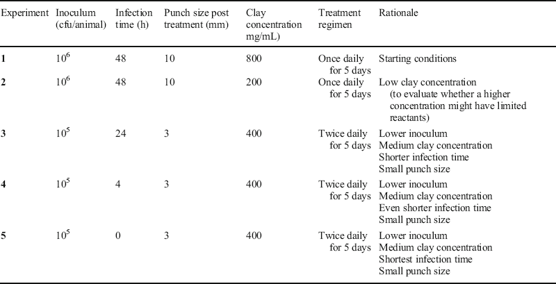
Experiments used 89 C57BL/6 mice, each weighing ~25 g. Five experimental groups were studied (Table 2). Infection was evaluated by standard tissue culture analysis reported in colony-forming units (cfu) per tissue. During the experiments, one animal died (experiment 2), and one tissue culture was contaminated (experiment 5); these were excluded from the study. A methicillin-resistant strain of S. aureus (IDRL-6169) was grown in a trypticase soy broth to a 0.5 McFarland standard (~1.5×10⁸ cfu/mL) and re-suspended in normal saline (final inoculum 105–106 cfu/animal). Animals were anesthetized with ketamine/xylazine (90/10 mg/kg). Buprenorphine SR (1 mg/kg) was given subcutaneously for pain management. Skin on the dorsum of the mouse was shaved and disinfected thrice, alternating between povidone-iodine and alcohol swabs. A full thickness wound was created using a 5 mm biopsy punch. A 10 μL suspension of S. aureus (IDRL-6169) was inoculated onto the wound bed. After 2 min, semi-occlusive transparent Tegaderm® (3M, St. Paul, Minnesota) was placed over the wound and secured with the liquid adhesive Mastisol® (Eloquest Healthcare, Ferndale, Michigan, USA). Animals were either treated immediately or the infection was allowed to become established for up to 2 days (Fig. 4) before treatment commenced.
List of experimental groups including: the inoculant, infection time, wound punch size used for culture at end of treatment, clay concentration used, frequency and length of treatment, and the rationale for each experiment

Photograph of established Staphylococcus aureus (IDRL-6169) wound infection.

The activity of the OMT Blue clay was assessed as a non-antibiotic method for treatment of wound infections and compared to OMT Red clay (the non-antibacterial, oxidized OMT Blue clay (as a negative control)), Bacitracin antibiotic ointment (166 U), Vaseline (carrier for the Bacitracin ointment), or no treatment. Treatments began after bacterial challenge and lasted for 5 days. Clay was hydrated by 24 h equilibration (rotary stirring) with deionized water (DIW) at the appropriate experimental dose and applied to a small square of sterile gauze (500 μL of 200–800 mg clay/mL) placed over the wound surface and allowed to oxidize in air for 2 min to begin oxidation of the clay. Approximately 500 μL of hydrogel (1.5 wt.% autoclaved xanthan gum in 100 mL DIW; Namaste Foods, LLC., Coeur d’Alene, Idaho, USA) was placed over the gauze and Tegaderm® used to affix the gauze. Daily, animals were anesthetized with isoflurane, Tegaderm® was removed, and the wound was cleansed by rinsing with sterile water. Wound sizes were measured at maximum diameters and documented photographically before adding new hydrated clay. Purulence of the wounds was measured using a grading scale from 0 to 5 (Kim et al., Reference Kim, Karau, Greenwood-Quaintance, Tilahun, Krogman, David, Pritt, Patel and Rajagopalan2015), where: 0 indicates no exudate in normal appearing wound bed; 1 indicates serous of slight turbid exudate; reddish wound clearly visible; 2 indicates a modest amount of whitish exudate; reddish wound bed visualized; 3 indicates a moderate amount of whitish exudate; wound bed not visualized; 4 indicates a moderate amount of yellowish exudate; exudate limited to wound bed; and 5 indicates a marked amount of gross pus; pus extended beyond wound edges.
After 5 days of treatment, animals were euthanized via CO2 asphyxiation and wound tissue harvested using a 3- or 10-mm biopsy punch. Wound punches were collected and placed into sterile tubes with 1 mL saline, homogenized with an Omni Tissue Homogenizer (TH)® (Omni International, Kennesaw, Georgia, USA), vortexed for 30 s, sonicated for 5 min, and vortexed for an additional 30 s. Quantitative culture of the wound tissue was performed on all animals from each group and log10 cfu/tissue compared among the groups. Given the small group sizes studied, analysis on the three animals tested in each trial was descriptive, including averages and standard deviations of the measurements.
Results
The results of the five animal experiments (Figs 5, 6, 7) showed the effect of OMT Blue clay compared to OMT Red clay and other controls. Compared to previous in vitro results (Caflisch et al., Reference Caflisch, Schmidt-Malan, Mandrekar, Karau, Nicklas, Williams and Patel2018), the same antibacterial action of the OMT Blue clay was not observed in vivo, showing only minimal reduction of S. aureus (IDRL-6169) in excised tissue from mice treated with OMT Blue clay, compared to untreated wound infections (Fig 5). Further, wound size did not decrease in OMT Blue clay-treated mice compared to untreated mice (Fig. 6). Purulence did, however, show a decrease over 5 days of treatment with OMT Blue clay compared to controls (Fig. 7). That both OMT Blue and Red clay-treated wounds had lower purulence than untreated wounds suggests removal of wound exudate by adsorption onto the clays.
Culture results of five animal experiments, where infected mice were treated with OMT-Blue clay, compared to controls and untreated infected wounds, over 5 days, with daily changes of wound dressing. Graphs a–e represent experiments 1–5 (Table 2). Black box = range; red lines = average; blue lines = standard deviation of each experiment (n = 3)

Animal wound sizes over 5 days of treatment with the OMT Blue clay, compared to controls and untreated infected wounds. Graphs a–e represent experiments 1–5 (Table 2). Black box = range; red lines = average; blue lines = standard deviation of each experiment (n = 3)

Purulence assessments over 5 days of treatment with the OMT Blue clay, compared to controls and untreated infected wounds. Graphs a–e represent experiments 1–5 (Table 2). Black box = range; red lines = average; blue lines = standard deviation of each experiment (n = 3)

Discussion
The in vivo results were unexpected given the significant reduction of S aureus (IDRL-6169) in prior biofilm experiments (Fig. 1). Several hypotheses were tested in vitro to explore the diminished antibacterial activity of the clay in vivo:
(1) the pH and Eh of aqueous suspensions of clay and bacteria in the wound may be out of the stability range for Fe2+ and Al3+ in solution, limiting the antibacterial reactions;
(2) reactants (Fe2+, Al3+) or products (e.g. H2O2, •OH) of Fenton reactions may be inactivated by complexation with components of wound fluids (i.e. speciation change);
(3) the antibacterial clay could be drying out such that aqueous reactions with mineral components were impeded; and
(4) the supply of oxygen to wounds may be diminished relative to that experienced by bacteria during in vitro testing, inhibiting Fenton reactions that ultimately lead to compromise of bacterial membranes and decomposition of intracellular proteins.
Aqueous Stability of Antibacterial Reactants
The longevity of mineral reactants involved in redox reactions within the hydrated clay was studied by monitoring the pH-Eh of an OMT Blue clay suspension in water (100 mg/mL) compared to the standard reference minerals illite (IMt-1) and smectite (SWy-1) over 100 h (Fig. 8). The OMT Blue clay is dominated by illite-smectite (rectorite), but the clay standards of the discrete end-member minerals (smectite and illite) are not antibacterial because they do not buffer the aqueous suspension to a pH-Eh range where Fe2+ and Al3+ are soluble. For each clay, there is a rapid increase in pH and Eh over the first 10 h in DIW. The pH then stabilizes, decreasing only slightly over 100 h. The Eh, however, declines by ~100 mV over that same time as aqueous oxidation reactions occur, reducing the minerals.
Comparison of pH-Eh trends over time for a 100 mg/mL OMT Blue clay suspension compared to standard reference minerals smectite (SWy-1) and illite (IMt-1) from the Source Clays Repository of The Clay Minerals Society (www.clays.org)

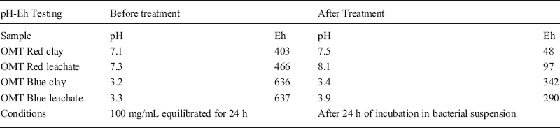
Changes in the pH and Eh of clay suspensions and aqueous leachates of the clays (containing soluble metals) were examined before and after incubation with bacteria over 24 h, the time between wound dressing changes. Table 3 shows that the Eh of both the clay and the aqueous leachate was reduced (by ~half) whereas the pH was not changed significantly. This suggested that antibacterial reactions that oxidize biomolecules leave metal hosts reduced over time. As the clay is reacting with other organic components in the wound in addition to bacteria, this could reduce the dose of H2O2 to bacterial cells and intracellular influx of Fe2+. This observation suggests that redox reactions occurring in the wound bed itself are altered, which are not observed in vitro. Nonetheless, because clay application to wounds was performed daily, the pH and Eh of the clay should remain in the stability field for Fe2+and Al3+; an alternative hypothesis, therefore, is that Fe2+ is complexing with wound fluids, changing the aqueous speciation of the reduced metal, and limiting the Fenton reaction progress.
Mineral suspension and leachate pH-Eh changes after incubation with Staphylococcus aureus (IDRL-6169) over 24 h at 37°C

Metal Complexation in Wounds
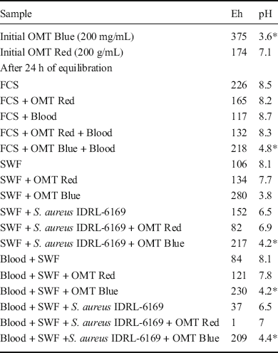
Previously, the aqueous speciation of metals released from OMT Blue clay were shown to be alterable by the concentration of salts in the Luria Bertani (LB) growth media used to establish log phase growth of bacteria tested in vitro (Morrison et al., Reference Morrison, Underwood, Metge, Eberl and Williams2014). Components of wound fluids were investigated, therefore, to determine how they might interact with hydrated OMT Blue clay. Simulated wound fluid (SWF) consisting of 50% fetal calf serum (FCS) and 50% physiological NaCl (0.9%) in 0.1% peptone (Werthén et al., Reference Werthén, Henriksson, Jensen, Sternberg, Givskov and Bjarnholt2010) and sheep blood were tested. Results of experiments with the OMT Blue and Red clays with SWF, combined with suspended S. aureus (IDRL-6169), showed an ~8-log reduction of planktonic bacteria in 10% LB, SWF, and SWF plus 5% sheep blood after 24 h (Fig. 9). For biofilms, however, (Fig. 10) bacterial reduction was only ~3 log units in the presence of FCS and blood; the NaCl and peptone components of SWF did not significantly impede antibacterial action. To explore these observations, the pH-Eh of OMT Blue and Red clays in aqueous suspension were compared to mixtures of the various wound components and S. aureus (IDRL-6169) equilibrated for 24 h at 37°C (Table 4). The OMT Blue clay suspensions with SWF, FCS, and blood were in the pH and Eh stability range for Fe2+ in solution (Morrison et al., Reference Morrison, Williams and Williams2017), though the Fe2+ and/or Al3+ involved in the antibacterial process may be complexing with components of the FCS and/or blood.
Results showing the effect of components of simulated wound fluid (SWF) on planktonic Staphylococcus aureus (IDRL-6169) reduction in the presence of the OMT Blue and Red (control) clays over 24 h

Results showing the effect of components of SWF in the presence of OMT Blue and Red (control) clays on reducing Staphylococcus aureus (IDRL-6169) biofilms over 24 h

Effect of fetal calf serum (FCS) simulated wound fluid (SWF) and blood on the pH-Eh of clay and bacterial suspensions incubated 24 h at 37°C

*Indicates antibacterial conditions
Wound Hydration
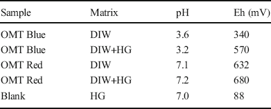
Clay applied to infected wounds in mice dried over the course of the experiments which could limit antimicrobial activity compared to in vitro conditions because water is required to transmit aqueous Fe2+ and Al3+ from clay to the bacteria. Various concentrations of clay and methods of stabilizing the dressing were tested, therefore. The potential use of hydrogel (1.5 wt.% xanthan gum in DIW) was used as a method to maintain hydration of the clay (Table 5) during in vivo testing. The pH of the clay suspensions did not change significantly, though the Eh of the OMT Blue clay increased slightly in hydrogel but remained within the stability field of Fe2+. In vitro testing was performed, comparing OMT Blue and OMT Red clay suspensions in DIW and hydrogel incubated with established S. aureus (IDRL-6169) biofilms (Fig. 11). Compared to the control, a 7-log reduction in bacterial biofilm growth was observed, showing that the hydrogel does not interfere with the antibacterial reactions.
Comparison of the pH-Eh of OMT Blue and Red clays equilibrated with hydrogel (HG) and deionized water (DIW) for 24 h

Comparison of OMT Blue and Red (control) clays reacted with Staphylococcus aureus (IDRL-6169) biofilms in deionized water and hydrogel for 24 h at 37°C

Wound Oxidation
Finally, Tegaderm® (a polyethylene-based film with adhesive backing) used to hold the clay on the wound has relatively low oxygen permeability (Marks et al., Reference Marks, Bucknor, Roussakis, Nowell, Kamali, Cascales, Kazei, Lin and Evans2020). If dissolved oxygen is limited, the Fenton reactions will be impeded, and this might account for lower Eh conditions after incubation with bacteria in wounds (Table 3). As noted previously, however, given that the OMT Blue clay dressing was re-applied to wounds daily minimizes the likelihood that the oxygen supply to the wound environment was diminished, instead suggesting that redox reactions in the wound account for the reduction in Eh during treatments, potentially limiting the Fenton reaction progress.
Ancillary in vitro Testing for Antibacterial Enhancement
Ancillary methods were tested in an attempt to improve the antibacterial efficiency of the OMT Blue clay in the wound environment. Intercalation of chitosan in the interlayers of a reduced-iron smectite showed that the clay surface charge could be made positive (Xia et al., Reference Xia, Wang, Zeng, Guo, Zhu, Chen and Dong2020), and thus, would be attracted to the negatively charged surface of most bacteria. If so, this might direct clay reactants that promote Fenton reactions toward bacteria and limit interactions with other wound components. Some reduced-Fe clays oxidize quickly in oxygenated water; thus, controlling the rate of the clay-mineral oxidation might be advantageous to the antibacterial process. Fe(II) in smectite was shown by Komadel et al. (Reference Komadel, Madejová and Stucki1999) to be partially stabilized by incorporating Li into vacant octahedral sites of the dioctahedral clay. The Komadel et al. (Reference Komadel, Madejová and Stucki1999) method was applied, therefore, to the OMT Blue clay to determine whether it enhanced the antibacterial effect. The results of these two manipulations of the OMT Blue clay (Fig. 12) showed no difference in the chitosan or lithium-treated clay in terms of antibacterial effect, showing ~3 log reductions in bacterial populations.
Treatment of the OMT Blue clay with chitosan or lithium incorporation in the octahedral sheet did not change the activity of the clay in reducing Staphylococcus aureus (IDRL-6169) biofilms

Other studies of natural antibacterial clays have shown similar in vivo reductions in effectiveness compared to in vitro experiments (Otto et al., Reference Otto, Kilbourne and Haydel2016) and suggested that topical application of natural clay is not capable of reaching bacteria in deep wound tissues. This is contrary to previous successful applications of reduced-Fe clay in the treatment of Buruli ulcer, a mycobacterial infection of the skin (Williams et al., Reference Williams, Holland, Eberl, Brunet and Brunet de Courssou2004, Reference Williams, Haydel, Giese and Eberl2008). That work showed successful treatment of infection and wound healing using a natural reduced-Fe clay (French Green Clay) applied daily over a period of ~3 months. While there are many reasons that diverse bacterial infections may respond differently to topical treatments by natural clays, successful medicinal applications clearly require better insights into the mineral reactions in the wound environment (wound fluids, bacteria, and tissue) to recapitulate the antibacterial activity observed in vitro.
Conclusions
Results of in vivo testing of the OMT Blue clay on MRSA (S. aureus (IDRL-6169)) infection in wounds did not reproduce previous in vitro findings over a 5-day treatment course. Four main hypotheses were tested to evaluate the differences observed between applications of the antibacterial clay to MRSA-infected wounds in mice and the successful in vitro experiments.
Hypothesis 1: The aqueous stability of antibacterial reactants (Fe2+, Al3+) from the clay was tested by measuring the pH-Eh conditions of the mineral reactants over time (Fig. 8) compared to the mineral-bacteria suspension (Table 3). The effect of SWF and blood on the pH-Eh of the mineral-bacterial suspension was also studied (Table 4). The OMT Blue clay buffered the SWF to a low pH (4.1 to 4.4), similar to OMT Blue clay in DIW, but the Eh of the suspension was reduced to nearly half in SWF compared to DIW. These values remain within the pH-Eh range where Fe2+ is stable in solution (Williams, Reference Williams2019), but the lower oxidation state of the suspension could impede Fenton reactions critical to membrane lipid and protein damage in the bacteria.
Hypothesis 2: The potential for metal complexation in wounds was tested by measuring the effect of the wound fluid components on S. aureus (IDRL-6169) planktonic and biofilm reduction in vitro. When mixed 1:1 with OMT Blue clay, wound fluids showed no inhibition of antibacterial effect of the clay on planktonic S. aureus, with an 8-log reduction in the bacterial population compared to the control (Fig. 9). The presence of fetal calf serum and blood reduced the antibacterial effect on the S. aureus biofilms by ~3 log units, however (Fig. 10), indicating that these wound components inhibit the antibacterial process. This may be due to the formation of metal complexes or speciation in the wound, removing the reactants or products from the Fenton reaction series and slowing the antibacterial activity.
Hypothesis 3: The hydration state of the wounds was improved by applying hydrogel, as it was previously shown that aqueous species of Fe2+ and Al3+ work in unison to attack bacterial cell membranes and intracellular proteins (Morrison et al., Reference Morrison, Misra and Williams2016). The antibacterial clay application was modified by addition of a xanthan gum-based hydrogel to keep the clay hydrated. The hydrogel showed no significant change in the pH or Eh of the hydrated antibacterial clay, and the natural clay was effective in vitro, showing a 7-log reduction in S. aureus biofilms compared to controls (Fig. 11). Therefore, applying hydrogel to the in vivo experiments did not inhibit the antibacterial effect of the OMT Blue clay.
Hypothesis 4: The reduced oxidation state of antibacterial clay after incubation with S. aureus (Table 3) and in wound fluids (Table 4) could be a limitation to the antibacterial efficacy. While pH was not greatly affected after 24 h of incubation with the bacteria or wound fluids, Eh was reduced by ~100 mV and the wound bed itself possibly plays a role in changing the Eh, making the wound environment less conducive to the antibacterial mechanisms exhibited in vitro. The wound dressing used here (Tegaderm®) may not be ideal for permeability of O2 into the wound (Sirvio & Grussing, Reference Sirvio and Grussing1989; Marks et al., Reference Marks, Bucknor, Roussakis, Nowell, Kamali, Cascales, Kazei, Lin and Evans2020) and thus alternative wound dressings that enhance oxidation (e.g. Ochoa et al., Reference Ochoa, Rahimi, Zhou and others2020) might be employed.
The evidence to date suggests that applications of antibacterial clays in vivo may require a different dosing regimen than considered in these preliminary animal trials. The results point to reduction of the oxidation state in the wounds; thus, enhanced oxidation of the mineral reactants in the wound, while keeping the clays hydrated, should be explored to possibly replicate the successful reduction of drug-resistant pathogens observed in vitro.
ACKNOWLEDGMENTS
The authors acknowledge Oregon Mineral Technologies Inc., for permission to study their clay deposit and thank Susan Selkirk at ASU for figure-drafting assistance. This work was funded by the U.S. National Science Foundation grant EAR-1719325 and by an ASU/Mayo seed grant for in vivo testing.
Funding
Funding sources are as stated in the Acknowledgments.
Compliance with ethical statements
Conflict of Interest
The authors declare that they have no conflict of interest.