Total anomalous pulmonary venous connection is a “congenital cardiovascular malformation in which none of the pulmonary veins connect to the morphologically left atrium.” Reference Franklin, Béland and Colan1 The supracardiac type is the most common; typical repair involves direct anastomosis of the pulmonary venous confluence to the left atrium. We report a unique repair: anastomosis of the vertical vein and left atrium, combined with a Warden procedure.
Case report
A 3 kg, 1-day-old infant presented with mild cyanosis. Echocardiography suggested a mixed form of supracardiac total anomalous pulmonary venous connection (Supplemental Fig 1). All pulmonary veins, except the right upper, drained into a pulmonary venous confluence that is connected to the superior vena caval–right atrial junction through a vertical vein. Multiple right upper pulmonary veins drained into the superior vena cava near the entry of the vertical vein.
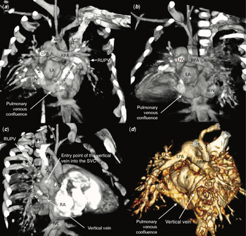
Upon computed tomography (CT) angiography (Fig 1) and cardiac catheterisation (Supplemental Fig 2), the pulmonary venous confluence and vertical vein appeared unobstructed. However, there was a mean pressure gradient of 5 mmHg from the pulmonary venous confluence to the superior vena cava. There was no indication of pulmonary venous obstruction. Due to intermittent, agitation-related serum lactate elevations, we decided on the surgical repair.
CT angiography with three-dimensional reconstruction. ( a ) The left upper (LUPV) and left lower (LLPV) pulmonary veins drained into the pulmonary venous confluence. The vertical vein ascended beside the left atrium and entered the superior vena cava (SVC). ( b ) The right middle (RMPV) and right lower (RLPV) pulmonary veins drained into the pulmonary venous confluence. ( c ) The right upper pulmonary vein (RUPV) entered the SVC separately from the vertical vein. ( d ) In the superior–posterior view, the pulmonary venous confluence is clearly visible. Atria (left, LA; right, RA); pulmonary arteries (left, LPA; right, RPA); inferior vena cava (IVC).

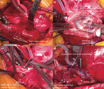
At 39 days old, we performed anastomosis of the vertical vein to the left atrium, and a modified Warden procedure. Segmental pulmonary veins from the right upper lobe entered the superior vena cava superior to the vertical vein (Fig 2a). The remaining pulmonary veins entered the pulmonary venous confluence posterior to the left atrium. The vertical vein ascended posterolateral to the left atrium and turned acutely into the superior vena caval–right atrial junction. Working through an enlarged secundum atrial septal defect, the posterior wall of the left atrium was anastomosed to the vertical vein (Fig 2b). A glutaraldehyde-fixed, autologous pericardial patch was used to baffle the internal orifice of the superior vena caval–right atrial junction to the atrial septal defect.
Intraoperative findings. ( a ) The vertical vein and right upper pulmonary veins entered the superior vena cava (SVC). ( b ) Anastomosis of the left atrium and vertical vein, accessed through an enlarged interatrial communication. ( c ) SVC division superior to the entry points of the right pulmonary veins. ( d ) The cavoatrial anastomosis was augmented with a bovine pericardial patch.

The superior vena cava was divided superior to the segmental pulmonary veins from the right upper lobe (Fig 2c). The cardiac end of the superior vena caval stump was closed with a glutaraldehyde-fixed, autologous pericardial patch. The transected superior vena caval anastomosis to the right atrial appendage was augmented with a bovine pericardial patch (Fig 2d). Hence, we allowed pulmonary venous flow into the left atrium, both through its anastomosis with the vertical vein and beneath the intracardiac baffle.
The patient was discharged with aspirin on post-operative day 9; echocardiography revealed unobstructed pulmonary venous return into the left atrium (Supplemental Fig 3). Two-month follow-up clinical and echocardiographic findings were satisfactory.
Comment
Echocardiography, CT angiography with three-dimensional reconstruction, and cardiac catheterisation were utilised to elucidate the anomalous pulmonary venous connections. Since all pulmonary venous return was to the superior vena cava, a Warden procedure alone might have allowed for adequate drainage of the total anomalous pulmonary venous connection. However, due to the acute turn of the vertical vein into the superior vena cava, and the gradient from the pulmonary venous confluence to the superior vena cava, it might increase the risk of post-operative pulmonary venous obstruction. We elected to perform anastomosis of the vertical vein to the left atrium, in addition to a Warden procedure, to allow for separate drainage pathways.
The Warden procedure was designed to repair partial anomalous pulmonary venous connection to the superior vena cava. Reference Warden, Gustafson, Tarnay and Neal2 Excellent outcomes employing the Warden procedure have been reported; Reference DiBardino, McKenzie, Heinle, Su and Fraser3 however, small infants are at high risk for systemic venous pathway obstruction. Reference Park, Kwak and Lee4 Despite our patient’s small size, the Warden procedure was determined as the best approach to address the pulmonary veins from the right upper lobe. To avoid potential post-operative systemic and pulmonary venous obstruction, repair included patch closure of the cardiac end of the superior vena caval stump, patch augmentation of the cavoatrial anastomosis, and enlargement of the atrial septal defect.
The mild gradient between the pulmonary venous confluence and superior vena cava might have been due to the acute turn of the vertical vein as it entered the superior vena caval–right atrial junction. We surmise that it became clinically significant upon agitation, which increased myocardial oxygen requirements, resulting in intermittent serum lactic acidosis. The latter did not recur after surgical repair.
Craig’s classification of total anomalous pulmonary venous connection is based on the anatomic level at which the pulmonary venous return is drained; Reference Craig, Darling and Rothney5 the supracardiac type is the most common, and mixed total anomalous pulmonary venous connection is the rarest. The latter is defined as total anomalous venous connections at two or more levels. Our patient’s diagnosis was supracardiac total anomalous pulmonary venous connection, as all pulmonary veins are connected to a supracardiac site. Reference Franklin, Béland and Colan1,Reference Craig, Darling and Rothney5 However, because there were two drainage sites at the same anatomic level, we concluded that our patient had a “mixed supracardiac” subtype.
Separate pulmonary venous drainage pathways decrease the risk of post-operative pulmonary venous obstruction. Although the unique combination of the separate pulmonary venous drainage pathways carried a possible risk of blood stagnation, our patient had an uneventful post-operative course and encouraging 2-month follow-up echocardiography. Careful follow-up is warranted to detect post-operative complications, including obstruction of the pulmonary venous and cavoatrial anastomoses.
Supplementary material
To view supplementary material for this article, please visit https://doi.org/10.1017/S1047951120003625
Financial support
This research received no specific grant from any funding agency, commercial, or not-for-profit sectors.
Conflicts of interest
None.