Introduction
Endospores are one of the most interesting life forms of the microbial world. They can survive boiling, vacuum, ionizing radiation (Heritage et al. Reference Heritage, Evans and Killington1995) and extreme acceleration (Mastrapa et al. Reference Mastrapa, Glanzberg, Head, Melosh and Nicholson2001). Culturable bacterial spores have been recovered after millions of years from the abdominal contents of extinct bees buried in Dominican amber (Cano & Borucki Reference Cano and Borucki1995). The extreme resilience of bacterial spores has piqued the interest of astrobiologists and led to considerable research in evaluating the resistance of spores against extreme environments of vacuum, temperature and ionizing radiation encountered in outer space or planets (Horneck et al. Reference Horneck, Rettberg, Reitz, Wehner, Eschweiler, Strauch, Panitz, Starke and Baumstark-Khan2001; Rettberg et al. Reference Rettberg, Rabbow, Panitz, Reitz and Horneck2002; Wassmann et al. Reference Wassmann2012; Noell et al. Reference Noell, Ely, Bolser, Darrach, Hodyss, Johnson, Hein and Ponce2015). The SPORES experiment of EXPOSE-R mission tested the survival of bacterial endospores to outer space on International Space Station (Panitz et al. Reference Panitz, Horneck, Rabbow, Rettberg, Moeller, Cadet, Douki and Reitz2015) and the PROTECT experiment of EXPOSE-E mission tested the survival of bacterial endospores to simulated trip to Mars on the International Space Station (Horneck et al. Reference Horneck2012). Further, many researchers are investigating the efficacy of various inactivating agents against spores of Bacillus anthracis and its surrogates since the ‘anthrax letter’ attacks in 2001 (Nicholson & Galeano Reference Nicholson and Galeano2003; CDC 2006; Pillet et al. Reference Pillet, Formosa-Dague, Baaziz, Dague and Rols2016a, Reference Pillet, Marjanovic, Rebersek, Miklavcic, Rols and Kotnikb; Raguse et al. Reference Raguse, Fiebrandt, Stapelmann, Madela, Laue, Lackmann, Thwaite, Setlow, Awakowicz and Moeller2016).
The dormant state is achieved by vegetative bacteria in a process known as sporulation, where a copy of the genetic material is enclosed in a thick multi-layered shell (Block Reference Block2001). Small acid-soluble proteins in the core protect the DNA from ionizing radiation, whereas the cortex provides heat resistance and the spore-coat layers protects the spore from chemical attack (Driks Reference Driks1999; Henriques & Moran Reference Henriques and Moran2000; Nicholson et al. Reference Nicholson, Munakata, Horneck, Melosh and Setlow2000; Riesenman & Nicholson Reference Riesenman and Nicholson2000; Block Reference Block2001; Atrih & Foster Reference Atrih and Foster2002). The presence of vegetative cells in spore suspensions can result in overly optimistic assessment of inactivating agents because vegetative cells are 10–25 000 times more sensitive to heat, radiation and chemical agents than spores (Atrih & Foster Reference Atrih and Foster2002). Therefore, it is imperative to use high purity suspensions of spores to obtain accurate results.
Several techniques are available for separating bacterial spores from a mixture of spores and vegetative cells (Zhao et al. Reference Zhao, Krishna, Moudgil and Koopman2008). Most of these techniques inactivate or destroy vegetative cells and achieve separation through multiple washes. Few researchers have considered the effect of these treatments on the susceptibility of spores after purification towards inactivating agents. Gorman et al. observed that addition of a lysozyme incubation step to a wash sequence increased the thermal susceptibility of B. subtilis spores (Gorman et al. Reference Gorman, Scott and Hutchinson1985), whereas Demidova and Hamblin have reported that inactivation of spores purified by a series of chemical washes and incubation with lysozyme was not significantly different when the lysozyme incubation step was removed (Demidova & Hamblin Reference Demidova and Hamblin2005).
The present study was undertaken to evaluate the effect of two widely used spore purification methodologies, heat shock (HS) and chemical wash-lysozyme-heat shock (CLH), on spore integrity and susceptibility to inactivating agents. Bacillus subtilis spores were chosen as the target microorganism as it is commonly used as surrogate for spores of B. anthracis. Ultraviolet A radiation (UVA; 320–400 nm) and free radicals generated by photocatalysis were chosen as the inactivating agents. UVA radiation generates free radicals, such as superoxide and hydroxyl-free radicals, that degrade the spore coats. In contrast, UVB (280–320 nm) and UVC (100–280 nm) radiations inactivate microorganisms directly by causing DNA damage (Nicholson et al. Reference Nicholson, Schuerger and Setlow2005). Titanium dioxide photocatalysis is of widespread interest for microbial inactivation because it is capable of completely mineralizing the microbe without generating toxic byproducts (Jacoby et al. Reference Jacoby, Maness, Wolfrum, Blake and Fennell1998; Bai et al. Reference Bai, Krishna, Wang, Moudgil and Koopman2012). Degussa P25 (commercial titanium dioxide containing mixture of anatase and rutile polymorphs) and multi-wall carbon nanotubes coated with anatase were chosen as photocatalyst for inactivation experiments. Degussa P25 is considered to be the best commercially available photocatalyst. Our previous study has shown that anatase coated multi-wall carbon nanotubes can inactivate bacterial spores faster than Degussa P25 (Krishna et al. Reference Krishna, Pumprueg, Lee, Zhao, Sigmund, Koopman and Moudgil2005). The sensitivity of spores towards photocatalysis and UVA should be similar as the mechanism for both photocatalysis and UVA inactivation is by free-radical generation (Zhao et al. Reference Zhao, Krishna, Hua, Moudgil and Koopman2009).
Experimental procedures
Chemicals
Chemicals were obtained from Fisher Scientific, except as noted. Titanium dioxide photocatalyst was obtained from Degussa. Solutions and deionized water for washing bacterial suspensions were autoclaved before use. Lysozyme and phenylmethylsulfonyl fluoride (PMSF) solutions were prepared using autoclaved buffers. Lysozyme powder was stored at 4°C and lysozyme solution was made from powder for each use. The PMSF powder was stored at 4°C. PMSF solution was prepared by dissolving the powder in 2–3 ml (the minimum possible) ethanol. TEP buffer containing 50 mM Tris–HCl at pH 7.2, 10 mM ethylenediaminetetraacetic acid (EDTA) and 2 mM PMSF was prepared fresh for each use.
Bacterial culture
Culture media was prepared according to the American Society for Testing and Materials (ASTM) E2111-00 standard (ASTM 2001) using Difco Columbia broth powder. Bacillus cereus (American Type Culture Collection, ATCC 2, Manassas, VA, USA) or B. subtilis (ATCC 6051, generously provided by Dr Sam Farrah, Department of Microbiology, University of Florida) colonies from Difco Tryptic soy agar plates were inoculated in 500 ml Erlenmeyer flasks containing 100 ml of 1/10th strength culture media fortified with 0.1 mM manganese sulfate (MnSO4.2H2O). The flasks were capped with foam plugs and wrapped in aluminium foil. The inoculated growth media was incubated for 72 h at 35 ± 2°C on an orbital incubator shaker (Model C24, New Brunswick Scientific, Edison, NJ, USA) with a speed of 200 rev min−1.
Spore purification methods
Heat shock
The HS procedure was modified from Peng et al. (Reference Peng, Tsai and Chou2001). A volume of 70 ml culture was transferred to an 85 ml centrifuge tube and centrifuged at 10 000 g for 10 min at 4°C using a Marathon 21000R centrifuge (Fisher Scientific, Hampton, NH, USA). The supernatant was poured off and the pellet was resuspended in 20 ml of deionized water. The spores and cells were washed three more times by repeated centrifugations and resuspensions in deionized water. Volumes of 20 ml suspension were then transferred to 125 ml flasks, which were subsequently immersed in an 80 ± 2°C water bath for 15 min. The flasks were then cooled to 4°C, and the suspensions were washed three times with deionized water. The final suspension was stored at 4°C for no more than 1 week. The purity (proportion of spores relative to the total number of spores and cells) of the final suspension was 67%.
Chemical wash-lysozyme-heat shock
The CLH method was carried out according to Xue and Nicholson (Xue & Nicholson Reference Xue and Nicholson1996). Culture was centrifuged at 10 000 g for 10 min at 4°C and the pellet was resuspended in 20 ml of 1 M KCl/0.5 M NaCl. This suspension was centrifuged and the pellet was resuspended in 20 ml of 50 mM Tris–HCl (pH 7.2) containing 50 µg ml−1 lysozyme and incubated at 37°C for 60 min. The suspension was then washed with, respectively, 1 M NaCl, deionized water, 0.05% (w/v) sodium dodecyl sulfate (SDS), TEP buffer and deionized water (three times). The washed suspension was heat shocked at 80 ± 2°C for 15 min and then stored at 4°C for no more than 1 week. The purity of the final suspension was 99%.
Counting
A spore suspension was serially diluted using phosphate-buffered saline amended with 2 mM SDS. At this low concentration, the SDS aids in dispersing spores, thereby reducing variability of subsampling (Krishna et al. Reference Krishna, Zhao, Pumprueg, Koopman and Moudgil2016). The spore suspension was serially diluted and aliquots of the three highest dilutions were plated in duplicate on Tryptic soy agar (100 × 25 mm2 Petri dishes). The plates were then incubated at 37°C for 24 h and then the colonies were counted manually. Counts between 30 and 300 were used for data analysis (Koch Reference Koch, Gerhardt, Murray, Costilow, Nester, Wood, Kreig and Phillips1981).
Transmission electron microscopy (TEM) sample preparation
TEM was done with a Hitachi H-7000. The samples were prepared on 300 mesh carbon-coated copper grids with formavar films (EMS). A 10 µL aliquot of spore suspension was added to a Petri dish and covered by a grid for 1 min. The grid was then removed and stained with 1% uranyl acetate.
Experimental setup
The UVA and photocatalytic inactivation studies were carried out in a UVA chamber with 16 solar UVA lamps (Southern New England Ultra Violet Company, Branford, CT, USA) with peak intensity at 350 nm. The UVA irradiance for inactivation experiments was maintained at 92 W m−2 by placing the reaction mixture at a distance of 10 cm from the UVA lamps. Suspensions were mixed using magnetic stirrers. Cooled air was passed over the Petri dish to limit the heating of the suspension.
UVA inactivation
The UVA lamps were stabilized for 30 min before each experiment. They were then turned off, a sterile plastic Petri dish was placed atop the magnetic stirrer in the UVA chamber, and 30 ml of spore suspension was added to the dish. Evaporation was limited by covering the dish with polyethylene film. After stirring in darkness for 10 min (to be consistent with photocatalysis experiments), four 0.25 ml aliquots of suspension were removed by pipette and placed in a sterile culture tube. The UVA lamps were then turned back on. The stirred suspension was sampled at predetermined times throughout the total irradiation period of 4 h. Each sample was serially diluted and plated immediately after it was collected.
Photocatalytic inactivation
The concentration of titanium dioxide (Degussa P25) employed was 0.01 wt% (Krishna Reference Krishna2007). The photocatalyst suspensions were prepared by transferring 3 mg of P25 to a sterile Erlenmeyer flask containing 20 ml of sterile deionized water. The suspension was sonicated for 30 min in a water-bath sonicator (EW-8890, Cole-Parmer, Vernon Hills, IL, USA). The photocatalyst suspension was then transferred to a sterile plastic Petri dish containing a sterile magnetic stirrer bar. Subsequently, 10 ml of spore suspension was added to the Petri dish and the mixture was stirred for 10 min in the dark, followed by exposure to UVA light for a total of 4 h. The spores were sampled and plated according to the procedure described previously.
Anatase-coated multi-wall carbon nanotubes were synthesized according to Krishna et al. (Reference Krishna, Pumprueg, Lee, Zhao, Sigmund, Koopman and Moudgil2005). The photocatalytic inactivation experiments were carried out as described above.
Data analysis
Survival ratio was calculated by normalizing the surviving population at any given time with the initial population. The pseudo first-order rate coefficients determined from first-order fits between the extent of spore inactivation and time were used to characterize the inactivation kinetics (Block Reference Block2001). The inactivation data were modelled with first-order fits using following equation
 $$\displaystyle{{C_0 - C_t} \over {C_0}} = \; 1 - e^{ - kt},$$
where C 0 and C t represent colony-forming units at times t = 0 and t = t, respectively, and k is the first-order rate coefficient, which was determined by least-squares linear regression analysis. Data were analysed by one-tailed Student's t test (Sokal & Rohlf Reference Sokal and Rohlf1997) with a 0.05 level of significance using data analysis in Microsoft Excel.
Results
UVA and photocatalytic inactivation
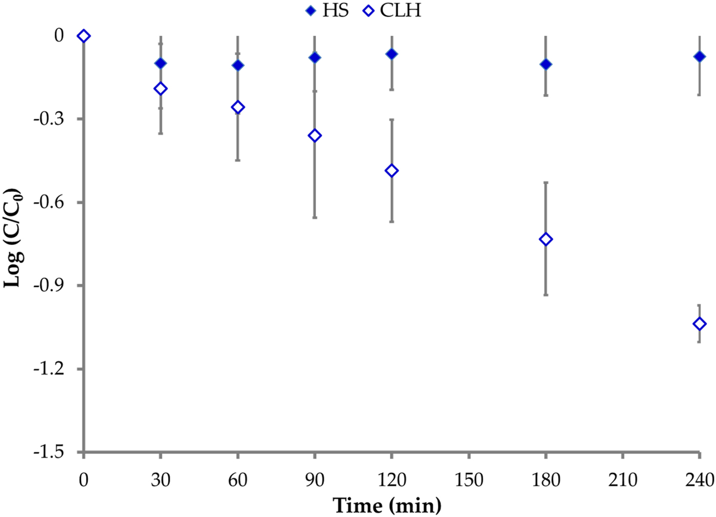
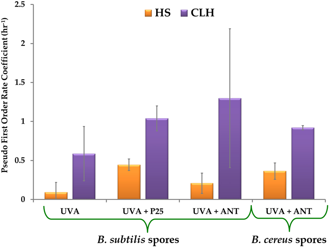
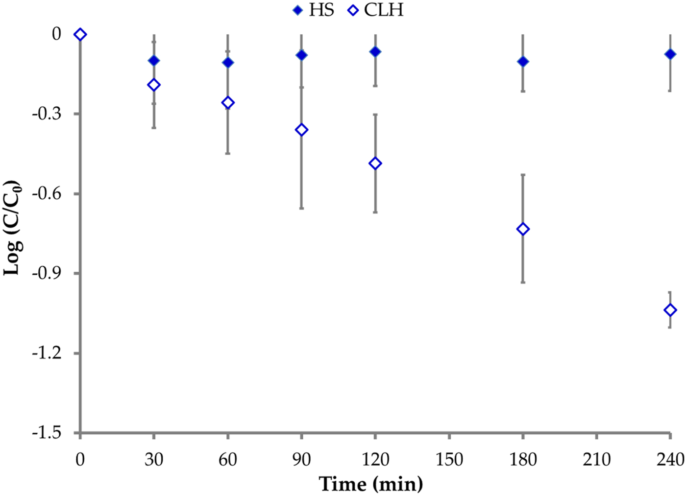
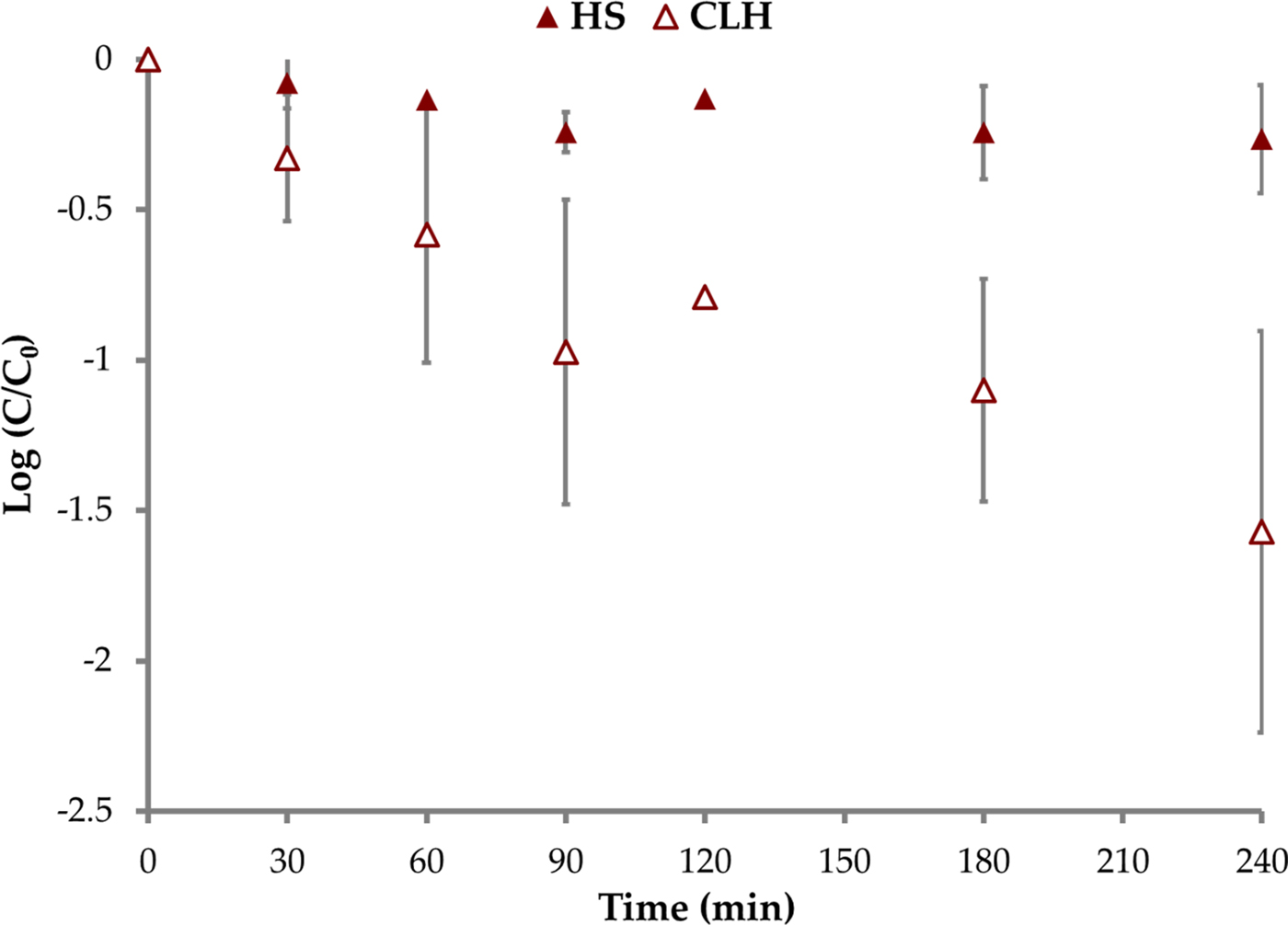
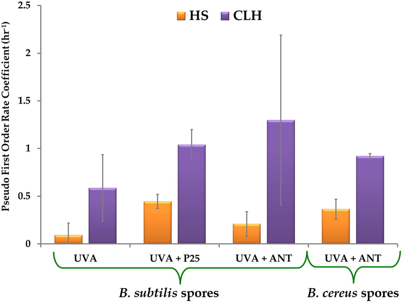
The UVA inactivation curves for HS- and CLH-treated B. subtilis spores are shown in Fig. 1. Each curve is based on averaged data from three individual trials. As can be seen from the plot, the HS-treated spores do not show any decrease in survival ratio over the length of the experiment. The pseudo first-order rate coefficient is 0.0918 ± 0.129 h−1. In contrast, the CLH-treated spores of B. subtilis have a significantly higher (α = 0.05) inactivation rate coefficient of 0.5870 ± 0.348 h−1.
Inactivation of HS- and CLH-treated Bacillus subtilis spores with UVA alone.
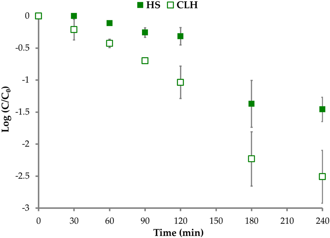
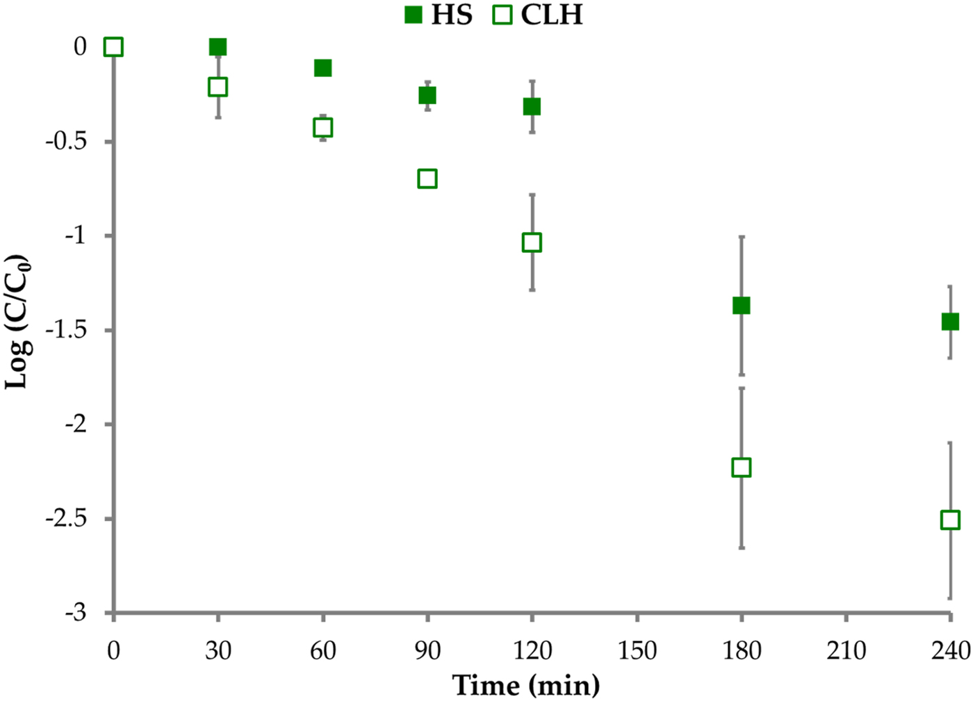
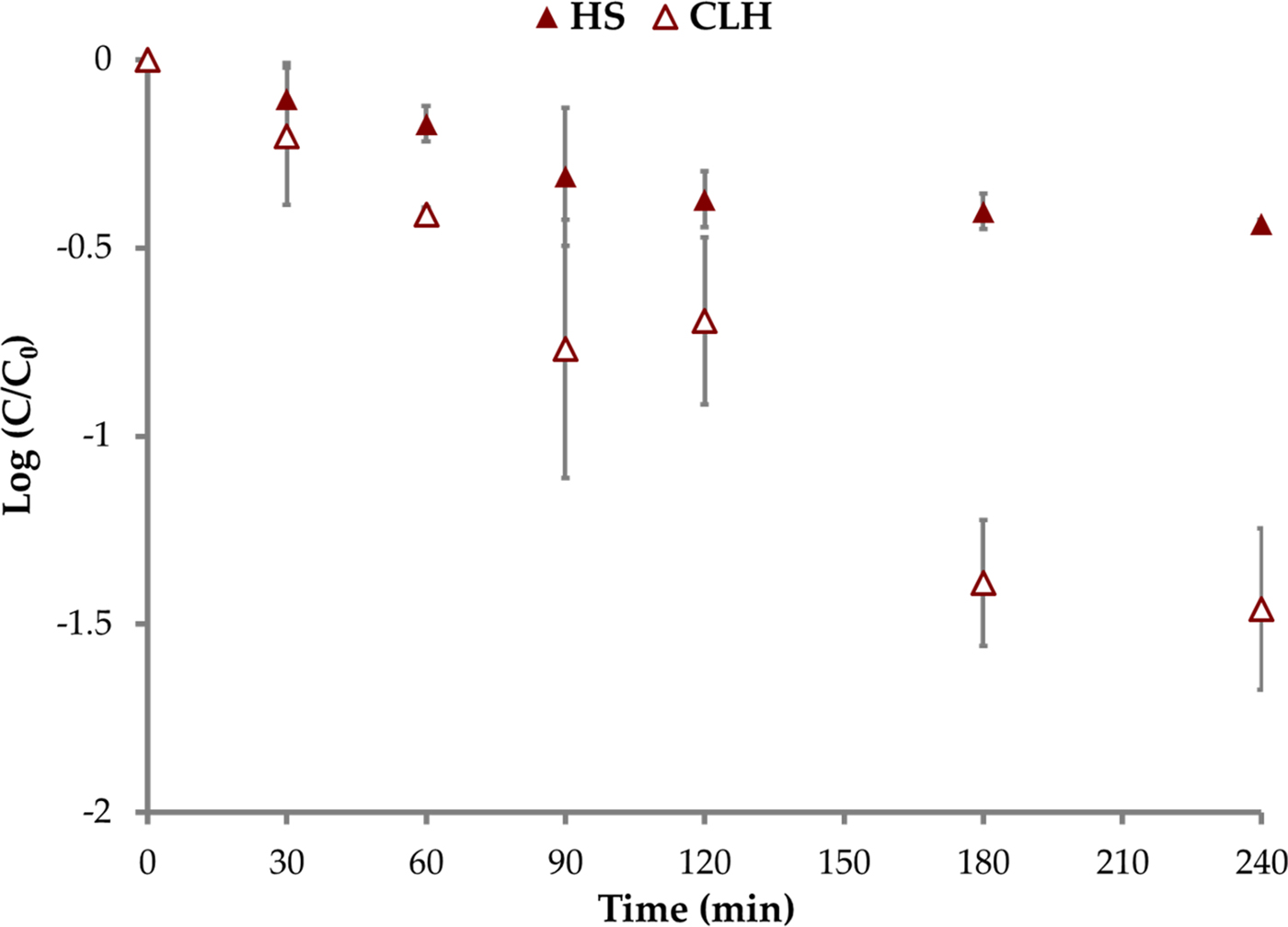
Photocatalytic inactivation curves with Degussa P25 for B. subtilis spores are presented in Fig. 2. Both the HS- and CLH-treated spores were sensitive to photocatalysis. However, the inactivation rate coefficient for CLH-treated spores (1.0393 ± 0.161 h−1) was significantly higher (α = 0.005) than the coefficient for HS-treated spores (0.4456 ± 0.074 h−1).
Inactivation of HS- and CLH-treated Bacillus subtilis spores with UVA and Degussa P25 photocatalyst.
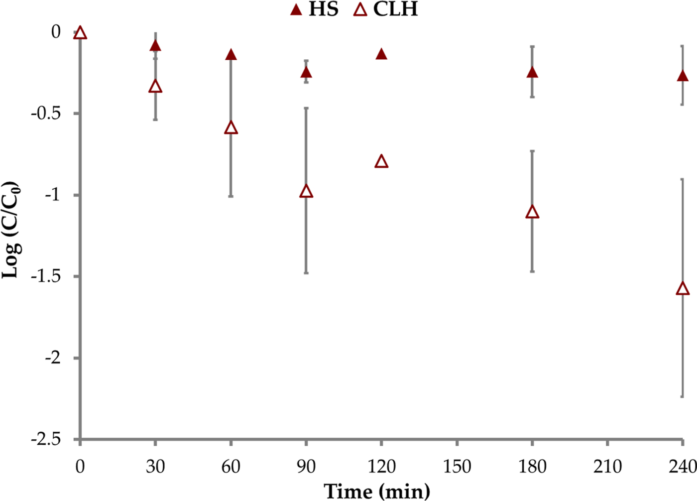
Photocatalytic inactivation curves with anatase-coated multi-wall carbon nanotubes for B. subtilis spores are presented in Fig. 3. The inactivation rate coefficient for CLH-treated spores (1.2989 ± 0.890 h−1) was again significantly higher (α = 0.1) than the coefficient for HS-treated spores (0.2091 ± 0.130 h−1).
Inactivation of HS- and CLH-treated Bacillus subtilis spores with UVA and anatase-coated multi-walled carbon nanotubes (ANT) photocatalyst.
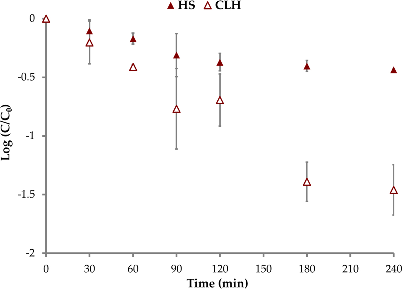
Photocatalytic inactivation curves with anatase-coated multi-wall carbon nanotubes for B. cereus spores are presented in Fig. 4. As before, the inactivation rate coefficient for CLH-treated spores (0.9209 ± 0.026 h−1) was significantly higher (α = 0.05) than the coefficient for HS-treated spores (0.3644 ± 0.105 h−1).
Inactivation of HS- and CLH-treated Bacillus cereus spores with UVA and anatase-coated multi-walled carbon nanotubes (ANT) photocatalyst.
The CLH treatment sensitizes B. subtilis spores to UVA or photocatalysis by six times, whereas the CLH treatment sensitizes B. cereus spores to photocatalysis by only 2.5 times as shown in Fig. 5. It is interesting to note that CLH treatment increases the inactivation rate coefficient by 0.389–0.495 h−1 for UVA inactivation and 0.556–1.089 h−1 for anatase-coated multi-walled carbon nanotubes inactivation with both bacterial species tested.
Comparison of pseudo first-order rate coefficients for HS- and CLH-treated spores against different inactivating agents.
TEM imaging
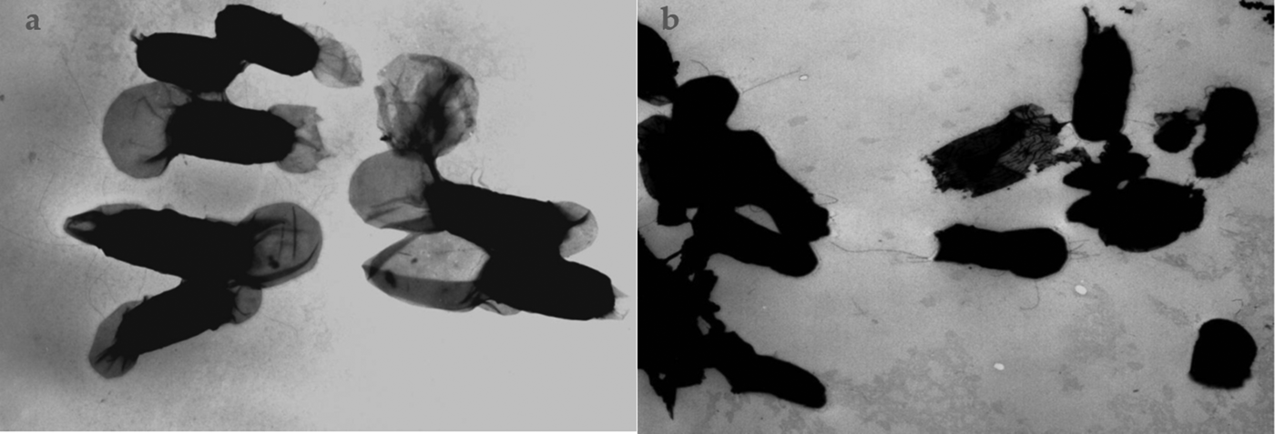
TEM images of B. cereus spores purified by HS and CLH treatment are shown in Fig. 6. The HS-treated B. cereus spores are characterized by presence of an exosporium as well as filaments (Bailey-Smith et al. Reference Bailey-Smith, Todd, Southworth, Proctor and Moir2005; Tauveron et al. Reference Tauveron, Slomianny, Henry and Faille2006). Both of these surface characteristics are clearly visible in TEM images of HS-treated B. cereus spores, suggesting that the spores suffered little or no damage in the purification process (Fig. 6(a)). In contrast, exosporium was not observed in CLH-treated spores (Fig. 6(b)).
Transmission electron microscope images of (a) HS- and (b) CLH-treated Bacillus cereus spores.

Discussion
Possible reasons for the increased sensitivity of CLH-treated spores in comparison to HS-treated spores include: (1) the disparity in the concentrations of spores yielded by the two different methods of purification and (2) damage to protective layers of the spore. The first possibility is not supported by the study results, as spore concentrations yielded by the HS procedure (8.1 × 10⁵–3.5 × 10⁶ cfu ml−1) were within the range of spore concentrations yielded by the CLH procedure (8.2 × 10⁴–1.35 × 10⁷ cfu ml−1). It is important to note that in spite of lower purity with HS-treated spores than CLH-treated spores (67 versus 99%), CLH-treated spores exhibited faster inactivation rates.
CLH treatment is a complex purification method in which spores are subjected to a total of nine washes, some with chemicals including high salt concentration, lysozyme, SDS and PMSF. Each of these chemicals alone or in combination damages the spore coats. Lysozyme inactivates bacteria by hydrolysing the peptidoglycan layer of cell walls. The peptidoglycan layer of spores is covered by two outer spore coats, which provide resistance to spores against lysozyme (Riesenman & Nicholson Reference Riesenman and Nicholson2000; Costa et al. Reference Costa, Serrano, Steil, Volker, Moran and Henriques2007). Suzuki and Rode have shown that certain strains of Bacillus megaterium spores are susceptible to lysozyme. Up to 90% reduction in optical density was observed after spores were exposed to 50 µg ml−1 lysozyme for 15 min (Suzuki & Rode Reference Suzuki and Rode1969). Images of damaged B. megaterium spores presented by Suzuki and Rode show loss of exosporium, similar to the images of damaged B. cereus spores shown in our study.
Gorman et al. observed that addition of a lysozyme incubation step (200 µg ml−1) to a wash sequence increased the thermal susceptibility of B. subtilis spores (Gorman et al. Reference Gorman, Scott and Hutchinson1985). In contrast, Demidova and Hamblin have reported that inactivation of spores purified with CLH treatment was not significantly different when the lysozyme incubation step was removed (Demidova & Hamblin Reference Demidova and Hamblin2005). However, no TEM images are provided to confirm these observations. It should also be noted that they report 5 log reduction in spore concentration within ca. 1.5–3 min based on the type of photosensitizer employed. The mechanism of inactivation with photosensitizer is similar to photocatalysis, i.e. generation of reactive oxygen species. Therefore, such high rate of inactivation may indicate that the spore coats are damaged and exposure of spores to lysozyme during purification may not be the cause of increased susceptibility of spores.
The multiple washes and associated chemicals used in the CLH process may also have a significant impact on spore integrity. Alderton and Snell reported that spores can undergo ion exchange, which affects their susceptibility to heat (Alderton & Snell Reference Alderton and Snell1963). Dipicolinic acid (DPA), which is usually chelated with divalent cations such as Ca2+, Mg2+ and Mn2+, is believed to play a role in spore resistance (Gorman et al. Reference Gorman, Scott and Hutchinson1985; Paidhungat et al. Reference Paidhungat, Setlow, Driks and Setlow2000; Block Reference Block2001). Ion exchange of divalent cations bound to DPA with monovalent cations increases the susceptibility of spores to heat (Gorman et al. Reference Gorman, Scott and Hutchinson1985; Nagler et al. Reference Nagler, Setlow, Li and Moeller2014). In the CLH process, washes with high sodium chloride and potassium chloride concentration (1–1.5 M) could similarly lead to ion exchange with DPA resulting in the observed damage to spore coats. SDS at a concentration of 1 wt% is used in conjunction with urea and dithiothreitol to remove spore coats (Setlow & Setlow Reference Setlow and Setlow1998; Block Reference Block2001). In the CLH process, SDS at a concentration of 0.05 wt% is used alone, and therefore its effect on spore coats is expected to be minimal. Adams et al. have reported that exposure of Clostridium perfringens spores to 10 mM EDTA for 1 h sensitizes the spores to lysozyme (Adams Reference Adams1974). Rassmussen and Labbe found that 50 mM EDTA decreased viability of B. cereus and Bacillus larvae spores, but had no effect on Bacillus alvei, Bacillus laterosporus, Bacillus thuringiensis and Bacillus polymyxa spores (Rasmussen & Labbe Reference Rasmussen and Labbe1996). At the concentration of EDTA used in lysozyme purification process (10 mM), we would expect EDTA to have minimal impact. Boschwitz et al. have reported that PMSF inhibits germination of B. cereus spores (Boschwitz et al. Reference Boschwitz, Milner, Keynan, Halvorson and Troll1983). Gorman et al. have shown that the number of water washes employed for purification of spores can affect the thermal resistance of spores. Fifteen washes had a consistently negative effect, whereas the effect of seven washes was not consistently different from that of a single wash. They also reported that the centrifugation speed (3000 versus 8000 rpm) had no significant impact on thermal resistance of spores (Gorman et al. Reference Gorman, Scott and Hutchinson1985). Accordingly, the nine washes of the CLH process may be expected to have a modest-to-severe impact on spore susceptibility.
We have demonstrated that the CLH process significantly alters the spore morphology and increases the susceptibility of B. subtilis and B. cereus spores to free radicals from UVA radiation and photocatalytic inactivation. Several of the steps in this process have the potential to damage spores. Comparison of process details with the literature suggests that it is the combination of a lysozyme incubation step with multiple washes that is responsible for observed alteration to spore morphology as well as increases in spore susceptibility to UVA, photocatalysis, photosensitizers and heat. Therefore, we recommend caution in employing CLH process for spore inactivation studies including extreme environment simulating outer space.
Acknowledgements
The authors acknowledge the financial support of the Particle Engineering Research Center (PERC) at the University of Florida, the National Science Foundation (NSF Grant EEC-94-02989), and the Industrial Partners of the PERC for support of this research. Any opinions, findings and conclusions or recommendations expressed in this material are those of the author(s) and do not necessarily reflect those of the National Science Foundation. Experiments were carried out at the University of Florida's Water Reclamation and Reuse Laboratory. The second author was supported by a University of Florida Alumni Fellowship. Titanium dioxide photocatalyst was a generous gift from Degussa.
Disclosure statement
No competing financial interests exist.