

 and strong ions give rise to a net osmotic force (~35 Torr) in venous end capillary plasma water that is coupled to ISF through pores in the endothelium. More importantly, diffusing
 and strong ions provide an incremental osmotic force (~150 Torr) that is essentially matched to any change in metabolic rate (e.g. muscular work) when CO
Introduction
The Starling equationReference Starling1 purports to explain the exchange of fluid between plasma and the interstitial space surrounding the capillary endothelium and its basement membrane (hereafter capillary endothelium). In his original experiment, Starling intermittently perfused the isolated hindlimbs of a dog with defibrinated blood (from the dog). The experimental leg was injected with 1% saline (subcutaneously). With the completion of intermittent perfusions (12–24 cycles), it was observed that the formed elements of the experimental leg were in lower concentration than that of the control leg; thus more interstitial fluid (ISF) had returned to the blood of the experimental leg despite the interstitial osmolality being greater than plasma. Interestingly, Starling discusses previous work showing that the osmotic pressure of the salts was ‘enormously higher’ than that of albumen, rendering albumen ‘insignificant in physiological processes’. Yet, Starling rejected this idea, noting that salts were permeable and their diffusion would minimize the effect of the original osmotic difference. The impermeable proteins would provide ‘the attraction of water’ and the salt would diffuse to re-establish the osmotic gradient dictated by the proteins. As will be discussed below, the proteins do not exert this effect. From this single publication of never-repeated experiments and based on Starling's interpretations, four pressures are said to determine whether fluid flows from plasma to ISF or from ISF to plasma. As plasma flows through a capillary, Starling recognized that capillary hydrostatic pressure (P c(x)) will normally exceed the ISF hydrostatic pressure (P t(x)) outside the capillary endothelium along the entire length of the capillary. These differing pressures cause extravasation of fluid into the ISF at the arterial end of the capillary, comprising the hydrostatic pressure term in the Starling equation. Starling, and subsequent interpreters of Starling's experimentReference Starling1, postulated another term, colloid osmotic pressure (COP; π) consisting of an osmotic pressure due to proteins in plasma (πc(x)) and ISF (πt(x)) at the same location along a capillary through which the plasma flows. These two terms are designated as the Starling forces in the Starling equation as follows:

The volume of fluid filtering through the capillary per unit time and unit length at (x) is J v(x). The hydraulic conductivity of the capillary at (x) is L p(x). The circumference of the capillary is S(x), and the reflection coefficient of the capillary endothelium for the colloids is σe(x).
The Starling second force or osmotic term (L p(x) σe(x)S(x) [πc(x) − πt(x)]) states that since the protein concentration in the plasma exceeds the protein concentration in the ISF, the associated difference in osmotic force returns fluid to the capillary when this osmotic force exceeds the hydrostatic force near the venous end of the capillary. The purpose of the present perspective is to show that this osmotic force due to proteins (πc) is not the force responsible for returning extravasated plasma and to suggest another osmotic force that may be the most important osmotic force in plasma–ISF exchange.
Physiological inconsistencies involving COP of plasma and fluid exchange
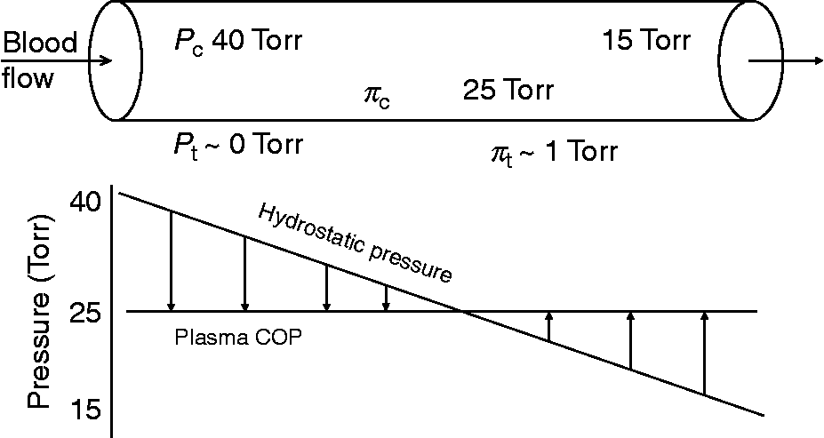
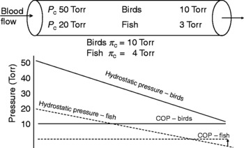
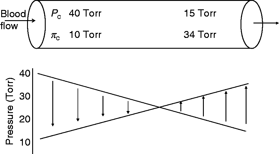
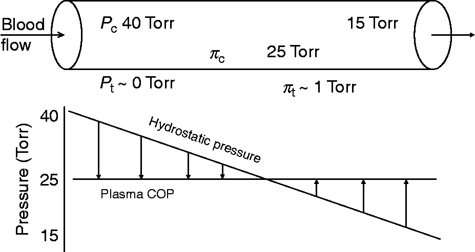
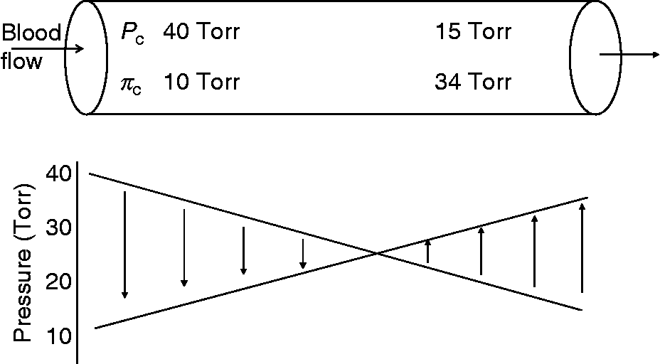
In mammals, the hydrostatic pressure at the arterial end of a capillary is 40 Torr, decreasing to 15 Torr at the venous end. The COP of plasma and ISF are 25 and 1 Torr, respectively. Thus, according to the Starling equation, the balance of forces at the arterial end of the capillary favours fluid extravasation (Fig. 1). As plasma moves along the capillary, hydrostatic pressure dissipates such that somewhere about the midpoint of the capillary the balances of forces reverse and the extravasated fluid is returned to the plasma (Fig. 1). Again, according to Starling, this reversal of force is primarily due to the influence of plasma COP. Birds have a high blood pressure relative to mammals, in some cases in excess of 200 TorrReference Schmidt-Nielsen2. Interestingly, birds have a relatively low COP (10 Torr). As blood flows along the capillary, the drop in hydrostatic pressure never reaches that of COP, and as there is no apparent reversal of pressures, the extravasated fluid cannot be recovered by means of COP (Fig. 2); therefore, another force must exist. Schmidt-NielsenReference Schmidt-Nielsen2 indicates that this is not well understood and suggests that avian capillaries must possess different properties from mammalian capillaries or birds must have a greater vascular resistance to keep capillary pressure low. He also concludes ‘that a higher resistance in arterioles seems meaningless, for why should birds then have a high arterial pressure to begin with?’Reference Schmidt-Nielsen2. This ‘problem’ with low COP is also observed in fish (Fig. 2). While lower blood pressures are typically associated with lower COP (3–8 Torr) in fish, reptiles and amphibians, this low COP probably contributes very little, if at all, to returning extravasated fluid and another force must exist. Indeed, fish capillary properties are different from mammals’ and birds’ and are highly permeable to proteins. Thus, as discussed below (see Essential role of plasma proteins section), the diffusion of protein through the membrane is the force that participates in fluid exchange, not its COP. This is further illustrated in amphibians, where the return of fluid to the vascular space is due primarily to lymphatic return and not explained by transcapillary forcesReference Hillman, Zygmunt and Baustian3.
Starling model for transcapillary fluid exchange in mammals at rest. Data are taken from Schmidt-NielsenReference Schmidt-Nielsen2
Starling model for transcapillary fluid exchange in birds and fish. Data are taken from Schmidt-NielsenReference Schmidt-Nielsen2 and OlsonReference Olson, Hoar, Randall and Farrell37

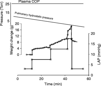
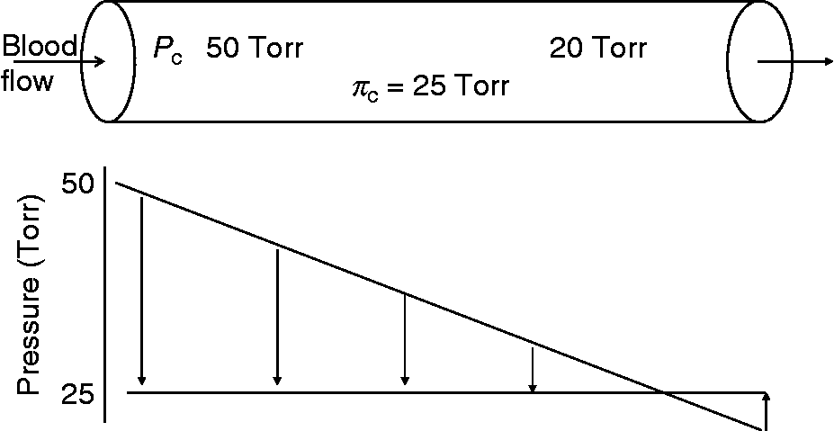
Applying Starling forces to transcapillary fluid exchange in the mammalian lung is also problematic. In the pulmonary circulation, P c is very low (~10 Torr), and it is assumed that extravasation of fluid is limited and the lung interstitial space remains ‘dry’ because plasma COP (~25 Torr) so greatly exceeds pulmonary P c. However, increases in pulmonary capillary P c (as little as 1 Torr) result in fluid filtration, despite P c being significantly less than plasma COP (Fig. 3)Reference Lunde and Waaler4. The clear conclusion is that plasma COP cannot directly oppose or, by inference, support fluid movement in and of itself. The transient filtration and attainment of a new steady state following the step change suggests that an additional force must be present, and while it does not oppose the initial extravasation following the perturbation, it can restore equilibrium at the higher P c (Fig. 3).
Starling model for transcapillary fluid exchange in mammalian lungs. The lower panel shows changes in lung weight with changes in left atrial pressure (LAP) in vitro. Data are taken from Lunde and WaalerReference Lunde and Waaler4

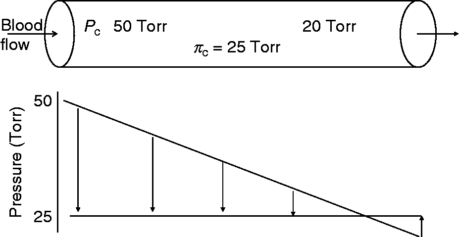
Another problem is the relative constancy in plasma COP. Increased blood pressure and local vasodilation associated with increased skeletal muscle work increases P c and favours increased plasma filtration (Fig. 4). In addition to the extravasated fluid, the increased production of cellular water associated with increased metabolic rate provides additional strain on cell volume regulation. Thus, the force responsible for moving fluid into the vascular space must address the increased water load of metabolism. Although plasma proteins have been shown to change during exercise in humansReference Sjogaard and Saltin5, Reference Mohsenin and Gonzalez6 and animalsReference Lindinger, McKeen and Ecker7, Reference Vengust, Staempfli, Viel and Heigenhauser8, the effect on plasma COP is minimal (3–5 Torr). Thus, plasma COP cannot contribute to plasma–ISF exchange more than it theoretically did in quiescence; thus, another force supporting plasma–ISF exchange must be present during periods of muscular work and increased metabolic rate.
Starling model for transcapillary fluid exchange during exercise
Another interesting conundrum regards aqueous humour dynamics. Aqueous humour is produced in the ciliary bodies and secreted into the posterior chamber of the eye, diffusing to the anterior chamber where intraocular pressure is about 20 Torr. Aqueous then exits the eye via the canal of Schlemm, which is a meshwork confluence with the episcleral veins. Interestingly, the venous pressure, venous plasma osmolality (20–30 Torr), [Na+] and [K+] are all greater than those of aqueousReference Duke-Elder9–Reference Maren11. With such differences in osmotic pressure, it was suggested that plasma water must diffuse into the eye at the venous endReference Davson and Davson12. The question then is ‘How does aqueous humour exit the anterior chamber against both the hydrostatic and osmotic gradient?’. Again, there must be another force responsible for the removal of aqueous from the eye to venous plasma.
Given these inconsistencies that question the role of proteins and their COP in fluid exchange, it appears that another physiologically relevant force must be identified to explain plasma–ISF exchange. We hypothesize that this force is an osmotic force related to the diffusion of bicarbonate ( ) and strong ions in plasma that is primarily responsible for the balancing of hydrostatic forces and sub-serving fluid movement from ISF to plasmaReference Hammel and Brechue13, Reference Brechue and Hammel14. The link between ion diffusion and plasma–ISF exchange is based on the concept of solute–solvent drag, the impact of diffusing ions on the water through which they are diffusing. Thus, the purpose of this study is to propose a conceptual framework supporting the hypothesized role of diffusing
 and strong ions as being the osmotic force for plasma–ISF exchange. For this, we must first reconsider the nature of osmosis of water.
Nature of osmosis in fluid equilibrium
The basis for the present problem is the supposition that proteins in plasma decrease the concentration of water in plasma, and that the water concentration of plasma is less than ISF water concentration. Therefore, water diffuses down its concentration gradient from ISF through the endothelium (impermeable to protein) and into plasma. On the contrary, colloids do not alter the concentration of waterReference Hammel and Scholander15 and therefore do not cause osmosis. Furthermore, one must consider the effect of solute on the concentration of water in a solution and its impact on osmosis. The basis for our hypothesis is Hulett's insight regarding the altered state of water in an aqueous solution with a free surfaceReference Hulett16, Reference Hammel17. Hulett recognized that a solute exerts a solute pressure against a distensible boundary of a solution and alters the solvent exactly like pure liquid solvent by lowering the pressure applied to it by the osmotic pressure of the solvent in solution. Thus, the chemical potential of the solvent in solution and the chemical potential of pure liquid solvent in solution are the same at the same temperature when the pressure applied to the pure liquid solvent is less by the osmotic pressure of the solvent. This then is the osmotic pressure of the solvent at a distensible boundary of the solution at the temperature and pressure applied to the solution. Solute molecules in a solution moving in random thermal motion (Brownian motion) at the boundary of the solution are reflected at that boundary, and this reflection changes their momentum. The molecules exert a force against the boundary that reflects them that is equal to the time rate of change in momentum normal to the surface of all reflected solute molecules. According to Hulett, this force per unit boundary is the internal solute pressure. In a free-standing solution, the force that opposes the solute pressure is the force that binds the water molecules together in the liquid phase. So the internal solute pressure alters the internal pressure tension in the cohesive force bonding the solvent. Let  (T,
 ,
 ,
 ) denotes the osmotic pressure of the water in the solution, where (T,
 ) are the temperature and external pressure applied to the solution, and (
 ,
 ) are the moles of solute B and water. Hulett recognized that every partial molar property of the water in the solution at (T,
 ) is exactly the same as the same molar property of pure water at (T,
 −
 ). For example, the chemical potentials of water, its molar free energy, in the solution and in pure liquid are equal when
 (T,
 ,
 ,
 ) =
 (T,
 −
 ), where the asterisk denotes pure liquid waterReference Hammel17–Reference Hammel19. Thus, solute alters the internal tension in the attractive force that bonds the water molecules in a solution with a free surface or with a distensible, impermeable boundary. Solute molecules are reflected at all boundaries; they change momentum and exert a pressure to distend a distensible boundary, thereby stretching the water in the solution. Similarly, reducing the pressure applied to pure liquid water by
 alters its internal tension by the same amount. Therefore, the chemical potential and all molar properties of the pure water at (T,
 –
 ) are now equal to the same partial molar properties of the water in the solution at (T,
 ).
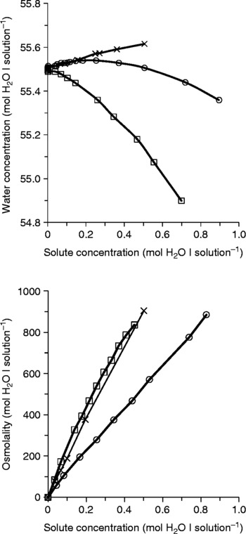
Given the above, this solvent tension rather than solvent concentration becomes the important factor in osmosisReference Mysels20, Reference Mysels21, and one must reconsider the effect of adding solute to water. The present understanding is that adding solute to water lowers the concentration of the water in the solution such that pure water diffuses from higher to lower concentration. Furthermore, this lowering of water concentration by solute causes osmosis. As shown in Fig. 5Reference Hammel18, Reference Miller and Knox22, the concentration of water decreases as small amounts of Na2SO4 are added to the water (Fig. 5, upper panel). However, the curvilinear relationship between molar concentration of water and the moles of Na2SO4 added does not correspond to the relationship between the osmolality of water and the moles of Na2SO4 added (Fig. 5, lower panel). In all cases, the osmolality of the water increases almost linearly as solute is added. For example, in NaF and MgSO4, the water concentration increases, not decreases, as these solutes are added (Fig. 5, upper panel), thus water flows out of the solution and into pure liquid water against the concentration gradient. Thus, the concentration of water does not cause or explain osmosisReference Hammel and Scholander15, Reference Hammel17–Reference Hammel19, Reference Hammel23.
Upper panel: water concentration versus molal concentration of solute at 0 °C. The concentration of pure liquid water is 55.500092 mol− 1 at 0 °C. The water concentrations of the aqueous solutions of NaF and MgSO4 increase with the addition of solute, whereas the water concentration of the Na2SO4 solution decreases. Lower panel: the osmolality of water in solutions of a 0.1 molal concentration of Na2SO4, MgSO4 and NaF is plotted as a function of solute concentration at 0 °C. Calculation of solution volume was based on the density of solutions measured by Miller and KnoxReference Mysels20, modified from HammelReference Hammel23

With respect to the present problem, the troublesome aspect of this is the idea that solute molecules lower the concentration of water in a solution at a semi-permeable membrane separating solution from pure liquid water. Frequently this happens, but this is not the reason that water moves into a solution across a membrane permeable only to water. Furthermore, this does not mean that water would diffuse out of the solution across the semi-permeable membrane into pure water on the other side, where the water concentration is less. It means that water concentration at the semi-permeable membrane does not cause osmosis, rather solute molecules lower the chemical potential of the water at the membraneReference Hammel17–Reference Hammel19, Reference Hammel23. Moreover, it means, as Hulett perceived, that the solute alters the chemical potential, vapour pressure and all other partial molar properties of the water at a free surface of the solution or at any distensible boundary of the solution. For these reasons, when the flow of plasma through a capillary is in steady state and the capillary diameter is constant (as a boundary), the colloidal proteins in the plasma and its COP can do very little to affect fluid exchange between plasma and ISF. Another force must do what has been hitherto been attributed to plasma proteins in Starling's equation. The question is ‘What force returns ISF to the plasma at the venous end of the capillary?’ To answer this cardinal question pursuant to our hypothesis, two issues must be addressed: (1) the presently proposed osmotic effect is induced by ‘solute–solvent drag’ based on Hulett's model and (2) which solutes in plasma are in highest concentrations, but more importantly, which concentrations change the most from the arterial to the venous end of the capillary. The solute(s) that yield the largest concentration difference (venous minus arterial) will be the dominant solute(s) affecting the osmotic pressure of water in the plasma flowing along the length of a capillary and will be the dominant solute(s) affecting fluid exchange.
The concept of ‘solute–solvent drag’
The osmotic effect of ‘solute–solvent drag’ is little known among physiologists. Therefore, it is appropriate to examine a few hypothetical situations to gain better insight into how it works. This force has not been recognized in the physiology of microcirculation and, to date, has not been acknowledgedReference Michel24, Reference Taylor and Moore25. The phrase ‘solute–solvent drag’ means the following: (1) solute pushes on and alters the solvent into which it is diffusing and will diffuse, (2) this altered solvent pulls on the solvent within the solution from which the solute is diffusing and (3) this pulled solvent within the solution is altered by the same amount as pure liquid solvent is altered by pulling on it an equal amount. The fact that solute drags on the water and alters the water through which it diffuses has been demonstrated experimentallyReference Hammel and Scholander15, Reference Hammel17, Reference Miller and Knox22.
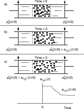
To illustrate, Fig. 6 shows how solute diffusing in water can drag on the water through which it diffuses and alter its internal tensionReference Hammel23. As shown in Fig. 6a, at time t < 0, the solute is retained in a solution confined at each end by a rigid membrane permeable only to the water and not to the solute. The solute molecules are in random, thermal motion. Some strike the membrane that reflects them, changing their momentum. The time rate of change in momentum is a thermal force acting on the membrane and the rigid membrane exerts an equal and opposing forceReference Hammel17, Reference Hammel18, Reference Mysels20, Reference Mysels21. This force exerted per unit area is a thermal pressure applied to the membrane. If the pressures  (t ≤0) applied by the pistons to the water at the two ends of the cylinder are the same, then no water moves in or out of the solution. The internal tension in the water is the same everywhere in the cylinder, i.e. it is the same in the solution as in the pure liquid water beyond the two membranes. The solute does not alter the internal tension of the water in the solution, as it would if the solution had a free surface or a distensible boundary impermeable to water. The pressure the solute exerts at its boundaries is already opposed by the two end membranes and by the wall of the cylinder. If the pressure applied by one piston exceeds the pressure applied by the other, then water will flow through the solution at a rate determined by the differing pressures and by the hydraulic conductivity (L p) of the membranes.
Thought experiment illustrating how solute induces an osmotic effect on water as it diffuses from a higher to a lower concentration in the water. The pistons are assumed to move freely through the cylinder without resistance. (a) Pressure,  (t), must be applied immediately as time = 0 to the right piston in (b) to maintain the piston above the arrow. Similarly, a pressure,
 (t), must be applied to the left piston in (c) The osmotic pressure,
 (t), changes in time as the solute concentration at the membrane decreases and as the solute concentration at the right piston increases. Note that the effect of gravity on the pressures applied to the fluids has been ignored. Modified from HammelReference Hammel23

Now at time 0, the membrane at the right end of the solution is removed, as in Fig. 6b or 6c. The solute molecules continue in thermal motion and they continue to be reflected. But now the water into which they are also diffusing (or will diffuse) reflects them. They exert a pressure (they push) on this water and may or may not alter its internal tension, depending on what pressures are applied by the left and right pistons.
When the membrane was removed at time 0 as in Fig. 6b, and as the solute molecules began to diffuse towards and were reflected by the water ahead, these solute molecules were pushing on the water ahead and into which they will diffuse. To prevent movement of either piston as in Fig. 6b, the pressure applied by the right piston to the liquid water between the piston and the solution must be increased immediately from  (t ≤0) to
 (t ≤0+
 (t>0); otherwise, the pistons would not have remained at their starting position (depicted by the arrows above the pistons). Increasing by
 (t>0), the counter-pressure applied to the right side of the pure water in the cylinder balances the pressure exerted by the diffusing solute molecules on the left side of the same water, i.e. applying an increased counter-pressure to the pure water between the right piston and the solution alters the internal tension in this pure water. This prevents the solute from altering the internal tension in the water within the solution that would have been induced by the drag of the solute molecules diffusing through it. More correctly, the pressure applied by the right piston to the pure water, which is also pushed by the solute molecules to its left, prevents it from pulling on the water in solution. Thus, there was no change in internal tension in the water within the solution, and there was no need to change the pressure applied by the left piston to the water between the piston and the remaining left membrane. The internal tension in this water remained as when the pressure applied to it remained at
 (t ≤0). Finally, the pressure applied by the right piston had to be lessened by about 1/2 the initial amount so as to retain both pistons at the stationary arrows. The graph in Fig. 6 illustrates how the osmotic effect of the solute on the solvent varies as a function of time as the solute is redistributed in Fig. 6b and 6c.
In the case shown in Fig. 6c, the pressure applied by the right piston on the water into which the solute will diffuse remains at  (t ≤0). Unless the pressure applied by the piston to the water left of the remaining membrane is decreased immediately by an amount equal to the osmotic pressure of the water in the solution in Fig. 6a, water will flow from left to right through the cylinder (not illustrated), i.e. when the pressure is decreased to
 (t ≤0-
 (t>0), the pistons remain stationary at the arrows (as illustrated). The pressure applied by the left piston alters the water left of the remaining membrane by the same magnitude as the diffusing solute alters the water in the solution near the right side of the remaining membrane. The water at the front of the diffusing solute molecules is beginning to be altered. The water into which the solute will diffuse has not yet been altered, so the only pressure applied to the water left of the right piston is
 (t ≤0). The pressure applied by the left piston must remain
 (t ≤0) −
 (t ≤0) as long as the highest solute concentration remains at the initial concentration and the lowest solute concentration in the solution remains 0, the solute concentration in pure liquid water. Moreover, this piston pressure must be applied immediately upon removal of the semi-permeable membrane, i.e. the moment there is no longer a rigid barrier to reflect the solute molecules so that they start to diffuse into the water ahead. Eventually, the solute concentration at the remaining membrane starts to decrease as the concentration at the right piston starts to increase above 0. To maintain the position of the piston (at the arrow) during this interval, the pressure applied by the left piston must gradually increase. Finally, when the solute concentration is the same throughout the solution, the piston pressure on the left must be increased to about 1 − 1/2x its initial value (not illustrated).
It is to be noted that a very important point is illustrated in Fig. 6c, which is central to our argument. During the interval between time 0 and final equilibrium, while the solute drags on the water through which it diffuses, the osmotic effect due to this drag is determined by the difference between the highest and the lowest solute concentrations. Another very important point is that this osmotic effect on the water due to solute drag is instantaneous. During the interval between time 0 and final equilibrium in Fig. 6b and 6c, the internal tension in the water in the solution differed from the internal tension in the water ahead into which the solute was diffusing and will diffuse. Finally, when the solute was uniformly distributed throughout the solution (not illustrated), the internal tension in the solution water was everywhere the same and was the same as in the water between the left membrane and the left piston in both Fig. 6b and 6c. Also, when the solute molecules are uniformly distributed throughout the solution, they exert their thermal pressure against the boundaries of the solution, namely the remaining rigid membrane and the right piston. These barriers oppose this pressure so that the internal tensions in the pure water and in the solution water are the same throughout the cylinder.
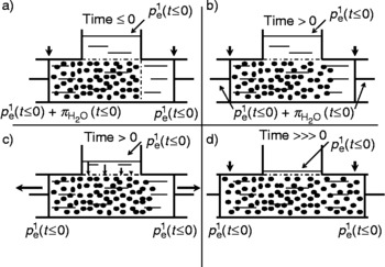
The concepts illustrated in Fig. 6 are now applied to a cylinder that is modified by placing liquid water in a reservoir above the solution and by separating it from the solution by a rigid, semi-permeable membrane as illustrated in Fig. 7Reference Hammel23. The pressure applied to this water is  (t ≤0), the same pressure as applied by the right piston to the water between the membrane and the piston. The internal tension in the pure water at the applied pressure
 (t ≤0) is also the same as the internal tension in the water in the solution (Fig. 7a). The reason for the equal internal tensions is that the thermal pressure exerted by the solute molecules in the solution is already opposed by the boundaries of the solution, namely the rigid membranes, the cylinder wall and the pressure at the left piston (
 (t ≤0)+
 (t < 0)), to maintain the piston position (at the arrow). When the right-hand membrane is removed (Fig. 7b), the solute molecules continue to push on the left piston as they did in Fig. 7a. To oppose this solute pressure, the piston continued to apply
 (t ≤0)+
 (t) to the solution. The solute molecules diffusing towards the right piston are now pushing on the water (between solution and piston) into which the solute will diffuse. To fix the right piston in position (at its arrow), the pressure it applied to the intervening water was increased immediately from
 (t ≤0) to
 (t ≤0)+
 (t ≤0). With these equal pressures applied by the left and right pistons, the internal tension of the water within the solution was unchanged and remained the same as the internal tension in the water in the reservoir at an applied pressure
 (t ≤0). For this reason, there was no flow of water across the remaining membrane.
Thought experiment illustrating how fluid in the upper chamber flows into the cylinder in response to changes in the internal tension of the water in the cylinder by diffusing molecules. This model represents how plasma–ISF exchange is caused by the osmotic effects of diffusing  from ISF into venous plasma and towards arterial plasma. Modified from HammelReference Hammel23

As shown in Fig. 7c, the right membrane is removed and the pressure applied to the right and left pistons is the same as applied to the reservoir on top ( (t ≤0)). Since the applied pressure to the pistons is not altered (as in Fig. 7b), they become distensible boundaries. The solute molecules press on the left piston, and at the same time the solute molecules diffusing into the pure water on the right press on and push the piston to the right. Thus, the intervening water in the solution becomes distended and pulls water from the reservoir. Note that the force pulling the water is not even along the length of the membrane (indicated by length of arrows in the reservoir). This is due to the fact that the solutes at the left piston are not yet diffusing to the right, and thus the water in this solution is the most distended because the solute molecules that are diffusing to the right are pressing on the water into which they will diffuse near the left piston. For the same reason, the water in the solution near the leading edge of the diffusing solute is least distended because this water is also pressed by solute molecules from the left. So, the flow of water across the reservoir membrane is greatest at the left end and least at the right end. Note that the left piston still moves to the left, because the distension of the water and its pull on the left piston remain a little less than that of the solute pressure exerted by the solutes against the piston. As this process continues (Fig. 7d; time >>>0), all the water will be removed from the reservoir and the solute molecules will be distributed homogenously throughout the solution. The solute molecules continue to exert a solute pressure that is now about 1/2 the initial
 (t ≤0). The internal tension of the water in solution is now altered by the same magnitude, as the internal tension of pure liquid is altered by reducing the pressure applied to it by ½
 (t ≤0). Hence, the pistons have shifted to the left and right of their original positions.
The thought experiments depicted in Figs 6 and 7 portray how diffusing solute can alter the water through which it diffuses giving rise to an osmotic effectReference Hammel and Scholander15, Reference Hammel18, Reference Hammel19. Figure 7 illustrates how the water altered by the diffusing solute can affect water movement across another membrane (from the reservoir) to which the solution was linked by the membrane. The most important conclusion from this model (Fig. 7c and 7d) is its direct application to the return of ISF (represented by the reservoir solution) to plasma space due to changes in the internal tension of water as ions diffuse from right to left. The remaining issue regards which ions in plasma have the appropriate venous–arterial concentration difference such that their diffusion would create the necessary osmotic force to return extravasated fluid to vascular space.
The COP of water in human plasma due to dissolved proteins is about 28 Torr (1 Torr = 133 Pa). If 1–2% of the plasma fluid is extravasated into the ISF at the arterial end of the capillary, plasma COP near the midpoint of the capillary increases by ~0.5 Torr, i.e. 1/50 of 28 Torr, an increase too small to be of importance. Changes in osmotic pressures due to changes in O2 and CO2 partial pressures at rest are, respectively, 1.3 and 3.6 TorrReference Dill, Edwards and Consolazio26. Changes in arterial and venous [Na+] in the capillary (from 152.3 to 153.1 mEq L− 1)Reference Dill, Edwards and Consolazio26 increase the osmotic pressure of the water in the plasma by 15.5 Torr. [Cl−] decreases through a–v (from 104.6 to 103.6 mEq L− 1)Reference Dill, Edwards and Consolazio26 decrease osmotic pressure of the water in the plasma by 19.3 Torr. Plasma potassium changes very little from a–v. Any increase in H+ has a negligible osmotic effect.
Plasma [ ] increases from an arterial concentration of 25.1 mEq L− 1 to a venous concentration of 27.1 mEq L− 1;Reference Dill, Edwards and Consolazio26. This 2.0 mEq L− 1 increase in a–v plasma flow amounts to an increase of 38.7 Torr in the osmotic pressure of water in plasma at 37 °C. The increase in negative charge due to the increase in
 ions is matched by an increase in positive charge due to the increase in strong ion difference, the change from 47.7 mEq L− 1 (152.3 − 104.6 mEq L− 1) to 49.5 mEq L− 1 (153.1 − 103.6 mEq L− 1), or a 1.8 mEq L− 1 increase in positive charge allowing the 2 mEq L− 1 increase in negative charge of the
 ion. Since in systemic capillaries
 and Na+ ions diffuse upstream (v–a) as Cl- ions diffuse downstream (a–v), the net diffusion of these ions amounts to a net change in osmotic effect of 34.9 Torr (38.7+15.5 − 19.3 Torr). Thus, the v–a difference in osmotic pressures obtained in human blood, as plasma flows from end to end in the capillary, causes a net increase of 34.9 Torr due to diffusion of
 , Na+ and Cl− ions. Similar conclusions can be made when comparing data from HendersonReference Henderson27, where the v–a ion differences show a net increase in osmotic pressure of 42.5 Torr. Collectively, these data, and data from more recent work in humansReference Kowalchuk, Heigenhauser, Lindinger, Obminski, Sutton and Jones28–Reference McKenna, Heigenhauser, McKelvie, MacDougall and Jones30 and animalsReference Stainsby and Eitzman31–Reference Brechue, Gropp, Ameredes, O'Drobinak, Stainsby and Harvey33, provide a calculated plasma osmotic pressure due to the
 and strong ion diffusion gradients ranging from 31 to 43 Torr.
Applying these changes in osmotic pressure due to net ion diffusion in the capillary and the model presented in Fig. 7, we propose the following model to represent Starling's second force for returning extravasated fluid to plasma. CO2 diffuses from cells through ISF and most of it is converted to  , a conversion hastened by endothelium-bound carbonic anhydrase (CA) as well as by CA in the red cells. At rest, [
 ] increases by about 8% (v–a) in human plasma, and this amounts to an increase of ~35 Torr in its osmotic pressure, as plasma leaves a systemic capillary (or a decrease of ~35 Torr as plasma leaves a pulmonary capillary). These changes in [
 ] and charge are allowed by corresponding changes in the concentration of strong ions and charge so that electroneutrality is maintained. Plasma H+,
 and strong ion concentrations are changing by association and dissociation with other plasma molecules, by exchange with ions in red cells and by exchange of fluid across the endothelium. However, during steady-state blood flow, these ions do not diffuse out of the capillary: the endothelium acts as a quasi-permeable membrane for them. Accordingly, each ion diffuses only from its highest to its lowest concentration within the capillary plasma.
 and Na+ ions diffuse upstream (v–a), whereas Cl− ions diffuse downstream (a–v) in a systemic capillary (in opposite directions in a pulmonary capillary). As these ions diffuse, they drag on the water through which they diffuse. For this reason, the net change in osmotic pressure of plasma water (~35 Torr) alters the internal tension in the attractive force bonding the water in its liquid phase as the tension in pure liquid water would be altered, where the pressure applied to it lessened by ~35 Torr. The internal tension of the plasma is altered immediately upon diffusion of the solutes and, as with hydrostatic pressure, the osmotic effects act regardless of the flow of plasma. The altered internal tension in plasma water is coupled to the ISF through the pores in the capillary endothelium and opposes the hydrostatic pressure in the plasma. Therefore, as plasma flows along the capillary, this net osmotic effect of diffusing
 and strong ions and the decreasing hydrostatic pressure are the primary determinants of exchange of fluid between plasma and ISF. The exchange rates are a function of arterial perfusion pressures and are proportional to the rates of O2 consumption and production of CO2 and obligatory metabolic water.
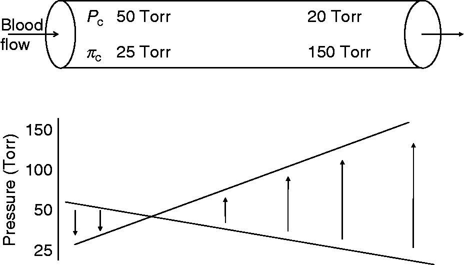
We conclude that this net osmotic force caused by ions diffusing through plasma, and not COP, is the force that explains the results obtained in Starling's classic experiment (Fig. 8). In accordance with Starling, the hydrostatic pressure is greater than the net osmotic effects of the diffusing ions and extravasates fluid into the ISF at the arterial end. At the venous end, the combined osmotic effect of  and strong ions diffusing upstream provides a force that exceeds the declining hydrostatic pressure returning extravasated ISF and moving metabolic water to the plasma by mass flow through the capillary endothelium. Moreover, at the venous end of the capillary, the pores in the capillary endothelium are more open than at the arterial end, thereby favouring the return of ISF to the plasma.
Theoretical model for transcapillary fluid exchange with Starling's second term, COP, replaced by the net osmotic pressure due to diffusing ions. Calculated osmotic pressures based on arterial and venous concentrations of  and strong ions from a variety of animalsReference Dill, Edwards and Consolazio26, Reference Henderson27, Reference Kowalchuk, Heigenhauser, Lindinger, Obminski, Sutton and Jones28–Reference Brechue, Gropp, Ameredes, O'Drobinak, Stainsby and Harvey33
Given tissue heat exchange, the effect of temperature on this net osmotic force must also be considered. Increasing the temperature of all solutes in a solution will increase the solvent's osmotic pressure and will also affect the drag of the solute on the solvent when the solutes are diffusing through the solvent. The heated end of an aqueous solution had fewer solutes per unit volume than at the cooler end, i.e. the osmotic pressure of the water in the solution from the heated end is less than the osmotic pressure of the water in the solution from the cooled end when both solutions are measured at the same temperature, the Soret effectReference Tyrrel34. To attain this Soret distribution in equilibrium, the solute had to diffuse from the warmer end of the solution to the cooler end. Depending upon where the plasma is heated as it traverses the capillary, there are two ways that thermal effects can alter fluid exchange between plasma and ISF. First, if the plasma is heated in the tissue before it enters the arterial end of the capillary, then all solutes will diffuse upstream through the plasma towards the cooler plasma upstream. These solutes drag on the plasma through which they diffuse and alter the internal tension of the water in the plasma by an amount equal to the difference between osmotic pressure of the warmer and cooler plasma. For example, the osmotic pressure of water in plasma is 150 mosm kg− 1 water at body temperature. If metabolically active tissue warms the plasma by 1 °C, the increased solute drag and the increased osmotic pressure of the plasma water is 9.4 Torr per °C. This Soret effect will both lessen extravasation of fluid from plasma and further increase the return of ISF to the plasma, as this warmed blood flows through the capillary without further warming.
The second possible thermal effect on the osmotic effects of diffusing  and the strong ions within plasma is dictated by further warming of the plasma as it flows a–v in the capillary. For example, increasing the temperature of the plasma by 1 °C as the plasma flows from end to end in the capillary increases the osmotic pressure of each ion by the factor 311/310. Applying this factor to the osmotic pressures of
 , Na+ and Cl− ions increases the amount of drag on the water through which they diffuse. Using concentrations of these ions from Dill et al. Reference Dill, Edwards and Consolazio26, the net osmotic effect of
 and the strong ions will increase from 38.7 to 52.5 Torr if the temperature of the plasma increases by 1 °C as plasma flows from one end of a capillary to the other. This thermal effect would lessen extravasation at the arterial end and enhance return of ISF at the venous end. In this case, the thermally enhanced return is greater than the thermally diminished extravasation, because the diffusion effect of metabolic
 is greatest at the venous end and least at the arterial end. On the other hand, if the plasma is already heated as it enters the tissue capillaries and if no further heating occurs as the plasma flows through the capillary, then there will be no further thermal enhancement of the diffusing ions and no increase in their osmotic effects. In highly active muscle (during muscle contraction), these two thermal effects may become important in altering plasma–ISF exchange within the capillary and the net effect will depend on where the plasma is heated as it enters the tissue and flows through the capillaries.
Altogether, there are sufficient osmotic effects from solutes diffusing upstream (or downstream) within the plasma to account for the plasma–ISF exchange (Fig. 8). These osmotic effects are also sufficient to account for negative ISF pressuresReference Mohsenin and Gonzalez6, Reference Guyton35. Moreover, unlike the Starling colloid force, these osmotic effects are load dependent, i.e. the rate of fluid exchange between plasma and ISF depends on the rate of CO2 and heat generated by the tissue. This relationship is important because metabolic rates vary among tissues and in some tissues can increase up to a factor of 80 from rest to maximal metabolic activity (discussed below). In all cases, plasma–ISF exchange remains relative to metabolic activity, thereby avoiding another problem inherent in the Starling hypothesis that utilizes a relatively constant colloid osmotic force to explain fluid exchange.
Essential role of plasma proteins
Although the plasma proteins cannot function as in Starling's second force, as hypothesized, they are nevertheless essential. The capillary endothelium and the surrounding tissue are not rigid. By their own random motion, the colloidal proteins exert a continuous pressure against the capillary wall and distend the wall in proportion to the protein concentration and its colloidal pressure. Therefore, capillary volume and total blood volume are a function of protein concentration. The relationship between protein concentration and the distension of the compliant capillary endothelium is important in regard to intermittent blood flow through the capillaries, a normal feature of the microcirculation, where proteins may contribute to plasma–ISF exchange under the appropriate circumstances. Following an interval of no blood flow, the pre-capillary sphincter relaxes, the hydrostatic pressure increases and blood flow is re-established. In addition, the high hydrostatic pressure at the arterial end of the capillary has two effects: (1) since it exceeds the hydrostatic pressure of the ISF, it forces a protein-free filtrate from the plasma in to the interstitial space and (2) further increases the distension of the capillary wall already distended by the colloid pressure exerted by plasma proteins. Note that the extent of the distension from the combined hydrostatic and colloid pressure is a function of the elasticity of the capillary endothelium and surrounding tissue. If these were not compliant, there would be no retention of plasma and no expansion of the capillary volume. The stretching and distension of the capillary wall increases the capillary surface area (S) and lowers the hydraulic conductivity (L p). Thus, when blood flow commences in a capillary, all plasma entering the capillary will flow through it; a small fraction will filter into the ISF and a fraction will be retained, expanding the capillary volume. The extent of the added distension is attributable to the increase in hydrostatic pressure at the arterial end of the capillary, which will depend on the distension already produced by the colloid pressure of the proteins. For a given increase in hydrostatic pressure, the distension that it causes and the amount of fluid extravasated will be less if the colloid pressure is already high. On the other hand, the distension and fluid extravasation caused by a given increase in hydrostatic pressure will be greater if the colloid pressure is low. Therefore, a normal concentration of protein and COP is essential for maintaining a normal fraction of protein-free filtrate from the capillary onto the ISF.
Once the capillary is fully distended, as the steady-state flow is attained, the plasma proteins play only a minor role in plasma–ISF exchange. Any extravasation of fluid into the ISF at the arterial end of the capillary concentrates the plasma proteins. These proteins can now diffuse down the concentration gradient and contribute an osmotic effect to plasma–ISF exchange by their longitudinal diffusion; however, the v–a concentration difference for proteins is small ( < 0.2 mg dl− 1), and therefore, it also contributes very little to the total osmotic effect ( < 0.5 Torr). Plasma proteins may accumulate over sites in the capillary endothelium where hydrostatic pressure forces plasma fluid into the interstitial space, e.g. over small openings in the junctional strand beneath the glycocalyx on the luminal side of the endotheliumReference Curry, Hu, Adamson, Liu and Weinbaum36. These concentrated proteins will diffuse away towards a lower concentration within the plasma and have a small osmotic effect to lessen extravasationReference Curry, Hu, Adamson, Liu and Weinbaum36. Moreover, at similar sites, at the venous end of the capillary where ISF returns to the plasma, plasma proteins will diffuse into this returning fluid and lessen the rate at which it enters the capillary. This latter osmotic effect of these diffusing plasma proteins is opposite to the return of ISF hypothesized by Starling's equation ( ). In actuality, this osmotic effect of plasma proteins at the venous end cancels the osmotic effect of protein molecules diffusing upstream within the capillary. Whenever proteins are diffusing along a concentration gradient, they can exert a force on fluid by dragging on the fluid through which they are diffusing. This is exhibited in fish, in which the capillary permeability to protein and the ratio of interstitial to plasma protein are highReference Olson, Hoar, Randall and Farrell37, and the return of extravasated fluid in fish can be accomplished by diffusing protein.
Thus, with steady-state flow or during intermittent flow, these contributions of protein and COP are very small compared with the role of the  ions. Note that when the flow rate of blood through a capillary is constant, the plasma proteins cannot influence plasma–ISF exchange significantly. If their 28 Torr were to have a steady-state influence, as stated by the Starling equation, this COP would be combined with the inevitable osmotic effects of the diffusing
 and strong ions. These combined osmotic effects would greatly exceed the hydrostatic pressure even at the arterial end of the capillary and prevent any extravasation of fluid into the ISF in tissue at rest and, perhaps also, during periods of increased metabolic flux in active tissue, i.e. skeletal muscle.
There are additional circumstances under which proteins do have a significant effect on fluid volume. When the COP of plasma is increased by the addition of albumin or other colloids to plasma, the capillary and total blood volumes expand. For example, increases in plasma volume associated with acute bouts of exercise have been related to increased albumin in the vascular spaceReference Gillen, Lee, Mack, Tomaselli, Nishiyasu and Nadel38, Reference Mack, Yang, Hargens, Nagashima and Haskell39. Again, this result happens only when plasma flow is intermittent and is associated with the increasing hydrostatic pressure following the relaxation of the arteriolar sphincter. For this reason, fluid is extravasated into the ISF along the entire length of the capillary. However, since the COP of the plasma has been elevated and these added colloids are also reflected by the endothelium, they press more against the endothelium. This greater pressure causes more outward displacement of the capillary wall, lessening the extravasation of fluid and therefore increasing retention of fluid and causing a greater expansion of plasma volume. Similarly, when plasma protein concentration and COP are insufficient, as in protein-deficient diseases, there is less reflection force on the capillary wall and extravasation is excessive, resulting in severe oedema and bloated spaces between tissues, as in kwashiorkor. When injecting an isotonic saline solution with a high protein concentration (COP) into the peritoneal space, water from the surrounding ISF flows into the space. The boundary of the injected solution containing the colloid is distensible. The colloid molecules reflect from this boundary and exert a pressure, equal to the COP of the injected solution. The boundary of the solution is pressed to enlarge, and this alters the internal tension of the water in the solution, as would the internal tension of pure water be altered by reducing the pressure applied to it by COP. Water from the ISF is pulled into the space until the COP of the solution equates with the surrounding ISF. Thus, it is clear from this discussion that our hypothesis does not necessarily preclude a role for the colloidal proteins in plasma–ISF exchange, but their role is clarified and limited to appropriate circumstances, intermittent flow and alteration in plasma protein concentration.
Linking HCO−3 and strong ion diffusion to fluid movement in physiological systems
While the primary focus of this discussion has been the role of diffusing  and strong ions in returning extravasated fluid to the vascular space, the role of
 -associated fluid movement (secretion and reabsorption) across a variety of biological systems and animal species has been established and will be briefly reviewed in support of our hypothesis. At the heart of this proposed role for
 is its direct link to CO2 chemistry and therefore involvement of the enzyme CA.
 is produced by the uncatalysed hydration (Equation 1) or hydroxylation (Equation 2) of CO2 characterized by individual rate constants that are independent (k 1) and dependent (k 2) of H+, as summarized belowReference Maren40:

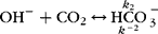
CA is a zinc-containing enzyme that, at the typical in vivo concentration, enhances the reversible rate of CO2 hydration by some 2800-fold (Equation 3). The basis for this catalytic augmentation is the protolysis of water and the extreme concentration of OH− at the zinc centre at the active site. H+ are shuttled away by a series of amino acids near the active site.
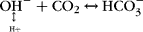
CO2– chemistry and the enzyme CA are ubiquitous in biological systems. CO2 conversion to
 in red blood cells, leaf and tissue fluid of the lens and rectal gland sub-serves CO2 transport and storage in blood plasma, muscle, plants, lens and the rectal gland of fish. Production and secretion of
 are linked to fluid secretion in the ciliary bodies (aqueous humour), choroid plexus (cerebrospinal fluid (CSF)), pancreas (pancreatic juice) and sweat and salivary glands. In contrast, the production and retention of
 in the cell lead to H+ secretion in renal tubules, stomach, gill and osteoclast. The hydroxylation of CO2 with 2 OH− results in the secretion of carbonate (
 ) in shell, coral, alkaline gland and mosquito larva.
While both the uncatalysed and catalysed reactions confer activity for the inter-conversion of CO2 and  , the cellular localization of the isoenzymic forms of CA determines the specific function (e.g. secretion, reabsorption, etc.) of
 . While there are at least 14 isoenzymes of CA with various species and tissue localization, only two isoforms will be considered in this study as they are of primary importance in supporting the present purpose. CA is found in the cytosol (CA-II) and bound to membrane (CA-IV); CA-IV exhibits significantly greater hydration–dehydration activity. Determination and significance of the physiological role of CA and pharmacological and clinical application have been bestowed by inhibition of CA activity. Sulfonamide inhibitors of CA have varying activity against the two forms of the enzyme, having some 20-fold less activity against CA-IV. Theoretical calculations and experimental results reveal that greater than 99% inhibition of the enzyme is required for a physiological effect, i.e. reduced fluid secretionReference Maren10, Reference Maren, Botre, Gros and Storey41, Reference Brechue42. Within the present context, membrane-bound CA-IV appears to play the primary role in the formation and secretion of
 from the cellReference Maren40, Reference Tinker, Coulson and Weiner43–Reference Ridderstralle, Wistrand and Brechue46 and facilitates the diffusion of CO2 across membranesReference Gros, Moll, Hoppe and Gros47, Reference Gutknecth, Bisson and Tosteson48. Similarly, the uncatalysed hydroxylation reaction appears to be physiologically significant at the secretory borders of cellsReference Maren40. The cytosolic form, CA-II, is responsible for rapid cellular equilibrium of CO2 and H2O. The choroid plexus is the only tissue where this relationship does not hold true, as there is no known membrane-bound CA. Support for this differential function based on isoenzymic localization is concluded from several physiological experiments. In the eye, aqueous humour secretion returns to baseline levels while CA-II (cytosolic form) is still 99.9% inhibited and the membrane-bound form CA-IV is below physiological inhibitionReference Brechue42, Reference Brechue and Maren45. Likewise in the kidney, the renal response to CA inhibition (increased Na+,
 and water excretion) is similar in control animals and animals in which only membrane-bound CA-IV has been selectively inhibitedReference Tinker, Coulson and Weiner43 or animals with intact membrane-bound CA-IV yet which are completely devoid of cytosolic CA-II (through site-directed mutagenesis)Reference Brechue, Kinne-Saffran, Kinne and Maren44.
A. Fluid secretion in secretory epithelia
The importance of  in fluid movement is further illustrated in the mechanism of fluid secretion among secretory epithelia. Na+ flux and fluid secretion are intricately linked to
 formation, either CA dependent or CA independent (hydroxylation of CO2 or ionization of carbonic acid H2CO3). Slowing the rate of
 formation by inhibiting CA results in decreased aqueous humour (30–40%)Reference Maren11, Reference Brechue42, Reference Brechue and Maren45, Reference Maren, Haywood, Chapman and Zimmerman49, Reference Brechue and Maren50, CSF (40–50%)Reference Maren, Botre, Gros and Storey41, Reference Vogh and Maren51, Reference Vogh, Godman and Maren52 or pancreatic juice (42–47%)Reference Rawls, Wistrand and Maren53 secretion. Concomitantly, Na+ flux is reduced by 25–30% in aqueous humourReference Maren11, Reference Maren40 and ~45% in CSFReference Vogh and Maren51, Reference Vogh, Godman and Maren52 after inhibition of CA. In each case, residual flow is related to CA-independent
 formation and Na+ flux. Blocking the non-catalysed formation of
 using acid salts of aluminium (AlCl3, Al2(SO4)3 or gallium (GaCl3)) reduces CSF secretion by an additional 33%Reference Vogh, Godman and Maren52. Hence, the tight linkage of fluid secretion to
 formation as
 accounts for ~80% of CSF secretion, while only ~20% of secretion is specifically linked to Na+ movement. Aqueous humour secretion is also be inhibited by adrenergic blockade, specifically β-blockers. The mechanism of action is also a reduction in
 flux that reduces aqueous flow by 30–40%Reference Brechue42, Reference Gregory54, Reference Brechue, Krumin and Godman55 but is dissociated from changes in Na+ fluxReference Maren, Botre, Gros and Storey41, Reference Maren and Vogh56, i.e. β-blockers do not alter Na+ flux into aqueous humour, yet secretion is significantly reduced. Fluid secretion in other tissues such as sweat gland, salivary gland, large intestine, etc. is also linked to
 secretionReference Maren10, Reference Maren40, Reference Maren, Tashian and Hewitt-Emmett57. The mechanism of using aluminium salts to inhibit
 formation is widely known for its effects on inhibiting sweat formation and secretion and is the basis for the function of antiperspirantsReference Maren40.
An interesting aspect of the proposed osmotic effect of diffusing  and Na+ would be the physiological relevance of the formation of ion pairs and anion–cation transport in fluid movement and/or cell volume regulation. Given the apparent importance of
 and Na+ in water diffusion and fluid secretion, it is interesting to note that these ions do not generally associate in solution. Within the high dielectric constant of water (ɛ = 78),
 and Na+ are only about 8% associated with a NaHCO3 flux rate that is quite low (0.07 × 10¹⁰ mol cm− 2 s− 1)Reference Conroy and Maren58. This rate of diffusion would preclude any physiological significance of NaHCO3 flux. However, when the low dielectric environment of the in vivo membrane region (ɛ = 6) is simulated with dioxane-impregnated cellulose, the association of
 and Na+ increases dramatically (98% associated)Reference Conroy and Maren58. The impact of this increased association is a 60-fold increase in the rate of NaHCO3 flux (4.3 × 1010 mol cm− 2 s− 1), a rate that approaches that of CO2. In contrast, NaCl has a low flux rate in water (0.03 × 10¹⁰ mol cm− 2 s− 1) compared with a lower dielectric constant environment (1.8 × 10¹⁰ mol cm− 2 s− 1), which is significantly lower than that achieved by the NaHCO3 pairReference Conroy and Maren58. As we hypothesized above, CO2 diffuses through the interstitial space and would be converted to
 at the membrane. It associates with Na+ in the membrane given the favourable lowering of dielectric constant in the vicinity of the cell membrane, which removes the water molecules from hydration and would facilitate the diffusion of
 across the membrane. After diffusing through the membrane, the ion pair would dissociate, rehydrate and diffuse along their physiological gradients. Ultimately, the concept of anion–cation transport and the role that ion pairs may play in determining the net osmotic effect of diffusing
 and Na+ are supported by:
(1)
 movement drives passive Na+ movementReference Duhm, Becker, Staub and Bolis59, an effect observed when the reduction in secretion by inhibition of carbonic anhydrase (CAI) results in decreased Na+ and fluid secretion in aqueous humour and CSFReference Maren11, Reference Maren40, Reference Vogh and Maren51, Reference Vogh, Godman and Maren52;
(2) the importance of the conversion of CO2 to
by the membrane-bound CA-IV isoenzyme serving effectively to ‘trap’ the CO2 and facilitate gradients, in apparent agreement with physiological data regarding the function of membrane-bound CA-IV in fluid movement/secretionReference Maren40, Reference Tinker, Coulson and Weiner43–Reference Ridderstralle, Wistrand and Brechue46;(3) the apparently limited role of NaCl in fluid secretion, accounting for ~20% of total aqueous humour and CSF secretionReference Maren11, Reference Vogh and Maren51, Reference Vogh, Godman and Maren52;
(4) while the divalent carbonate anion (
) contained within may actually be the anion driving this transportReference Weith60, it ultimately facilitates the transport of , which would affect fluid exchange as predicted by our proposed net osmotic effect model.
 movement drives passive Na+ movement
 secretion by inhibition of carbonic anhydrase (CAI) results in decreased Na+ and fluid secretion in aqueous humour and CSF
 by the membrane-bound CA-IV isoenzyme serving effectively to ‘trap’ the CO2 and facilitate
 gradients, in apparent agreement with physiological data regarding the function of membrane-bound CA-IV in fluid movement/secretion
 ) contained within
 may actually be the anion driving this transport
 , which would affect fluid exchange as predicted by our proposed net osmotic effect model.
Lastly, the role of anion–cation transport and ion pairs has received increasing attention in relation to Na– cotransport in renal basolateral membranesReference Soleimani and Aronson61 and thick ascending limb of the Loop of HenleReference Krapf62, gastric oxyntic cellsReference Curci, Debellis and Fromter63, corneal epithelial cellsReference Jentsch, Keller, Koch and Wiederholt64 and pH regulationReference Boron65. Thus, ion pairs would appear to be an important aspect of our hypothesized model.
Clearly, fluid secretion and movement is directly linked to the formation of  . However, the transport of
 becomes a consideration as it is a charged, membrane-impermeant species. With respect to the present discussion, two important issues emerge. First, given the relatively high membrane permeability of CO2, the cellular localization of CA and conversion of CO2 to
 would appear problematic, slowing the movement of CO2 out of the cell. Secondly, and important to the present hypothesis, any direct osmotic effect of CO2 could be minimized by counter-diffusion of O2 in certain settings (discussed above and in Section B below); thus
 is the important moiety. Specific transmembrane transport of
 is facilitated by a series of integral membrane transport proteins linked to Na+ Reference Gross, Pushkin, Abuladze, Fedotoff and Kurtz66–Reference Loiselle, Morgan, Alvarez and Cadey68, Cl− Reference Vince and Reithmeier69, Reference Sterling, Reithmeier and Casey70 and anions in generalReference Mount and Romero71. The electrogenicity and stoichiometry of these transporters is dependent upon the specific tissue and cellular localization. The flux of
 is enhanced by the formation of transport metabolon complexes and the specific binding of CA to
 transport proteins that maximizes
 movement. Metabolon complexes with cellular CA-II and membrane-bound CA-IV have been demonstratedReference Vince and Reithmeier69, Reference Sterling, Reithmeier and Casey70, and disruption of the complexes reduces
 transport independent of CA activityReference Alvarez, Vilas and Casey72. Metabolon complexes have been shown to be operative in proximal tubule
 reabsorption, pancreatic juice secretion and red blood cell and myocardial cell acid–base regulationReference Alvarez, Loiselle, Supuran, Schwartz and Casey67, Reference Alvarez, Vilas and Casey72. Thus, the ultimate transport of
 and its cardinal role in fluid movement are driven by the formation (and perhaps removal) of
 , the rate of which is dictated by CA and directly linked to
 transport proteins. The linkage of CA and
 transporters appears to be a general phenomenon in
 chemistry and transport.
Muscular work
The energetic demand of resting tissue is met by oxidative metabolism (oxygen uptake;  ). CO2 is produced during the consumption of oxygen at a rate that is determined by the substrate metabolized (mixture of fat, glucose and protein). For instance, oxidation of 1 mol of glucose requires 6 mol of O2 and produces 6 mol of CO2. For fat, the ratio is 0.7. Oxidative metabolism also results in the production of water (0.25 ml l− 1 O2 consumed). Both CO2 and water (extravasated and metabolic) must be removed to maintain tissue fluid volume and acid–base status; the removal of both is linked in the present model.
When the respiratory quotient is < 1, fewer molecules of CO2 (dissolved and as  ) are removed from the cells than molecules of O2 delivered to the cells. This would mean that that CO2 and
 as such cannot function in removing water from cells, i.e. the osmotic effect of the O2 difference across the ISF exceeds the opposite osmotic effect of CO2 and
 differences. Since O2 molecules are combined with haemoglobin and sequestered within the red cells,
 ions can function in returning ISF to plasma. Removing most of the O2 molecules from the plasma minimizes its osmotic effect in the plasma to near 0 as blood flows from end to end in a capillary.
Carbonic anhydrase inhibitor (CAI), with the sulfonamide acetazolamide, decreases CO2 output in humans without a change in  Reference Brechue and Stager73–Reference Kowalchuk, Heigenhauser, Sutton and Jones75. The diuretic effect of CAI results in reduction of total body water (1.7 l) and extracellular fluid (ECF; 3.3 L)Reference Brechue, Stager and Lukaski74. The loss of ECF is partitioned between reductions in ISF (2.98 l or 27%) and plasma volume (0.32 l or 8.8%), while increasing intracellular volume (1.6 l). Given the proposed model linking
 diffusion within plasma with flux of ISF to plasma, increasing tissue CO2 and slowing of the conversion of CO2 to
 lessen
 movement from the cell, resulting in accumulated intracellular waterReference Brechue, Stager and Lukaski74. Furthermore, in the context of the present model, the effects of CA inhibition on cell water accumulation are self-limiting. Re-establishing CO2 gradients, despite continued CA inhibition (albeit at a higher plasma and tissue CO2 tension), restores baseline rate of CO2 removal and fluid flux. Thus, ISF would decrease with increased CO2/
 gradients in plasma and the initial accumulation of intracellular water is maintained. Hypercapnia, independent of CA inhibition, similarly increases tissue water as
 accumulates in the interstitial spaceReference Dell, Lee and Winters76.
Intense exercise can increase whole-body  and CO2 production 10- to 15-fold in untrained humans, ~25-fold in highly trained humansReference Astrand, Rodahl, Dahl and Stromme77 and ~60-fold in various animal speciesReference Taylor, Maloiy, Weibel, Langman, Kamau and Seeherman78–Reference Kearns, McKeever, John-Adler, Abe and Brechue80. Intense stimulation (in vivo or isolated muscle) and muscle contractions increase metabolic rate across skeletal muscle as much as 80-fold in a variety of animal models and fibre typesReference Brechue, Gropp, Ameredes, O'Drobinak, Stainsby and Harvey33, Reference Dobson, Parkhouse, Weber, Stuttard, Harman and Snow81–Reference Brechue, Barclay, O'Drobinak and Stainsby83. Muscular work is associated with increased cell water production, incrementing with oxidative metabolism plus release of water from the hydrolysis of glycogen (2.7 g water per g glycogen). Despite these large changes in metabolic water production and increased extravasated water, the change in ISF in humans and animals is relatively small (3–6%) while the change in ECF appears slightly larger (10–20%).
During whole-body exercise in humansReference Sjogaard and Saltin5 and animalsReference Lindinger, McKeen and Ecker7, extracellular volume decreases primarily due to decreases in plasma volume ranging from 10 to 18% and dependent upon exercise intensityReference Sjogaard and Saltin5, Reference Lindinger, McKeen and Ecker7, Reference Brechue and Stager73, Reference Lundvall, Mellander, Westling and White84–Reference McKeever, Hinchcliff, Reed and Robertson87. Intracellular fluid does not appear to changeReference Sjogaard and Saltin5, Reference Lindinger, McKeen and Ecker7. ISF increases as indicated by the 3–6% increase in tissue volumes in humansReference Sjogaard and Saltin5, Reference Lundvall, Mellander, Westling and White84 and animal modelsReference Kjellmer88–Reference Drahota90 without changes in ICFReference Lindinger, McKeen and Ecker7; although in instances of exercise, thermal or diuretic dehydration, ICF can decrease to sustain blood volumeReference Lundvall, Mellander, Westling and White84, Reference Nose, Mack, Shi and Nadel91, Reference Lindinger, Heigenhauser, McKelvie and Jones92. In animals, increased tissue volume is associated with fibre type, increasing with the percentage of slow-twitch fibresReference Sreter and Woo93. Supporting for tissue volume changes is reported by increased tissue pressure in human (2.5 cmH2O)Reference Mohsenin and Gonzalez6 and cat muscle (6 cmH2O)Reference Kjellmer89. Consequently, the changes in ISF and plasma volume have been attributed to increases in both P c and tissue osmolality, which limits the return of extravasated fluid to the plasma space, although the latter mechanism is questioned here. Additionally, ISF increases due to increased metabolic water associated with increased metabolic rate.
There are changes in the Starling forces during muscular work. P c increases with muscular work due to increased mean arterial pressure and/or capillary recruitment; the intensity-dependent magnitude ranges from 40 to 80 Torr in humansReference Folkow, Haglund, Jodal and Lundgren94 and 10 to 18 Torr in isolated muscle modelsReference Kjellmer88, Reference Lundvall95, Reference Jacobsson and Kjellmer96. Capillary permeability is believed to change during muscular work in humansReference Senay97 and only slightly in animal modelsReference Artuson and Kjellmer98. As indicated in the Introduction, it is not likely that a constant osmotic force due to COP can explain fluid flux during muscular work. However, what fluid is moved to the plasma has been attributed to the increase in [protein] observed during exerciseReference Sjogaard and Saltin5–Reference Lindinger, McKeen and Ecker7, Reference Brechue and Stager73, Reference Greenleaf, Van Beaumont, Brock, Morse and Mangseth85, Reference Miles, Sawka, Glaser and Petrofsky86. [Protein] increases about 1 g dl− 1 during exercise, accounting for a change in COP of ~5 Torr (calculated and observed)Reference Mohsenin and Gonzalez6. While [protein] and COP increase with the intensity of muscular work, the magnitude of change appears minimal in comparison with both the increased volume of extravasated and obligatory metabolic water. Examination of  and strong ion changes associated with muscular work leads to a very different conclusion.
During intense muscular work in humans, the v–a differences in  (5-6 × rest levels), Na+(4-6 × rest levels) and Cl− (2-3 × rest levels) increase significantly compared with rest levelsReference Sjogaard and Saltin5, Reference Kowalchuk, Heigenhauser, Lindinger, Obminski, Sutton and Jones28, Reference Kowalchuk, Heigenhauser, Lindinger, Sutton and Jones29 and in one case
 and Na+ changed ten fold from restReference McKenna, Heigenhauser, McKelvie, MacDougall and Jones30. With muscular work, changes in lactic acid (L−) become a significant contributor to the total strong ion osmotic effect, which is not a significant contributor in quiescence. L− output increases with exercise intensity and, given the v–a gradient would diffuse upstream in plasma, the same as
 and Na+. Increased tissue L− levels have been implicated in changing tissue osmolality and contributing to the accumulation of ISF, which is unlikely under the present model. The increased production of L− certainly increases its concentration in the cell. The three to five fold increase in v–a difference indicates that L− is diffusing through the ISF and into plasma. Despite the increase in intracellular and interstitial osmolality, the greatest proportion of water accumulation is in the interstitial space, where [L−] is relatively low. Furthermore, the minimal change, if any, in ICF is associated with the highest [L−]. Given the L− and net osmotic gradient for diffusing ions, ISF water is being dragged into plasma.
Ion exchange across non-working muscle during exercise presents a slightly different picture. ICF and ISF of non-working muscle decrease during exercise, purportedly to redistribute ECF and limit losses in plasma volumeReference Sjogaard and Saltin5, Reference Lundvall, Mellander, Westling and White84, Reference Lindinger, Heigenhauser, McKelvie and Jones92. Unlike contracting muscle, Na+ is taken up by non-working muscle during exercise and the gradient (~4 mEq L− 1) is a–vReference Kowalchuk, Heigenhauser, Lindinger, Obminski, Sutton and Jones28, Reference Jacobsson and Kjellmer96. This Na+ would translate into an individual osmotic force of ~80 Torr, favouring tissue water uptake. In addition, non-working muscle serves as a sink for L−, so the uptake of lactate would again favour water uptake. What is most interesting is the four-fold increase (over rest) in v–a  difference across non-working muscleReference Kowalchuk, Heigenhauser, Lindinger, Obminski, Sutton and Jones28, Reference Jacobsson and Kjellmer96. While this
 would serve to restore plasma levels lost to buffering L− Reference Kowalchuk, Heigenhauser, Lindinger, Obminski, Sutton and Jones28, an additional function of this v–a gradient would be to provide the osmotic gradient that explains water efflux from tissue to plasma.
Experiments performed in isolated hindlimb muscle show very much the same  and strong ion response to whole- animal experiments. In experiments conducted in dog gastrocnemius muscle isolated in situ with an intact circulation, there was a net release of
 (3-4 × rest levels), Na+(3-4 × rest levels) and L− (2-3 × rest levels) from contracting muscle at peak
 , the magnitude of which was related to CO2 outputReference Stainsby and Eitzman31–Reference Brechue, Gropp, Ameredes, O'Drobinak, Stainsby and Harvey33, Reference Brechue and Stainsby99. K+ output is very small and insignificant. There is a large net uptake of Cl− by the contracting muscle that is in proportion to
 Reference Stainsby and Eitzman31–Reference Brechue, Gropp, Ameredes, O'Drobinak, Stainsby and Harvey33, Reference Brechue and Stainsby99. CO2 is converted to
 in the cell and diffuses from the cell; hence it appears that
 and Cl− exchange at the sarcolemma to maintain electrical neutrality.
The integral role of  in fluid movement is supported by results of experiments where CA is inhibited. In isolated dog muscle, CAI reduced CO2 output and v–a
 , L− and Cl− without changing
 , compared with controlsReference Beekely and Brechue100. Na+ and K+ output were not altered by CAI. There was a slight increase in tissue volume (2%), not observed in controls, following 30 min, and repetitive contractions were associated with the impaired CO2–
 movement. Similar results were observed in exercising humans, but tissue water compartments were not specifically studied. CAI reduced CO2 output without a change in
 75. The loss of plasma volume was similar with CAI (13.9 ± 1.4%) and controls (11.6 ± 0.6%). While CAI reduced CO2 output and v–a
 , L− and Cl− differences, Na+ and K+ output from the muscle remained the same as controls.
Taken together, these experiments reveal a strong relationship between  diffusion and water flux in contracting and non-working skeletal muscle. Surprisingly, v–a ion differences across muscle in exercising humans and animals and contracting isolated skeletal muscle are remarkably similar. On average, the maximal v–a
 , (~6 mEg l− 1), Na+(~6 mEg l− 1) and L− (~4 mEg l− 1) would provide an upstream gradient for ion diffusion in plasma, while v–a Cl− differences (~8 mEg l− 1) would provide a downstream gradient. The net osmotic effect of these ions diffusing in plasma would render a maximal osmotic pressure equivalent to 150 Torr. In all cases, the magnitude of the v–a differences is relative to the intensity of the work, so ~150 Torr represents a maximal response to intense work and maximal metabolic rate. In the extreme case with ten fold changes in v–a
 and Na+ differencesReference McKenna, Heigenhauser, McKelvie, MacDougall and Jones30, the calculated osmotic impact may reach 500 Torr. Inhibition of CA would reduce the net osmotic effect of diffusing
 and strong ions by ~40–50 Torr and would appear to explain the changes in body water compartments.
In conclusion, the changes in  and strong ions associated with whole-body exercise or across active skeletal muscle (in vivo or isolated) clearly support the concept that these diffusing ions are of the magnitude and direction to contribute a significant osmotic force that accounts for movement of fluid from the intracellular and interstitial spaces to plasma. The relationship of the v–a change in
 and strong ions is the same as observed in resting tissue. However, it is important to note the production of CO2, conversion to and the concentration of
 , and strong ions in plasma increase with the intensity of work. Thus, the net osmotic effect of diffusing
 and strong ions is not constant, but matches increasing demand for water movement. The role of
 is further supported by impairing the inter-conversion of CO2 to
 by CAI, which reduces the diffusion of
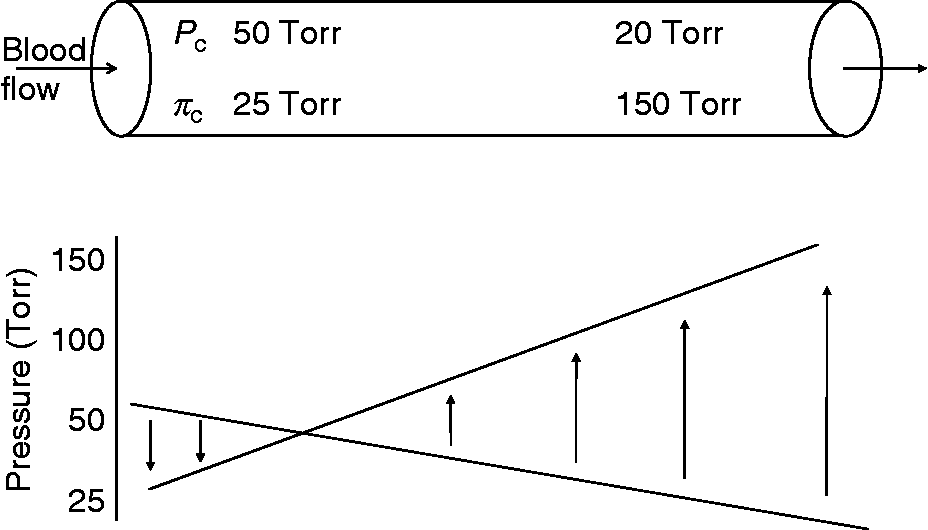
 and leads to increases in tissue water accumulation until new, higher CO2 gradients can be established. We conclude that this net osmotic force caused by ions diffusing through plasma, and not COP, is the force that explains the results obtained in Starling's classic experiment (Fig. 9). In accordance with Starling, the hydrostatic pressure is greater than the net osmotic effects of the diffusing ions and extravasates fluid into the ISF at the arterial end. At the venous end, the combined osmotic effect of
 and strong ions diffusing upstream provides a force that exceeds the declining hydrostatic pressure, returning extravasated ISF and moving metabolic water to the plasma.
Theoretical model for transcapillary fluid exchange during muscular work with Starling's second term, COP, replaced by the net osmotic pressure due to diffusing ionsReference Kowalchuk, Heigenhauser, Lindinger, Obminski, Sutton and Jones28–Reference Brechue, Gropp, Ameredes, O'Drobinak, Stainsby and Harvey33
B. Autoregulation of blood flow and cell volume regulation
Increasing metabolic rate by repetitive skeletal muscle contractions or uncoupling of oxidative phosphorylation (e.g. dinitrophenol) leads to hyperaemia, i.e. increasing blood flow to the tissue. Occlusion of blood flow results in a post-occlusion reactive hyperaemia that increases with the duration of occlusion. These changes in blood flow occur through locally controlled autoregulatory mechanisms operating to alter vasomotor tone; a number of local changes signal the local vascular changes. Local changes in  and strong ion concentrations may also serve as a signalling mechanism to alter vasomotor tone locally. Undoubtedly, blood flow impacts cell volume regulation, and thus autoregulation probably plays an important role in transcapillary water exchange and maintenance of tissue water volume/pressureReference Stainsby and Renkin101–Reference Jones and Berne103. Indeed, it is recognized that changes in plasma and blood osmolality alter blood flow to tissue, i.e. increasing plasma osmolality causes vasodilationReference Lundvall95, Reference Senay and Pivarnik104.
Linking diffusion of  and strong ions within plasma to the flux of ISF and intracellular water into plasma also links the autoregulation of cell volume to ion exchanges that maintain acid–base balance and the intracellular to extracellular [H+] difference. The attraction of our model, based on the osmotic effects caused by diffusion of
 and strong ions within plasma, is that CO2 production,
 and strong ion concentrations change in concert with metabolism and, therefore, water production when oxidative rate increases. Diffusion of
 and strong ions within plasma and the flux of water from cells and across the interstitial space will be variable and matched to metabolic demand. Thus, there is a common, physicochemical link between regulation of metabolism and local control of blood flow, autoregulation of cell volume and acid–base balance. More importantly, this physicochemical link provides an incrementing force matched to metabolism. This would solve another problem with the assumption that a constant COP due to plasma proteins could meet the increasing demands of water production and removal to maintain cell volume during periods of increased work.
C. Renal function
Acidification of the urine is accomplished by reabsorption of  and water through CA in mammalsReference Maren10, Reference Maren105, birdsReference Wolbach106 and frogsReference Hober107. Inhibition of CA in mammals and birds is characterized by an extremely consistent response that includes increased renal excretion of
 , Na+, K+ and waterReference Maren10, Reference Brechue, Kinne-Saffran, Kinne and Maren44, Reference Brechue, Stager and Lukaski74, Reference Maren, Wadsworth, Yale and Alonso108, Reference Brown and Brechue109. The link between
 and Na+ and water loss at the kidney is the basis for the diuretic effect of CAI; acetazolamide was developed as the first non-mercurial diuretic. As in the eye, the aderenergic nervous system has been observed to regulate
 flux and water reabsorption in the proximal tubule of the kidney as wellReference Garg110. While acetazolamide results in a vigorous diuretic effect, pharmacologically, it is considered a ‘poor’ diureticReference Brechue, Stager and Lukaski74, e.g. compared with loop diuretics. In the present context, the limited diuretic effects can be explained by the fact that the diuresis is linked to the initial perturbation of renal reabsorption of
 and strong ions that are relatively quickly recovered despite continued inhibition of CA. For instance, Na+,
 and water excretion increase initially with CA inhibition but return to baseline within 6 h (Na+) and 72 h (
 and water), despite continued CA inhibition in animalsReference Maren10, Reference Brechue, Kinne-Saffran, Kinne and Maren44, Reference Maren, Wadsworth, Yale and Alonso108 and humansReference Brechue, Stager and Lukaski74, Reference Brown and Brechue109. Thus, it is the re-establishment of CO2/
 gradients that return of
 and strong ion reabsorption–excretion to baseline levels, that limits the magnitude of the diuresis and total body water loss, as well as the magnitude of intracellular water accumulation (discussed above) despite continued CA inhibition. Ultimately, the magnitude of the diuretic effect is linked in magnitude to initial losses in
 and water that were unrecoverable with continued CA inhibitionReference Maren10, Reference Maren, Wadsworth, Yale and Alonso108, Reference Brown and Brechue109.
Another example of solute diffusion from a higher to a lower concentration is the standing gradient modelReference Diamond and Bossert111. Na+, Cl−, and  ions and CO2 accumulate in the clefts between tubular cells constituting the tubule of the proximal convolution of a kidney nephron. These ions and CO2 and O2 diffuse from their respective higher to lower concentrations, drag on the water through which they diffuse and alter its internal tension. Since O2 diffuses in the opposite direction and in about an equal amount to the diffusion of CO2 and
 , their osmotic effects on the water in the cleft are cancelled. Only the diffusion of Na+ and Cl− ions in the cleft pulls water out of the adjacent cells and from the lumen of the tubule and returns it to the ISF surrounding peritubular capillaries.
Summary
Several physiological inconsistencies question the role of proteins and COP in returning extravasated fluid to the vascular space, Starling's second force. Based on Hulett's explanation of osmosis, the altered chemical potential of water and its altered internal tension link solute diffusion to solvent drag; diffusion of  and strong ions along physiological concentration gradients provides a net osmotic force that can impact compartmental fluid exchange. Considering the v–a differences in
 and strong ions, a net osmotic force of ~35 Torr present in the venous end capillary water is coupled to the ISF through the pores in the capillary endothelium and opposes the hydrostatic pressure in the plasma. The important role of
 and Na+ in fluid movement is supported by the mechanistic link to fluid secretion in a variety of secretory epithelia. Perhaps more importantly, diffusing
 and strong ions provide an incremental osmotic force (~150 Torr) that is essentially matched to any increase in metabolic rate (e.g. muscular work) when CO2 output and water production are increased. This proposed model does negate the role for protein in water movement, which under appropriate circumstances can impact water movement when diffusing along its concentration gradient and/or by expanding the fluid space through its thermal motion and pushing on distensible membranes (e.g. intermittent blood flow and/or capillary recruitment). Lastly, this model links cell volume regulation with acid–base balance through CO2 chemistry.
 formation in aqueous humor: mechanism and relation to the treatment of glaucoma
 synthesis related to fluid secretion, pH control, and CO2 elimination
 cotransport in rabbit renal basolateral membrane vesicles
 cotransporter by carbonic anhydrase II and PKA
 exchanger