Introduction
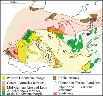
The Variscan Ibero-Armorican Arc comprises two branches that represent two Cambrian lateral margins of West Gondwana (Pouclet et al., Reference Pouclet, Álvaro, Bardintzeff, Imaz, Monceret and Vizcaïno2016) (Fig. 1): the SW branch comprises the Iberian Massif, and the NE branch the Armorican Domains, the northern Massif Central, the Occitan Domain (or southern Massif Central, including the Montagne Noire), and its lateral equivalent through southern Corsica and Sardinia, and the Pyrenean Domain. In the Pyrenees, the Ediacaran–Lower Ordovician (pre-Sardic) stratigraphic succession is laterally variable owing to structural complexity and related metamorphism. As a result, radiometrically dated Cambrian deposits are variously affected by the Variscan metamorphism and, except for two single findings of poorly preserved acritarchs (Casas and Palacios, Reference Casas and Palacios2012; Laumonier et al., Reference Laumonier, Calvet, Wiazemsky, Barbey, Marignac, Lambert and Lenoble2015), are mostly depleted of fossil record (Perejón et al., Reference Perejón, Moreno-Eiris and Abad1994; Laumonier et al., Reference Laumonier, Abad, Alonso, Baudelot, Bresiére, Besson, Centéne, Barnolas, Chiron and Guérangé1996, Padel et al., Reference Padel, Álvaro, Clausen, Guillot, Poujol, Chichorro, Monceret, Pereira and Vizcaïno2017, Reference Padel, Clausen, Álvaro and Casas2018b; Casas et al., Reference Casas, Álvaro, Clausen, Padel, Puddu, Sanz-López, Sánchez-García, Navidad, Castiñeiras, Liesa, Quesada and Oliveira2019). Nevertheless, the Ediacaran–Cambrian lithological successions of the eastern Pyrenees share close similarities with those from the Iberian Chains and the Cantabrian Zone (Spain), the Montagne Noire (France), and SW Sardinia (Italy), supporting reliable lithostratigraphic correlations (Laumonier et al., Reference Laumonier, Abad, Alonso, Baudelot, Bresiére, Besson, Centéne, Barnolas, Chiron and Guérangé1996, Reference Laumonier1998, Reference Laumonier, Autran, Barbey, Cheilletz, Baudin, Cocherie and Guerrot2004; Padel et al., Reference Padel, Clausen, Álvaro and Casas2018b). In the eastern Pyrenees, some volcanosedimentary complexes have been recently constrained by U-Pb zircon dating, in which the Ediacaran-Cambrian boundary interval has recently been precisely identified (Padel et al., Reference Padel, Álvaro, Clausen, Guillot, Poujol, Chichorro, Monceret, Pereira and Vizcaïno2017, Reference Padel, Álvaro, Casas, Clausen, Poujol and Sánchez-García2018a). However, although paleo(bio)geographic relationships among most of the Cambrian domains of the Ibero-Armorican Arc are now relatively well established (e.g., Álvaro et al., Reference Álvaro, Monceret, Monceret, Verraes and Vizcaïno2010, Reference Álvaro, Casas, Quesada, Murphy, Strachan and Quesada2021; Pouclet et al., Reference Pouclet, Álvaro, Bardintzeff, Imaz, Monceret and Vizcaïno2016), the Cambrian setting of the tectonostratigraphic Pyrenean subunits remains uncertain (Laumonier et al., Reference Laumonier, Abad, Alonso, Baudelot, Bresiére, Besson, Centéne, Barnolas, Chiron and Guérangé1996, Reference Laumonier, Autran, Barbey, Cheilletz, Baudin, Cocherie and Guerrot2004; Ballèvre et al., Reference Ballèvre, Bosse, Ducassou and Pitra2009; Álvaro et al., Reference Álvaro, Bauluz, Clausen, Devaere, Gil Imaz, Monceret and Vizcaïno2014b; Pouclet et al., Reference Pouclet, Álvaro, Bardintzeff, Imaz, Monceret and Vizcaïno2016). As a result, the paleogeographic position of the Pyrenees is often questioned or even omitted in many Ediacaran–early Paleozoic paleogeographic reconstructions of Gondwana (e.g., Murphy et al., Reference Murphy, Pisarevsky, Nance and Keppie2004; Nance et al., Reference Nance, Murphy, Strachan, Keppie, Gutiérrez-Alonso, Fernández-Suárez, Quesada, Linnemann, d'Lemos and Pisarevsky2008; but see Álvaro et al., Reference Laumonier, Marignac and Kister2010; Pouclet et al., Reference Pouclet, Álvaro, Bardintzeff, Imaz, Monceret and Vizcaïno2016).
Sketch map of the tectonostratigraphic units of the westernmost Variscan realm (modified after Martínez-Catalán et al., Reference Martínez Catalán, Arenas, Díaz García, González Cuadra, Gómez-Barreiro, Abati, Castiñeiras, Fernández-Suárez, Sánchez Martínez, Andonaegui, González Clavijo, Díez Montes, Rubio Pascual, Valle Aguado, Hatcher, Carlson, McBride and Martínez Catalán2007; Ballèvre et al., Reference Ballèvre, Bosse, Ducassou and Pitra2009; Álvaro et al., Reference Álvaro, Monceret, Monceret, Verraes and Vizcaïno2010; Pouclet et al., Reference Pouclet, Álvaro, Bardintzeff, Imaz, Monceret and Vizcaïno2016). AM = Armorican Massif; C = Cornwall; CELA = Cantabrian-Ebroan Land Area; CIZ = Central Iberian Zone; CZ = Cantabrian Zone; GTMZ = Galicia-Trás-os -Montes Zone; MC = French Central Massif; MN = Montagne Noire; OM = Ossa-Morena Zone; PZ = Pyrenean Domain; RHZ = Rheno-Hercynian Zone; SAD = South-Armorican Domain; OD = Occitan Domain; SPZ = South-Portuguese Zone; STZ = Saxo-Thuringian Zone; WALZ = West Asturian-Leonese Zone.

The Terrades inlier, located in the southeastern Pyrenees (Gerona province, Spain) (Fig. 2), stands apart from this general scheme. It has provided the only significant and indisputable ‘early Cambrian’ fossil record from the Pyrenees. Broad correlations between the Terrades inlier and the Iberian Peninsula, Morocco, Siberia, Sardinia, South China, and Montagne Noire have been suggested based on the described Cambrian Epoch 2, Age 3 archeocyaths (late Ovetian according to the Iberian chart, and early Botoman according to the Siberian chart; Abad, Reference Abad1988; Perejón et al., Reference Perejón, Moreno-Eiris and Abad1994; Laumonier et al., Reference Laumonier, Abad, Alonso, Baudelot, Bresiére, Besson, Centéne, Barnolas, Chiron and Guérangé1996; Menéndez et al., Reference Menéndez, Perejón and Moreno-Eiris2015). The presence of skeletonized microfossils, usually known as ‘Small Shelly Fossils’ (SSF), has been mentioned from the described reefal and perireefal deposits, but never formally studied (Abad, Reference Abad1988; Perejón et al., Reference Perejón, Moreno-Eiris and Abad1994). Cambrian SSF are highly common and diverse worldwide in earliest Cambrian carbonate platforms and have been therefore intensely studied for their major chronostratigraphic and paleobiogeographic implications (Devaere et al., Reference Devaere, Clausen, Steiner, Álvaro and Vachard2013; Babcock et al., Reference Babcock, Peng, Zhu, Xiao and Ahlberg2014; Clausen et al., Reference Clausen, Álvaro, Devaere, Ahlberg and Babcock2015; and references therein).
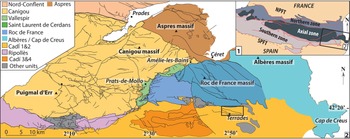
Geological sketch maps of the Pyrenees: (1) location and main structural domains of the Pyrenees, southwestern Europe; (2) main Alpine tectonostratigraphic units of the Pyrenees (after Baudin et al., Reference Baudin, Autran, Guitard and Laumonier2008; Laumonier, Reference Laumonier2015), with location of the Terrades (or La Salut) unit studied herein, 42°19′17.30″N, 2°50′42.20″E (inset box, magnified in Fig. 3); NPFT and SPFT: North Pyrenean Fault Thrusts and South Pyrenean Fault Thrusts.

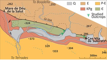
Simplified geological map of the Terrades inlier (La Salut unit) and surrounding area with the GI-504 road and location of the studied outcrops (after Berastegui Batalla and Losantos Sistach, Reference Berastegui Batalla and Losantos Sistach1997). Numbers are altitudes in meters. E = Eocene (Ypresian; Sagnari Formation); P-E = Paleocene-Eocene (Thanetian-Ypresian, Cadi Formation); Q = Quaternary; Kpg = Cretaceous-Paleogene (Garumnian); ɛ = Cambrian.

This paper is aimed at describing for the first time the SSF assemblage from the Terrades inlier to further assess advanced correlations of the pre-Variscan successions in the Pyrenees, and in particular to clarify the paleobiogeographic affinities between the Pyrenees and neighboring areas of West Gondwana.
Geologic setting
The Pyrenean Ranges (France–Spain), bounded by the North- and South-Pyrenean fault Thrusts, are structurally subdivided into three major E-W trending zones (Barnolas et al., Reference Barnolas, Chiron and Guérangé1996): the Northern, Axial, and Southern zones (Fig. 2). The Ediacaran–Cambrian sedimentary record of the Pyrenees mainly crops out in the eastern part of the ranges, from the Mediterranean Sea to the Noguera-Pallaresa Valley. The eastern Axial Zone mostly consists of Ordovician (Sardic-related) and Variscan granitic intrusions encased in a thick Ediacaran–Ordovician sedimentary succession that was consequently affected by polyphasic metamorphic and deformation events (Laumonier et al., Reference Laumonier, Autran, Barbey, Cheilletz, Baudin, Cocherie and Guerrot2004, Reference Laumonier, Marignac and Kister2010; Olivier et al., Reference Olivier, Gleizes, Paquette and Munoz-Saez2008; Aguilar et al., Reference Aguilar, Montserrat, Castiñeiras and Navidad2014; Denèle et al., Reference Denèle, Laumonier, Paquette, Olivier, Gleizes and Barbey2014; Pereira et al., Reference Pereira, Castro, Chichorro, Fernández, Díaz-Alvarado, Martí and Rodríguez2014; Cochelin et al., Reference Cochelin, Chardon, Denèle, Gumiaux and Le Bayon2017, Reference Cochelin, Lemirre, Denèle, De Saint Blanquat, Lahfid and Duchêne2018; Lemirre et al., Reference Lemirre, Cochelin, Duchene, de Saint Blanquat and Poujol2019). The Southern Pyrenean Zone, where the Terrades inlier is located (Figs. 2, 3), mostly consists of Paleogene successions interrupted by Mesozoic klippes. The Southern Pyrenean Zone overthrusts, through the South-Pyrenean Frontal Thrust, the variably deformed strata of the Cenozoic Ebro Foreland Basin.
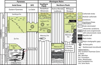
The Ediacaran–lower Paleozoic stratigraphic scheme of the Pyrenees was defined based on the successions cropping out in the Axial Zone (Cavet, 1957; Laumonier et al., Reference Laumonier, Abad, Alonso, Baudelot, Bresiére, Besson, Centéne, Barnolas, Chiron and Guérangé1996, Reference Laumonier, Autran, Barbey, Cheilletz, Baudin, Cocherie and Guerrot2004). It has traditionally been divided into the Ediacaran Canaveilles Group and the Cambrian–Lower Ordovician Jujols Group (Fig. 4), recently revised by Padel et al. (Reference Padel, Clausen, Álvaro and Casas2018b). The Canaveilles Group consists of a heterogeneous set of shales and marbles with tuffs and volcanosedimentary complexes that record the Ediacaran-Cambrian transition (Padel et al., Reference Padel, Álvaro, Casas, Clausen, Poujol and Sánchez-García2018a). The siliciclastic Err Formation forms the lower part of the Jujols Group; it is overlain by the Valcebollère Formation, which consists of massive carbonates overlain and grading laterally into shale/carbonate alternations and green shales bearing carbonate nodules. This formation is estimated to be Cambrian Age 3–early Age 4, according to the local occurrence of acritarchs reported by Laumonier et al. (Reference Laumonier, Calvet, Wiazemsky, Barbey, Marignac, Lambert and Lenoble2015). As a consequence, the Ediacaran–Cambrian succession of the Axial Zone has been traditionally compared to the chronostratigraphically constrained and lithologically similar successions from the Montagne Noire (southern France; Cavet et al., Reference Cavet1957; Laumonier et al., Reference Laumonier, Abad, Alonso, Baudelot, Bresiére, Besson, Centéne, Barnolas, Chiron and Guérangé1996; Álvaro et al., Reference Álvaro, Bauluz, Clausen, Devaere, Gil Imaz, Monceret and Vizcaïno2014b; Padel et al., Reference Padel, Álvaro, Casas, Clausen, Poujol and Sánchez-García2018a) (Fig. 4). In addition, the Ediacaran-Cambrian boundary has been geochronologically constrained in both areas using U-Pb dating of zircon in volcanosedimentary rocks (Padel et al., Reference Padel, Álvaro, Clausen, Guillot, Poujol, Chichorro, Monceret, Pereira and Vizcaïno2017, Reference Padel, Álvaro, Casas, Clausen, Poujol and Sánchez-García2018a).
Stratigraphic comparison between the Ediacaran-Cambrian (part.) successions of the eastern Pyrenees and the Montagne-Noire (modified from Padel et al., Reference Padel, Clausen, Álvaro and Casas2018b after Devaere et al., Reference Devaere, Clausen, Steiner, Álvaro and Vachard2013, Reference Devaere, Clausen, Monceret, Tormo, Cohen and Vachard2014a, Reference Tateb).

The exact relationship between the Terrades inlier (also referred to as La Salut unit; Laumonier et al., Reference Laumonier, Autran, Barbey, Cheilletz, Baudin, Cocherie and Guerrot2004; Baudin et al., Reference Baudin, Autran, Guitard and Laumonier2008; Padel et al., Reference Padel, Clausen, Álvaro and Casas2018b) and surrounding formations (in the Pyrenees and elsewhere) is unclear owing to a lack of structural and lithostratigraphic continuity. Moreover, the Cambrian limestones of the Terrades inlier display delicate thromboid textures that are unknown from time-equivalent carbonates in the region (e.g., the Valcebollère Formation; see Padel et al., Reference Padel, Clausen, Álvaro and Casas2018b for a complete review of the Ediacaran-Lower Ordovician stratigraphic framework of the Pyrenees), which have been transformed into marbles during the Variscan orogeny. The inlier is located in the Southern Zone, near Figueres (Gerona province) (Fig. 3). The Cambrian carbonate rocks of the Terrades inlier crop out in two places (Abad, Reference Abad1988; Perejón et al., Reference Perejón, Moreno-Eiris and Abad1994): near Can Vila de Subirats (F11 section of Perejón et al., Reference Perejón, Moreno-Eiris and Abad1994) and along the road GI-504 from Terrades to Boadella (F12/F13 sections of Perejón et al., Reference Perejón, Moreno-Eiris and Abad1994). The succession outcropping near Can de Vila de Subirats is dominated by limestone strata, whereas the outcrop aside and below from the GI-504 is siliciclastic-dominated but contains scattered archeocyathan-microbial patch reefs. The latter can be further subdivided into two outcrops located on the right and left banks of the Salt del Barral stream underlying the GI-504 road. The left bank (F12 section of Perejón et al., Reference Perejón, Moreno-Eiris and Abad1994) presents meter-sized calcimicrobial reefs overlain by decimeter-sized archeocyath-microbial patch reefs surrounded by onlapping shales (Fig. 5). The right bank is siliciclastic-dominated and contains decimeter-sized archeocyath-microbial patch reefs at its base.
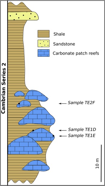
Lithological log of the Cambrian succession outcropping on the left bank of the Salt del Barral stream, north of Terrades (Gerona Province, Spain), with location of studied samples (modified from Perejón et al., Reference Perejón, Moreno-Eiris and Abad1994).

Material and methods
Samples were recovered from the left bank of the Salt del Barral stream close to the GI-504 road (F12 section of Perejón et al., Reference Perejón, Moreno-Eiris and Abad1994; 42°19′17.30″N, 2°50′42.20″E; Fig. 3). Most samples were extracted from flanks onlapping archeocyathan-microbial patch reef cores (Fig. 5), preserved as limestones and not as marbles. Phosphatized microfossils were etched from their carbonate matrix by maceration in ~10% acetic acid. Acid solution was renewed and residues sieved (using 50 μm, 100 μm, 250 μm, 1 mm, and 2 mm meshes) every 48 h. Microfossils were hand picked from the dried residues under binocular microscope. Specimens were imaged using a FEI Quanta 200 scanning electron microscope (SEM) at the Oceanography and Geosciences Laboratory (LOG, UMR 8187 CNRS) of the University of Lille I, France.
Repository and institutional abbreviations
All specimens are housed in the paleontological collections of the University of Lille (USTL). Other cited repositories are: GIN = Geological Institute of the Russian Academy of Sciences, Moscow; NIGP = Nanjing Institute of Geology and Palaeontology, China; CAMSM = Sedgwick Museum of Earth Sciences, University of Cambridge, UK.
Systematic paleontology
Phylum Arthropoda Siebold and Stannius, Reference Siebold and Stannius1845
Class indet.
Order Bradoriida Raymond, Reference Raymond1935
Family indet.
Genus indet., sp. indet.
Remarks
Bradoriids are described below following the terminology of Siveter and Williams (Reference Siveter and Williams1997), Hou et al. (Reference Hou, Siveter, Williams and Xiang-Hong2001), and Vannier et al. (Reference Vannier, Williams, Álvaro, Vizcaïno, Monceret and Monceret2005). They are oriented according to the criteria given by Hinz-Schallreuter et al. (Reference Hinz-Schallreuter, Gozalo and Liñán2007): when present, the dorsal cusp and/or the oblique ridge between one node and the dorsum are considered anterior.
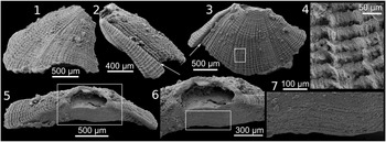
Specimens recovered herein are deformed with crinkled valves (Figs. 6.1, 6.13, 7.5, 7.9, 7.19, 7.10), which are consistent with a flexible, weakly mineralized to unmineralized shield construction, which would have been secondarily phosphatized (Siveter and Williams, Reference Siveter and Williams1997; Vannier et al., Reference Vannier, Williams, Álvaro, Vizcaïno, Monceret and Monceret2005; Zhang, Reference Zhang2007; Devaere et al., Reference Devaere, Clausen, Monceret, Vizcaïno, Vachard and Genge2014b).
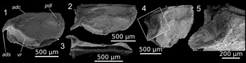
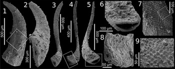
Bradoriida sp. indet. A, sample TE2F, articulated, partly broken valves of specimen USTL 3235-3: (1) left lateral view, (2) oblique dorsal view, (3) ventral view, (4) oblique anterior view, (5) detail of boxed area (spine) in (4). adc = anterodorsal cusp; ads = anterodorsal spine; pdl = posterodorsal lobe; vr = ventral ridge.

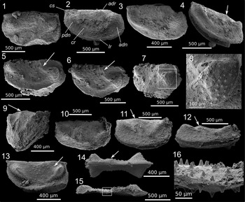
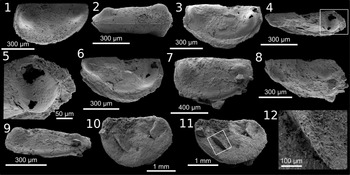
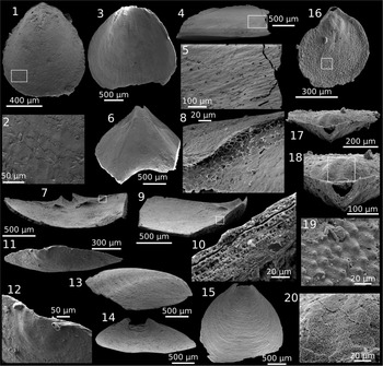
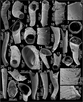
Bradoriida sp. indet. B, sample TE2F: (1) deformed, subamplete, articulated valves of specimen USTL 3235-2, lateral view; (2–4) articulated, partly opened valves of specimen USTL 3236-2: (2) right lateral view; (3) oblique dorsal view; (4) oblique ventral view; (5–8) partly preserved, articulated valves of specimen USTL 3235-7: (5) right lateral view; (6) oblique dorsal view; (7) anterior lobe; (8) detail of boxed area (pustulose and polygonal ornaments) in (7); (9–12) articulated valves of specimen USTL 3235-11: (9) oblique posterior view; (10) oblique dorsal view; (11) right lateral view; (12) ventral view; (13, 14) articulated valves of specimen USTL 3242-8: (13) right lateral view; (14) dorsal view showing highly projecting nodes; (15, 16) articulated valves of specimen USTL 3242-6: (15) dorsal view; (16) detail of boxed area (spinose ornamentation) in (15). Arrows point to the anterodorsal ridge. adn = anterodorsal node; adr = anterodorsal ridge; cr = connecting ridge; cs = cardinal spine; lr = lateroadmarginal ridge; pdn = posterodorsal node.

Bradoriida sp. indet. A
Figure 6
Occurrence
Sample TE2F.
Description
The valves are ~1.12 mm in length and ~741 μm in height, with a length/height ratio of ~1.5. The anterodorsal spine is ~362 μm in length. The carapace consists of two valves articulated at the dorsum. The subamplete valves are connected through a well-defined hinge line anteriorly interrupted to form a faint cusp (Fig. 6.1, 6.2). No cardinal spines were observed. The lobation consists of a slightly developed posterodorsal lobe, subparallel to the dorsal margin, extending from below the anterior cardinal corner to approximately one-third the length of the valve. The weak anterodorsal lobe is prolonged by an elongated spine (Fig. 6.4, 6.5), inclined anteriorly and bent toward the ventral margin at midheight (the orientation could be taphonomically constrained; Fig. 6.2, 6.3). Its base is extended ventrally into a narrow, well-defined ridge extending to the midlength of the valve (Fig. 6.1, 6.2). Admarginal ridges and furrows are absent. The surface does not show any other ornamental feature.
Materials
One articulated specimen, broken ventrally, preserved as a phosphatized carapace (USTL 3235-3).
Remarks
The lack of diagnostic features precludes a definite assignment for this specimen at the family level. Nondiagnostic spines are present in a number of bradoriid species (Topper et al., Reference Topper, Skovsted, Harper and Ahlberg2013). However, their overall shape, size, and location strongly differ from the prominent, elongated anterodorsal spine observed on the specimen studied here. Its curvature toward the ventral margin seems to be taphonomically constrained, given the crinkle line observed in ventral view (Fig. 6.3). A similar anterodorsal spine connected to a ventrally extending ridge is also found in the monasteriid Monasterium oepiki Fleming, Reference Fleming1973, yet it is of Miaolingian age and lacks a posterior lobe and cusp (Hinz, Reference Hinz1992; Topper et al., Reference Topper, Skovsted, Harper and Ahlberg2013). An anterodorsal cusp characterizes only two bradoriid families, the Cambriidae and Kunmingellidae (Hou et al., Reference Hou, Siveter, Williams and Xiang-Hong2001; Williams et al., Reference Williams, Siveter, Popov and Vannier2006). However, each of these show extremely short cardinal spines or prominent ventral spines (e.g., Kunmingella typica Huo and Shu, Reference Huo and Shu1985; Hou et al., Reference Hou, Siveter, Williams and Xiang-Hong2001). Nonetheless, some enigmatic genera showing elongate spines on the cardinal area were tentatively assigned to those families (e.g., Isoxys Walcott, Reference Walcott1890 and Gladioscutum Hinz-Schallreuter and Jones, Reference Hinz-Schallreuter and Jones1994; Siveter et al., Reference Siveter, Williams, Peel and Siveter1996; Vannier et al., Reference Vannier, Williams, Álvaro, Vizcaïno, Monceret and Monceret2005). Their extremely fine and elongated shape is nonetheless clearly different from that observed on the specimen here.
Family Cambriidae? Lee, Reference Lee1975
Bradoriida sp. indet. B
Figures 7–9
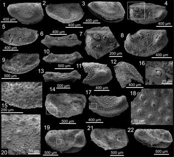
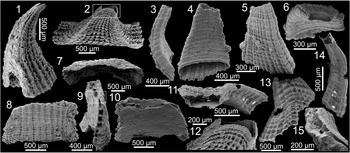
Bradoriida sp. indet. B, sample TE2F: (1–3) articulated, slightly open valves of specimen USTL 3236-6: (1) right lateral view; (2) oblique dorsal view; (3) oblique ventral view; (4–7) articulated, closed valves of specimen USTL 3236-12: (4) left lateral view; (5) oblique dorsal view, (6) dorsal view; (7) detail of boxed area in (4); (8–10) articulated valves (anterodorsal corner broken) of specimen USTL 3236-13: (8) left lateral view; (9) oblique dorsal view; (10) dorsal view; (11, 12, 16) broken valve (USTL 3235-6): (11, 12) lateral views; (16) detail of a broken spine; (13–15) articulated valves with broken ventral margin of specimen USTL 3235-4: (13) dorsal view; (14) right? lateral view; (15) detail of boxed area (spinose and polygonal ornaments) in (14); (17, 18) incomplete specimen USTL 3236-7: (17) anterolateral view showing well-developed admarginal convex area; (18) detail of boxed area (spinose ornament) in (17); (19–22) deformed, crinkled specimen USTL 3235-12 with slightly broken articulated valves: (19) lateral view; (20) detail of boxed area pustulose and polygonal ornament(in (19); (21) oblique dorsal view, (22) oblique ventral view.

Bradoriida sp. indet. B, sample TE2F: (1, 2) internal mold of articulated valve of specimen USTL 3235-8: (1) left lateral view; (2) dorsal view; (3–6) articulated, subamplete valves of specimen USTL 3236-14: (3) right? lateral view; (4) oblique dorsal view; (5) detail of boxed area (superficial polygonal meshwork) in (4); (6) oblique view; (7–9) articulated valves of specimen USTL 3236-11: (7) left lateral view; (8) oblique dorsal view; (9) ventral view; (10–12) strongly flattened specimen USTL 3234-5: (10) lateral view; (11) oblique view; (12) detail of boxed area (flattened posterior lobe) in (11).

Occurrence
Sample TE2F.
Description
The valves range 679 μm−2.4 mm in length, and 338 μm−1.6 mm in height; length/height ratio ranges 1.22–2. The carapace is constituted of two subovate valves articulated at the dorsum. The orientation is inferred by the presence of a faint to well-developed, ridge-like swelling between a node and the dorsum that is considered to be the anterodorsal ridge (Fig. 7, arrow). The valves are amplete (Figs. 7.1, 7.5, 7.10, 8.14, 9.6, 9.7, 9.10) to weakly postplete and elongate (Figs. 7.2, 8.1, 8.21, 9.1). The hinge line is well defined, straight (Figs. 7.3, 7.6, 7.10, 7.13, 8.2, 8.4, 9.1) to faintly curved dorsally (Figs. 7.1, 8.1), and slightly shorter than or equal to the length of the valve. The ventral margin is rounded anteriorly, with dorsal extremities approximately perpendicular to the dorsal margin, forming nearly right cardinal corners (Figs. 7.1, 7.2, 7.11, 7.13, 8.1, 8.7). The cardinal corners, when preserved, have short cardinal spines (Figs. 7.1–7.4, 7.10, 7.11, 7.13, 8.21). The lateroadmarginal ridge is often missing (damaged?), although better preserved specimens show a discrete to well-developed, lateroadmarginal broad swelling of the ventral margin (Figs. 7.4, 8.1, 8.8, 8.17, 9.7, 9.9), in some cases bordered ventrally by a narrow flat strip (Figs. 8.3, 8.17, 9.7, 9.9). The anterodorsal and posterodorsal nodes are strongly convex, rounded, boss-like (Figs. 7.1, 7.2, 7.6, 8.1, 8.11, 8.14, 8.19, 8.21, 9.1, 9.6, 9.7) to ridge-like (Figs. 7.9, 7.11, 7.13, 8.4, 8.8), and extend from cardinal regions to the approximate midheight of the valve. They are conjoined ventrally by a slightly narrower connecting ridge, which tends to reduce or become faint at midlength (Figs. 7.2–7.6, 7.10, 7.13, 8.1, 8.17). The maximum elevation of nodes is reached toward the cardinal corners (Figs. 7.4, 7.11, 7.13, 8.3, 8.4, 8.8, 8.14). The anterodorsal node tends to be slightly better developed (Fig. 7.2–7.6). The anterodorsal ridge is a faint to well-developed, ridge-like swelling emerging dorsal to or from the anterodorsal node and extending obliquely to the approximate mid-dorsal area (Fig. 7.2, 7.6, 7.12, 7.14). On the exterior side of the anterior and posterior nodes, a large, broadly spaced, pustulose to spiny ornament (often distally abraded) tends to be distinct (Figs. 7.2, 7.3, 7.5–7.8, 7.13, 8.4, 8.6–8.9), but covers the entire surface of the valve in better-preserved and larger specimens (Figs. 7.13–7.15, 8.14, 8.17, 8.19, 8.21). The maximum length of the ornamentation spines is ~25 μm, and their maximum diameter ~35 μm. A polygonal surface ornament can be observed on thin layers overlying the spinose ornament (Figs. 7.8, 8.14–8.16, 8.19, 8.20, 9.5). Smaller specimens (Fig. 9) have a slightly different aspect. One specimen preserved as an internal mold exhibits a clear, straight hinge line (Fig. 9.1, 9.2). Otherwise, phosphatized small specimens have faint to absent connecting ridges and their surfaces are smooth, except for a fine ornament of polygonal ridges visible on one anterior node (Fig. 9.5).
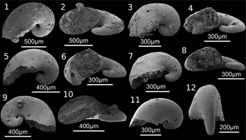
Pelagiella aff. P. subangulata? Tate, Reference Tate1892, sample TE2F: (1, 2) specimen USTL 3239-3: (1) basal view; (2) apertural view; (3, 4) specimen USTL 3239-4: (3) basal view; (4) apertural view; (5, 6) specimen USTL 3239-8: (5) basal view; (6) apertural view; (7, 8) specimen USTL 3239-7: (7) basal view; (8) apertural view; (9, 10) specimen USTL 3239-17: (9) dorsal view; (10) apertural view; (11, 12) specimen USTL 3239-14: (11) basal view; (12) lateral view.

Materials
Approximately 30 articulated valves, more or less fragmentary, preserved as phosphatized valves and/or phosphatic internal molds, including the figured specimens USTL 3234-5, 3235-2, 3235-4, 3235-6 to 3235-8, 3235-11, 3235-12, 3236-2, 3236-7, 3236-11 to 3236-14, 3242-6, and 3242-8.
Remarks
Although granulose, pustulose, and reticulate ornamentations occur in many bradoriid taxa (Hou et al., Reference Hou, Siveter, Williams and Xiang-Hong2001; Vannier et al., Reference Vannier, Williams, Álvaro, Vizcaïno, Monceret and Monceret2005; Williams et al., Reference Williams, Siveter, Popov and Vannier2006; Zhang, Reference Zhang2007), broadly spaced, hollow spines have been described only in a few species (Skovsted, Reference Skovsted2005; Skovsted et al., Reference Skovsted, Brock and Paterson2006; Topper et al., Reference Topper, Skovsted, Brock and Paterson2007; Peel et al., Reference Peel, Skovsted and Wallet2021). Among those, the recovered specimens most closely resemble Spinospitella coronata Skovsted, Brock, and Paterson, Reference Skovsted, Brock and Paterson2006 in being covered by randomly distributed surface spines overlying a reticulate pattern. As in Spinospitella Skovsted, Brock, and Paterson, Reference Skovsted, Brock and Paterson2006, the spinose ornamentation on the studied specimens seems to develop in preferential parts of the carapace depending on ontogeny (Skovsted et al., Reference Skovsted, Brock and Paterson2006), being absent in the smallest specimens, restricted to the node areas in medium-sized ones, and covering the entire shield in the largest specimens. The length and shape of the spines also appear to vary throughout ontogeny, being smaller and more rounded in juvenile specimens (Fig. 8.15, 8.18, 8.20; Topper et al., Reference Topper, Skovsted, Brock and Paterson2007). Adult specimens of Spinospitella coronata also have clear connecting ridges, although this feature is only known from a single 3.4 mm long valve from North Greenland (Peel et al., Reference Peel, Skovsted and Wallet2021). Our specimens differ from Spinospitella in having boss-like rather than spine-shaped nodes occupying a more cardinal position, and in having spines with a single base lacking a distinct crown of smaller spines on their surface (Skovsted et al., Reference Skovsted, Brock and Paterson2006).
The overall morphology and position of nodes and ridges in the recovered specimens compare more closely with the cambriid genera Auriculatella Tan, Reference Tan, Yin, Ding, He, Li and Shen1980 and Cambria Neckaya and Ivanova, Reference Neckaya and Ivanova1956, which each exhibit a long commarginal ridge connecting the anterodorsal and posterodorsal nodes, a faint ridge and/or node linking the anterodorsal node to the mediodorsal margin, and a lateroadmarginal ridge between the cardinal corners (Williams et al., Reference Williams, Siveter, Hinz-Schallreuter, Melnikova, Athersuch, Horne, Lord, Siveter and Whittaker1994, Reference Williams, Siveter, Popov and Vannier2006; Siveter et al., Reference Siveter, Williams, Peel and Siveter1996; Hou et al., Reference Hou, Siveter, Williams and Xiang-Hong2001; Vannier et al., Reference Vannier, Williams, Álvaro, Vizcaïno, Monceret and Monceret2005). Despite these broad similarities, our specimens differ from these cambriid genera in having spinose ornamentation and lacking the diagnostic anterodorsal cusp of the Cambriidae (sensu Hou et al., Reference Hou, Siveter, Williams and Xiang-Hong2001), or an anterior cardinal corner passing into a more or less abrupt anterodorsal downslope that terminates at the anterodorsal corner (sensu Hinz-Schallreuter et al., Reference Hinz-Schallreuter, Gozalo and Liñán2007). The faint unevenness of the hinge line observed in some lateral views is either due to breakage (Figs. 7.2, 8.8), compression (Fig.7.12), or to the projection of the anterodorsal ridge beyond the dorsal margin (Fig. 7.11, 7.13). The presence of faint, straight hinge lines has already been reported in Cambriidae (Hou et al., Reference Hou, Siveter, Williams and Xiang-Hong2001; Hinz-Schallreuter et al., Reference Hinz-Schallreuter, Gozalo and Liñán2007), but only in specimens preserved as open valves, which necessarily involves postmortem compression resulting in the removal of the cusps (e.g., Petrianna Siveter et al., Reference Siveter, Williams, Peel and Siveter1996, fig. 6j; Cambria danvizcainia Vannier and Siveter in Vannier et al., Reference Vannier, Williams, Álvaro, Vizcaïno, Monceret and Monceret2005, fig. 4b). Considering the very small size of the studied specimens compared to described adults (6–17.5 mm, according to Hou et al., Reference Hou, Siveter, Williams and Xiang-Hong2001), it is also likely that the Pyrenean specimens represent early growth stages that have not yet developed the diagnostic features of the family. However, the ontogeny of the Cambriidae and their morphological variations are poorly known (Williams et al., Reference Williams, Siveter, Hinz-Schallreuter, Melnikova, Athersuch, Horne, Lord, Siveter and Whittaker1994), and only highly disputable hypotheses can be made by analogy with the closest families. For instance, the appearance of the dorsal cusp in Kunmingellidae is generally mentioned in preadult stages (instar 4 of Zhang, Reference Zhang2007), at the same time as the lobation. If one considers a similar pattern among the Cambriidae, a dorsal cusp should be also present in our specimens, which exhibit distinct lobation.
Despite the absence of observable cusps, the broad resemblance between Bradoriida sp. indet. B specimens and cambriid genera (i.e., Cambria and Auriculatella) justifies their tentative attribution to Cambriidae. The absence or weak development of a connecting ridge, ornamentation, and cardinal spines in some small specimens recovered here (Figs. 8.4, 9.1, 9.7) is interpreted as being ontogenetic in origin, and tends to suggest that the differentiation of nodes precedes the development of the connecting ridge, cardinal spines, and surface sculptures in cambriid ontogeny.
Phylum Mollusca Cuvier, Reference Cuvier1797
Class indet.
Order Pelagiellida Mackinnon, Reference Mackinnon1985
Family Pelagiellidae Knight, Reference Knight1956
Genus Pelagiella Matthew, Reference Matthew1895
Type species
Cyrtolites atlantoides Matthew, Reference Matthew1894; New Brunswick, Canada; Cambrian Stage 3?.
Diagnosis
See Kouchinsky et al. (Reference Kouchinsky, Bengtson, Clausen, Gubanov, Malinky and Peel2011).
Pelagiella aff. P. subangulata? Tate, Reference Tate1892
Figure 10
Occurrence
Sample TE2F.
Description
The univalve conch is dextrally coiled with approximately one rapidly expanding whorl (Fig. 10.1, 10.3, 10.5, 10.7, 10.9, 10.11). The conch is asymmetrical; the spire is flat to concave on the adapical side and convex on the abapical side (Fig. 10.2, 10.4, 10.6, 10.8, 10.10). The width of the cross section rapidly expands toward the apertural region where it reaches its maximum. There, the cross section is suboval (Fig. 10.2, 10.4) to subtriangular with a slight angle on the abapical apertural margin (Fig. 10.8). The apex is slightly bulbous (Fig. 10.3, 10.5, 10.7, 10.9, 10.11). The external surface of the internal mold is smooth (Fig. 10.8, 10.12). Maximum diameter ranges 518−1.12 mm, apertural width 385−733 μm, and shell height 245−467 μm.
Materials
More than 100 internal molds, including the figured specimens USTL 3239-3, 3239-4, 3239-7, 3239-8, 3239-14, and 3239-17.
Remarks
The specimens described above are assigned to the genus Pelagiella due to their dextrally coiled conchs composed of one to two rapidly expanding whorls with a flattened, slightly convex or concave spiral surface (Kouchinsky et al., Reference Kouchinsky, Bengtson, Clausen, Gubanov, Malinky and Peel2011). The genus is subdivided into > 20 species, sometimes highly similar in shape and bearing intraspecific variations, which emphasizes the necessity of a systematic revision (Parkhaev, Reference Parkhaev2004; Devaere et al., Reference Devaere, Clausen, Monceret, Vizcaïno, Vachard and Genge2014b). Based on the lack of distinct ornamental features, any specific affiliation can only be tentative (Skovsted, Reference Skovsted2004). The above description could correspond either to Pelagiella madianensis Zhou and Xiao, Reference Zhou and Xiao1984 or to Pelagiella subangulata, yet these species bear strongly similar characters and their differences are subtle to almost nonexistent in juveniles (Parkhaev in Gravestock et al., Reference Gravestock, Alexander, Demidenko, Esakova, Holmer, Jago, Lin, Melnikova, Parkhaev, Yu, Rozanov, Yu, Ushatinskaya, Zang, Zhegallo, Zhuravlev and Yu2001). Their conspecificity has already been suggested (Parkhaev, Reference Parkhaev2004). The reduced rate of whorl coiling along with the absence of carena better recalls the shell of Pelagiella madianensis (synonymous with Pelagiella adunca Missarzhevsky in Rozanov and Missarzhevsky, Reference Rozanov and Missarzhevskiy [Missarzhevsky]1966; Bengtson et al., Reference Bengtson, Conway Morris, Cooper, Jell and Runnegar1990; Parkhaev in Gravestock et al., Reference Gravestock, Alexander, Demidenko, Esakova, Holmer, Jago, Lin, Melnikova, Parkhaev, Yu, Rozanov, Yu, Ushatinskaya, Zang, Zhegallo, Zhuravlev and Yu2001; Parhaev, Reference Parkhaev2004). However, these two characters present considerable intraspecific and ontogenetic variation (Parkhaev, Reference Parkhaev2004). Furthermore, Pelagiella madianensis differs from Pelagiella subangulata in having a concave supraperipheral surface, which has not been noticed in the recovered specimens. Based on its apertural cross section, less extended than in Pelagiella madianensis, and its coplanar upper apertural margin and spiral, our specimens better resemble Pelagiella subangulata.
Phylum Brachiopoda Duméril, Reference Duméril1806
Subphylum Linguliformea Williams et al., Reference Williams, Carlson, Brunton, Holmer and Popov1996
Class Lingulata Gorjansky and Popov, Reference Gorjansky and Popov1985
Order Lingulida Waagen, Reference Waagen1885
Superfamily Linguloidea Menke, Reference Menke1828
Family Eoobolidae Holmer, Popov, and Wrona, Reference Holmer, Popov, Wrona and Gazdzicki1996
Genus Eoobolus Matthew, Reference Matthew1902
Type species
Obolus (Eoobolus) triparilis Matthew, Reference Matthew1902; Cape Breton, Canada; Wuliuan.
Diagnosis
See Balthasar (2009).
Eoobolus sp. indet.
Figures 11–13
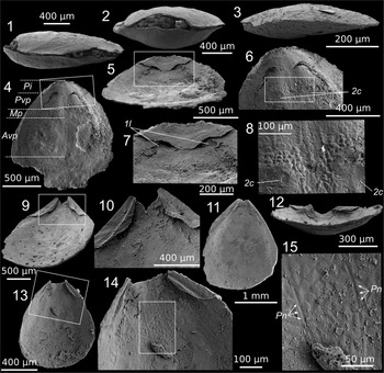
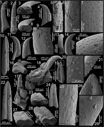
Eoobolus sp. indet.: (1, 2) specimen USTL 3250-2, sample TE1E: (1) lateral view; (2) apical view; (3) specimen USTL 3239-16, sample TE2F, lateral view; (4–8) dorsal valve of specimen USTL 3231-4, sample TE1E: (4) inner view; (5) anterior view; (6) detail of boxed area (pseudointerarea) in (4); (7) detail of boxed area (elevated pseudointerarea) in (5); (8) detail of boxed area (polygonal structures) in (6); (9–11) ventral valve of specimen USTL 3232-2, sample TE1E: (9) anterior oblique view; (10) detail of boxed area (pseudointerarea) in (9); (11) inner view; (12–15) ventral valve of specimen USTL 3232-5, sample TE2F: (12) anterior view; (13) inner view; (14) detail of boxed area (apical region) in (13); (15) detail of boxed area (polygonal structures) in (14). Avp = anterior portion of visceral platform; Mp = median projection of visceral platform; Pi = pseudointerarea; Pn = scars of pedicle nerves; Pvp = posterior portion of visceral platform; 1l = first lateral muscle scars; 2c = second central muscle scars.

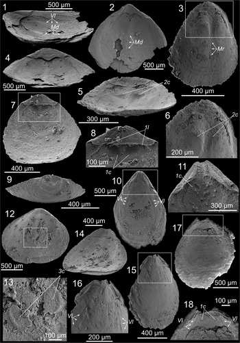
Eoobolus sp. indet.: (1, 2, 4) dorsal valve of specimen USTL 3253-11, sample TE1E: (1) lateral oblique view; (2) inner view; (4) anterior view; (3, 5, 6) dorsal valve of specimen USTL 3232-5 7, sample TE1E: (3) inner view; (5) oblique anterior view; (6) detail of boxed area in (3); (7–9) dorsal valve of specimen USTL 3232-6, sample TE1E: (7) inner view; (8) detail of boxed area in (7); (9) external apical view showing rounded larval shell; (10, 11) ventral valve of specimen USTL 3240-2, sample TE2F: (10) inner view; (11) detail of boxed area in (10); (12–14) dorsal valve of specimen USTL 3244-5, sample TE1E: (12) inner view; (13) detail of boxed area in (12); (14) anterior oblique view; (15, 16) ventral valve of specimen USTL 3254-1, sample TE1D: (15) inner view; (16) detail of boxed area in (15); (17, 18) ventral valve of specimen USTL 3232-10, sample TE1E: (17) inner view; (18) detail of boxed area in (17). Md = median depression; Mr = median ridge; Vl = vascular lateralia; 1c = first central muscle scar; 1l = first lateral muscle scar; 2c = second central muscle scar; 3c = third central muscle scar.

Eoobolus sp. indet.: (1, 2) dorsal valve of specimen USTL 3245-15, sample TE1E: (1) inner view; (2) detail of boxed area (radiating faint ridges) in (1); (3–5) dorsal valve of specimen USTL 3250-3, sample TE1E: (3) inner view; (4) lateral view; (5) detail of boxed area (radiating fine ridges) in (4); (6–10) ventral valve of specimen USTL 3247-1, sample TE1E: (6) inner view; (7) anterior view showing emarginature; (8) detail of boxed area (polygonal structures between pseudointerarea and shell floor) in (7); (9) lateral view; (10) detail of boxed area (columnar microstructure) in (9); (11, 12) dorsal valve of specimen USTL 3247-11, sample TE1E: (11) oblique posterior view; (12) larval shell; (13–15) specimen USTL 3250-1, sample TE1E: (13) lateral view; (14) posterior view showing median sulcus; (15) external view; (16–20) juvenile articulated specimen USTL 3239-19, sample TE2F: (16) dorsal view; (17) posterior view; (18) detail of U-shaped emarginature; (19) detail of boxed area (pustulose ornament on postlarval shell) in (16); (20) detail of boxed area (pitted ornament on larval shell) in (18).

Occurrence
Samples TE2F, TE1D, TE1E.
Description
The shells are inequivalved, slightly ventribiconvex (Figs. 11.1, 11.2, 13.17) or more rarely slightly dorsibiconvex (Fig. 11.3), with the maximum width anterior to midlength. The dorsal valves are subovate to subtriangular in outline (Figs. 12.2, 12.3, 12.7, 12.12, 13.1, 13.3), and the ventral valves have an acuminate apex conferring a teardrop shape (Figs. 11.11, 11.13, 12.10, 12.15, 13.16). The pseudointerareas of both valves are subtriangular, moderately elevated above the shell floor and bear longitudinal folds or growth lines (Figs. 11.5–11.7, 11.10–11.14, 12.1, 12.4, 12.6–12.8, 12.11, 12.16, 12.18, 13.7, 13.8). Ventral pseudointerareas are bisected by strong flexure lines on each side of a narrow, deep median groove slightly widening anteriorly (Figs. 11.9–11.11, 12.10, 12.11, 13.6, 13.7). The partial preservation of the pseudointerarea shortens the extension of the pedicle groove that can be almost indistinguishable, but it can be expressed as a U-shaped emarginature on ventral valves (Figs. 11.9–11.14, 12.17, 12.18, 13.7). On dorsal valves, the groove is shallow and broad, sometimes indistinct, and the flexure lines are weak to almost absent (Figs. 11.4–11.7, 12.1–12.8). Immediately in front of the pseudointerarea lies a visceral platform that can be extended anteriorly to approximately one-third of the valve. In ventral valves, the visceral platform and its proximal projection are rarely preserved but can be inferred from scars of pedicle nerves (Fig. 11.14, 11.15) or umbonal muscles (first central muscles according to Balthasar, Reference Balthasar2009; Fig. 12.11, 12.18). In dorsal valves, the visceral area is divided into a proximal and a distal portion, yet the latter is rarely preserved or tends to disappear proximally (Fig. 11.4). It can be bisected by a median depression or ridge (Fig. 12.1–12.3).
Subovate muscle scars can be present on the posterior portion of the dorsal visceral platform, immediately beyond the flexure lines of the pseudointerarea (first lateral muscles; Fig. 11.7), or beyond the median groove (first central muscles; Fig. 12.8, 12.18). Larger, subcircular muscle scars can also occur on both sides of the median projection of the visceral platform (second central muscle scars; Figs. 11.6, 11.8, 12.5, 12.6). One specimen shows subovate, slightly diverging muscle scars on the anterior portion of the visceral platform (third central muscles; Fig. 12.12, 12.13). Some characters in vascula lateralia are weak, arcuate to straight, and begin from the proximal end of the pseudointerarea to approximately midlength of the valve, following its margins (Fig. 12.1, 12.10, 12.16, 12.18). A radiating network of fine depressions is present on the internal surface of some valves and tends to be more prominent on their peripheral parts (Fig. 13.1–13.5). Both valves can also show growth lines paralleling the valve margin on their anteriormost parts (Fig. 12.3, 12.17). Polygonal structures can be present on the posterior halves of well-preserved valves (Fig. 11.15). They tend to be best developed in dorsal valves at the junction between the second central muscle scars and the proximal margins of the posterior part of the visceral platform (Fig. 11.6, 11.8), but they can also occur between the pseudointerarea and the valve floor (Fig. 13.8). The shell microstructure consists of fine laminae separated by a dense network of transverse columns (Fig. 13.10).
The outer surface of juvenile valves shows a subcircular, strongly convex larval shell on which a lobation is tentatively inferred from weak sublinear depressions (Fig. 13.16–13.18). This structure is ornamented with a dense network of tiny pits, ~ 0.7–1.5 μm in diameter (Fig. 13.20). The postlarval shell of juvenile specimens is covered by fine, closely spaced pustules, ~7–10 μm in diameter (Fig. 13.19). Adult shells are devoid of any ornamental features and only bear fine growth lines parallel to the valve margin (Fig. 13.15). A larval shell is suggested by a rounded convex area located on the posterior end of the valve, but lacks the pitted ornament observed on juvenile specimens (Figs. 12.9, 13.11, 13.12). This area seems to consist of at least two lobes located on its proximal margin. Adult shells can also bear a shallow, broad sulcus better developed proximally (Fig. 13.13, 13.14). Measurements are provided in Table 1.
Measurements of ventral and dorsal valves of Eoobolus sp. indet. Il = length of pseudointerarea; Iw = width of pseudointerarea; L = length of valve; Ls = length of larval shell; MAX = maximum value; MIN = minimum value; N = number of values; SD = standard deviation; Vl = length of visceral platform; W = width of valve.

Materials
More than 100 articulated and disarticulated valves, including figured specimens from sample TE1D (USTL 3254-1); sample TE1E (USTL 3231-4, 3232-2, 3232-6, 3232-7, 3232-10, 3244-5, 3245-15, 3247-1, 3247-11, 3250-1 to 3250-3, 3253-11); and sample TE2F (USTL 3239-16, 3232-5, 3239-19, 3240-2).
Remarks
The pustulose and pitted ornament covering adult and larval shells, respectively (Fig. 13.19, 13.20), is a diagnostic feature of the family Eoobolidae (Holmer et al., Reference Holmer, Popov, Wrona and Gazdzicki1996). Balthasar (Reference Balthasar2009) suggested abolition of the family Eoobolidae and reassigned Eoobolus to the family Zhanatellidae, pointing out the importance of preservational biases on the diagnostic features of the Eoobolidae. However, Zhang et al. (Reference Zhang, Zhang, Holmer and Li2015) noticed that most specimens of Eoobolus lack the apical emarginature on ventral valves that characterize the Zhanatellidae. As a consequence, our specimens share the diagnostic characters of the family Eoobolidae despite the lack of ornamental features on adult shells and the presence of an emarginature on most ventral valves. The specimens described above are assigned to the genus Eoobolus because of: (1) the elevated position of the pseudointerarea and its flexure lines; (2) the deep, narrow pedicle groove on the ventral valve; (3) the presence of a median projection of the visceral platform commonly bisected by a median ridge (in some cases abraded into a median depression in our specimens); and (4) the shape and position of muscle scars (Balthasar, Reference Balthasar2009). Notable differences with the genus Eoobolus include: (1) the presence of a fine network of radiating ridges on the inner side of some valves (Fig. 13.1–13.5); and (2) the diverging morphology of the third central muscle scars in one dorsal valve (Fig. 12.12, 12.13). The latter differs from the specimens of Balthasar (Reference Balthasar2009) on which exceptionally well-preserved dorsal valves show subparallel, closely spaced muscle scars separated by a prominent median ridge. Moreover, in contrast to Eoobolus, the Pyrenean specimens have a very slightly elevated pseudointerarea.
Any assignment at the specific level relies on the apical angle of the ventral valve, which seems to represent the only constant feature during ontogeny (Balthasar, Reference Balthasar2009). The ventral valves described above show apical angles ranging 50–140°, preservational biases accounting for extreme values. This high variability likely reflects the presence of multiple species in the assemblage. Of the 50 ventral valves, almost half show apical angles ranging 80–90°. This small range has already been reported for E. aff. E. viridis Cobbold, Reference Cobbold1921 from South China (Li and Holmer, Reference Li and Holmer2004; Balthasar, Reference Balthasar2009; Zhang et al., Reference Zhang, Zhang, Li and Holmer2016). Angles between 70° and 80° are also common (eight valves) and can also support assignment to either E. elatus Pelman in Pelman and Pereladov, Reference Pelman, Pereladov and Zhuravleva1986 and E. priscus Poulsen, Reference Poulsen1932, for which apical angles range 70–90° (Balthasar, Reference Balthasar2009). Finally, apical angles < 70° are also reported for seven valves, which could indicate the presence of E. triparilis among the previously listed species.
The specimens described above are different from all known species of Eoobolus in being ventribiconvex, having subequal length and width, and a short pseudointerarea, especially on dorsal valves (Table 1). The smallest length/width ratio fits E. incipiens Zhang, Popov, Holmer, and Zhang in Zhang et al., Reference Zhang, Ghobadi Pour, Popov, Holmer, Chen, Chen, Brock and Zhang2021 (averaging 1.09) and the synonym E. aff. E. viridis from Australia (averaging 1.15; Ushatinskaya and Holmer in Gravestock et al., Reference Gravestock, Alexander, Demidenko, Esakova, Holmer, Jago, Lin, Melnikova, Parkhaev, Yu, Rozanov, Yu, Ushatinskaya, Zang, Zhegallo, Zhuravlev and Yu2001), yet the typical apical angle is much more gentle (100–110°). Dorsal pseudointerareas of E. cf. E. triparilis from Canada occupy 14% of the valve length and 50% of the valve width (Balthasar, Reference Balthasar2009), which is identical to our mean measurements (Table 1). However, the pseudointerareas on ventral valves are far more extended and each bears a strongly acute apical angle. The valves described above are more similar to E. aff. E. viridis from China in having an apical angle of mostly 80–90°, a short pseudointerarea (compared to specimens from Australia), a diverging median groove, weak to indistinct flexure lines on dorsal valves, and a median sulcus (Li and Holmer, Reference Li and Holmer2004). The pustulose ornamentation of the Chinese material, large and coarsely distributed, is also a distinctive feature shared with the specimens from the Terrades inlier. The valves described above differ from Chinese specimens in having a less-elevated median ridge, yet this feature probably derives from abrasion. The shallow, poorly defined radiating ridges observed here are also seen in specimens from South Australia, referred to E. priscus (see Paterson et al., Reference Paterson, Skovsted, Brock and Jago2007, fig. 3C), but their sporadic occurrence questions their taxonomic significance. Polyspecificity is not excluded given the high variability of the studied specimens, for instance shown by the broad range of apical angle values.
Polygonal patterns (Figs. 11.8, 11.15, 13.7) have already been reported for a number of brachiopod taxa including Eoobolus (Balthasar, Reference Balthasar2009). Similar structures on the valve interiors of acrotretid brachiopods have been interpreted as molds of various types of epithelial cells distributed across distinct areas of the shell (Zhang et al., Reference Zhang, Zhang, Li and Holmer2016).
Phylum indet.
Class Hyolitha Marek, Reference Marek1963
Order Hyolithida Sysoev, Reference Sysoev1957
Family indet.
Genus Microcornus Mambetov, Reference Mambetov1972
Type species
Microcornus parvulus Mambetov, Reference Mambetov1972; Kazakhstan; Cambrian Age 3.
Diagnosis
See Kouchinsky et al. (Reference Kouchinsky, Bengtson, Clausen and Vendrasco2015).
Microcornus cf. M. petilus Bengtson in Bengtson et al., Reference Bengtson, Conway Morris, Cooper, Jell and Runnegar1990
Figure 14
Occurrence
Sample TE2F.
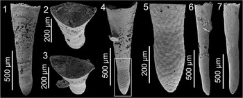
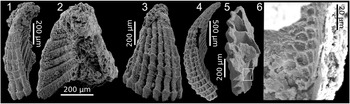
Microcornus cf. M. petilus Bengtson in Bengtson et al., Reference Bengtson, Conway Morris, Cooper, Jell and Runnegar1990, sample TE2F: (1, 2) specimen USTL 3238-10: (1) dorsal view; (2) apertural view; (3–5) specimen USTL 3238-1: (3) apertural view; (4) dorsal view; (5) detail of boxed area (weak polygonal patterns on surface of internal mold) in (4); (6) specimen USTL 3238-16, lateral view; (7) specimen USTL 3238-4, lateral view.

Description
The conch is broadly orthoconic, 1–1.25 mm in length, with straight lateral margins and small apical angle (Fig. 14.1, 14.4, 14.6, 14.7). The ventral side is flattened to weakly convex, and the lateral sides are rounded, forming two longitudinal ridges (Fig. 14.2, 14.3). The dorsal side is strongly convex bearing a weak median longitudinal ridge, producing a triangular cross section with rounded edges (Fig. 14.2). The protoconch is bulbous, tapering into a nearly straight, rounded tip (Fig. 14.1, 14.4–14.7). It is separated from the mature conch by a shallow constriction (Fig. 14.1, 14.4, 14.5). Three specimens exhibit a weak polygonal network on the surface of the bulbous protoconch, gradually disappearing after the constriction (Fig. 14.4–14.7). Neither opercula nor ligula were observed. The apertural width and height range 350–435 μm and 170–215 μm, respectively. The apical angle is ~15°.
Materials
Several tens of internal molds, including the figured specimens USTL 3238-1, 3238-4, 3238-10, and 3238-16.
Remarks
The specimens described above are assigned to the genus Microcornus based on their overall shape with a bulbous protoconch, a median ridge on the dorsal side, and the resulting rounded triangular cross section (Bengtson et al., Reference Bengtson, Conway Morris, Cooper, Jell and Runnegar1990; Malinky and Skovsted, Reference Malinky and Skovsted2004). The narrow conical morphology of the conchs produced by a small apical angle, along with the weakly convex ventral side, conforms to Microcornus petilus (see Bengtson et al., Reference Bengtson, Conway Morris, Cooper, Jell and Runnegar1990; Demidenko in Gravestock et al., Reference Gravestock, Alexander, Demidenko, Esakova, Holmer, Jago, Lin, Melnikova, Parkhaev, Yu, Rozanov, Yu, Ushatinskaya, Zang, Zhegallo, Zhuravlev and Yu2001; Malinky et al., Reference Malinky, Wilson, Holmer, Lardeux, Webby, Paris, Droser and Percival2004; Topper et al., Reference Topper, Brock, Skovsted and Paterson2009). However, the specimens recovered here differ from Microcornus petilus in having a poorly defined dorsal ridge and in lacking an ornament of fine wrinkles parallel to the apertural margin. Moreover, three specimens exhibit an unusual polygonal network on the bulbous protoconch. Such a pattern has never been mentioned on internal molds of hyolithids, and probably reflects the ornamentation of the inner surface of the conch.
Order indet.
Family Cupithecidae Duan, Reference Duan1984
Genus Cupitheca Duan in Xing et al., Reference Xing, Ding, Luo, He and Wang1984
Type species
Paragloborilus mirus He in Qian, Reference Qian1977; Maidiping, Emei County, Sichuan Province, South China; Fortunian.
Diagnosis
See Parkhaev and Demidenko (Reference Parkhaev and Demidenko2010).
Cupitheca sp. indet.
Figure 15
Occurrence
Sample TE2F.
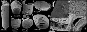
Cupitheca sp. indet., sample TE2F: (1, 2) specimen USTL 3238-19: (1) lateral view; (2) apical view; (3, 5, 6, 9, 11) specimen USTL 3238-9: (3) lateral view; (5) detail of larger boxed area (faint transverse striations) in (3); (6) detail of smaller boxed area (pits on external coating) in (3); (9) apical view; (11) detail of boxed area (phosphatic rods and corresponding pits) in (9); (4, 10) specimen USTL 3238-15: (4) apical view; (10) detail of boxed area (phosphatic rods) in (4); (7, 8) specimen USTL 3238-14: (7) lateral view; (8) apical view.

Description
The conchs slowly and uniformly expand from a closed termination to an invariably broken abapical part (Fig. 15.1, 15.3). The maximum preserved length ranges 426 μm−1.15 mm, the maximum abapical diameter of the conch 224–813 μm, and the diameter of the rods 2.5–6 μm. The cross section is subcircular to oval. The closed termination (apex) is slightly (Fig. 15.1) to strongly (Fig. 15.7) convex and a shallow but well-defined constriction separates it from the conch (Fig. 15.1–15.3, 15.7–15.9). At the apex, internal and external coatings of the conch wall connect with transverse phosphatic rods expressed as tiny pits on the surface of the external coating (Fig. 15.4, 15.9–15.11). The external coating exhibits faint transverse striations (Fig. 15.3, 15.5).
Materials
Ten specimens, including USTL 3238-9, 3238-14, 3238-15, and 3238-19.
Remarks
The recovered conchs are assigned to the genus Cupitheca based on their gently tapering conical shape with a convex apex separated by a marked constriction. Their identification at the specific level remains open because the ornamentation of faint transverse striations is not typical of any species of Cupitheca. The evidence of faint annular sculpture on the external surface, together with the presence of rod-like microstructure, is comparable to one specimen of Cupitheca holocyclata Bengtson in Bengtson et al., Reference Bengtson, Conway Morris, Cooper, Jell and Runnegar1990 described by Malinky and Skovsted (Reference Skovsted2004, fig. 12A).
Phylum indet.
Class indet.
Order Tommotiida Missarzhevsky, Reference Missarzhevsky1970
Family indet.
Genus Kelanella Missarzhevsky in Rozanov and Missarzhevsky, Reference Rozanov and Missarzhevskiy [Missarzhevsky]1966
Type species
Kelanella altaica Missarzhevsky in Rozanov and Missarzhevsky, Reference Rozanov and Missarzhevskiy [Missarzhevsky]1966; Altai Mountains, Siberian Platform, Russia; Cambrian Age 5.
Diagnosis
See Devaere et al. (Reference Devaere, Clausen, Monceret, Vizcaïno, Vachard and Genge2014b)
Kelanella altaica Missarzhevsky in Rozanov and Missarzhevsky, Reference Rozanov and Missarzhevskiy [Missarzhevsky]1966
Figures 16–19
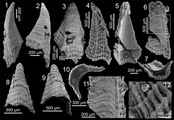
Type A sclerites of Kelanella altaica Missarzhevsky in Rozanov and Missarzhevsky, Reference Rozanov and Missarzhevskiy [Missarzhevsky]1966: (1–3) specimen USTL 3242-13, sample TE2F: (1, 2) lateral views; (3) upper view; (4–6, 10–12) specimen USTL 3248-2, sample TE1E: (4) lateral view; (12) detail of boxed area (gridded ornament) in (4); (5, 6) lateral views showing duplicature; (11) detail of boxed area in (6); (10) apertural view; (7–9) specimen USTL 3242-3, sample TE2F: (7) apertural view; (8, 9) lateral views.

Type B sclerites of Kelanella altaica Missarzhevsky in Rozanov and Missarzhevsky, Reference Rozanov and Missarzhevskiy [Missarzhevsky]1966, specimen USTL 3248-8, sample TE1E: (1–3) lateral views; (4) detail of boxed area in (3); (5) apical view; (6) detail of boxed area in (5); (7) detail of boxed area (weak growth lines) in (6). Arrows point to deep sulcus.

Type C sclerites of Kelanella altaica Missarzhevsky in Rozanov and Missarzhevsky, Reference Rozanov and Missarzhevskiy [Missarzhevsky]1966: (1, 2, 7, 12) specimen USTL 3254-2, sample TE1D: (1) lateral view; (2) oblique apical view; (7) apertural view; (12) detail of boxed area in (2); (3–6) specimen USTL 3248-1, sample TE1E: (3) lateral facet; (4) duplicature; (5) gridded facet; (6) apical view; (8–10, 15) specimen USTL 3248-5, sample TE1E, lateral views: (8) gridded facet; (9) transverse septa; (10) duplicature; (15) detail of boxed area in (9); (11, 13, 14) specimen USTL 3243-7, sample TE2F: (11) apical view; (13) lateral view showing gridded facet; (14) lateral view showing lateral facet.

Sclerites of Kelanella altaica Missarzhevsky in Rozanov and Missarzhevsky, Reference Rozanov and Missarzhevskiy [Missarzhevsky]1966: (1–4) Type E: (1–3) specimen USTL 3249-2, sample TE1E, lateral views: (1) gridded and single lateral facets; (2) duplicature and lateral facet; (3) gridded facet; (4) specimen USTL 3243-8, sample TE2F, lateral view showing gridded facet and single lateral facet; (5, 6) specimen USTL 3248-6 (unrecognizable type), sample TE1E: (5) internal cavity with transverse septa; (6) detail of boxed area (polygonal network on septa) in (5).

Holotype
GIN 3470/95 (Rozanov and Missarzhevsky, Reference Rozanov and Missarzhevskiy [Missarzhevsky]1966, p. 95, pl. 11, fig. 6, pl. 13, fig. 9); Altai Mountains, Siberian Platform, Russia; Kameshkovsky horizon.
Occurrence
Samples TE2F, TE1D, and TE1E.
Description
Following the nomenclature of Devaere et al. (Reference Devaere, Clausen, Monceret, Vizcaïno, Vachard and Genge2014b), four morphological types (A−C, E) were identified herein. Type A sclerites (Fig. 16) are bilaterally symmetric to subsymmetric, the apex slightly bending over the duplicature (Fig. 16.1, 16.2). The cross section is crescent-shaped (Fig. 16.7, 16.10). The broad convex facet shows a gridded ornament that consists of a radiating network of irregularly spaced longitudinal folds and more discrete transverse ribs (Fig. 16.3, 16.4, 16.8, 16.12). This ornamentation is well developed in the apertural region and gradually disappears toward the apex (Fig. 16.3). The opposite, concave duplicature shows fine subparallel transverse growth lines slightly curving to the apical region following the shape of the apertural margin (Fig. 16.5, 16.6, 16.11). They seem to be continuous with the transverse ridges of the gridded facet (Fig. 16.11). Reduced lateral facets are present between the gridded facet and the duplicature (Fig. 16.5).
Type B sclerite (Fig. 17) is asymmetrical, polyhedron-shaped, and wider than Type A (Fig. 17.3, 17.5). The centrally situated apical area (apex not preserved) gently curves over the duplicature (Fig. 17.1, 17.2). The weakly convex gridded facet displays a subparallel, closely spaced network of transverse ridges overlapping irregularly spaced longitudinal folds radiating from the apical region (Fig. 17.3, 17.4). The asymmetry of the specimen is based on a deep sulcus situated on the convex gridded facet, extending from the apical region to the aperture, where it is expressed as a notch (Fig. 17.2, 17.3). The slightly convex duplicature bears fine subparallel growth lines that are prolonged into more elevated ridges in two lateral facets (Fig. 17.5–17.7). The suboval cross section is visible in the apical region due to breakage (Fig. 17.5, 17.6).
Type C sclerites (Fig. 18) are slightly asymmetrical (Fig. 18.4, 18.5) with a subrectangular cross section (Fig. 18.2, 18.6, 18.7, 18.11). The apertural region is more or less strongly curved over the duplicature (Fig. 18.1, 18.2, 18.6, 18.11, 18.14). The gridded facet is concave to weakly convex (Fig. 18.2, 18.6). The opposite duplicature varies from concave toward the aperture to planar in the apical region (Fig. 18.6, 18.7, 18.11). It can bear a fine network of subparallel growth lines, gradually curving toward the apical region (Fig. 18.4, 18.10). The gridded facet and the duplicature are separated by two concave-to-planar lateral facets (Fig. 18.1, 18.3, 18.11, 18.14). The ornamentation of the gridded facet consists of well defined, broad longitudinal folds that tend to disappear toward the apical region (Fig. 18.1, 18.2, 18.12). These irregularly spaced radiating lines are overlapped by weaker transverse ridges in a gridded arrangement. In some specimens, the lateral facets bear a transitional ornamentation consisting of discrete to absent longitudinal folds and well-defined transverse ridges gradually attenuating toward the duplicature margin (Fig. 18.3, 18.11, 18.14). Our largest specimen shows, nonetheless, a strongly gridded ornamentation on its lateral facets (Fig. 18.1, 18.2). Transverse internal septa have been observed in broken specimens (Fig. 18.6, 18.9, 18.10, 18.15).
Type E sclerites are asymmetrical and slightly twisted (Fig. 19.1, 19.2). The apical area is strongly curved toward the duplicature (Fig. 19.2, 19.3). The gridded facet is partially separated from the opposite concave duplicature by a single, subplanar to concave lateral facet (Fig. 19.1, 19.2, 19.4). The transition between the lateral facet and the duplicature can be marked by a weak longitudinal crest (Fig. 19.2). The ornamentation on the gridded facet consists of broad longitudinal folds radiating from the apex, superimposed by thinner transverse ridges (Fig. 19.3). The lateral facet bears only well-developed transverse ribs (Fig. 19.1, 19.2, 19.4).
In one broken specimen that cannot be assigned to any type, the internal cavity shows slightly concave transverse septa, one of which possesses a polygonal network of fine ridges (Fig. 19.5, 19.6).
Measurements follow the stated dimensions of Devaere et al. (Reference Devaere, Clausen, Monceret, Vizcaïno, Vachard and Genge2014b). The maximum height ranges 1.2–1.9 mm for Type A, 1.6 mm for Type B, 1.2–2.1 mm for Type C, and 870 μm−2.1 mm for Type E. The length ranges 93–225 μm for Type A, unavailable for Type B, 555–875 μm for Type C, and 93–300 μm for Type E. The maximum width ranges 600 μm−1 mm for Type A, 2.4 mm for Type B, 2.7 mm for Type C, and 475 μm−1 mm for Type E.
Materials
Approximately 80 specimens, mostly fragmentary; 20 sclerites identified as belonging to Type A (5), Type B (1), Type C (9), and Type E (5). Figured specimens are from sample TE1D (USTL 3254-2); sample TE1E (USTL 3248-1, 3248-2, 3248-5, 3248-6, 3248-8, 3249-2); and sample TE2F (USTL 3242-3, 3242-13, 3243-7, 3243-8).
Remarks
The sclerites described here are assigned to the genus Kelanella on the basis of their overall polyhedral shape, the presence of transverse septa and facets, one of which bears gridded ornamentation. They are specifically assigned to the Types A, B, C, and E of the species Kelanella altaica as defined by Devaere et al. (Reference Devaere, Clausen, Monceret, Vizcaïno, Vachard and Genge2014b). As far as the original scleritome of Kelanella altaica is unknown, the reason for the absence of Type D sclerites in the recovered assemblage remains uncertain. Sampling bias could be invoked (Type D sclerites might not be dominant in the scleritome), but potential ontogenetic and ecophenotypic effects on Kelanella altaica assemblages should also be considered. The specimens assigned to Types A, B, C, and E in the material from the Pyrenees are very similar to specimens assigned to the same morphotypes in the material from the Montagne Noire described by Devaere et al. (Reference Devaere, Clausen, Monceret, Vizcaïno, Vachard and Genge2014b), and comparison to other representatives of Kelanella altaica figured in the literature detailed in this publication can be followed. The single specimen assigned to Type B shows a noticeable difference with the specimens described by Devaere et al. (Reference Devaere, Clausen, Monceret, Vizcaïno, Vachard and Genge2014b) in the presence of fine, discrete, closely spaced growth lines on the duplicature (Fig. 17.5–17.7), along with prominent and elevated folds on the gridded facet (Fig. 17.3, 17.4). Although the affiliation of this peculiar type to Kelanella altaica was questioned by Devaere et al. (Reference Devaere, Clausen, Monceret, Vizcaïno, Vachard and Genge2014b), the recovery of the Type B sclerites together with other sclerites undoubtedly assigned to Kelanella altaica supports the assumption that they belong to the same scleritome. One specimen assigned to Type C, due to its quadrangular cross section, shows a strongly gridded pattern on its lateral facets (Fig. 18.1, 18.2) that had not been described for this type by Devaere et al. (Reference Devaere, Clausen, Monceret, Vizcaïno, Vachard and Genge2014b). The gridded pattern of the facet of some Pyrenean specimens assigned to Type A is slightly faint in way similar to those of specimens described by Rozanov and Missarzhevsky (1966). The numerous, well-illustrated specimens of Type E sclerites from China by Yuan and Zhang (Reference Yuan and Zhang1983) compare well with the material from the Pyrenees and from the Montagne Noire. Except for the left/right specimen differentiation, they show little variation. Only the transverse ribs of the lateral facets are thicker in the specimens from the Pyrenees than in those described by Yuan and Zhang (Reference Yuan and Zhang1983) and Devaere et al. (Reference Devaere, Clausen, Monceret, Vizcaïno, Vachard and Genge2014b).
Polygonal structures were observed on one specimen of type E in the material from the Pyrenees and have already been described for the same sclerite type in the material from the Montagne Noire (Devaere et al., Reference Devaere, Clausen, Monceret, Vizcaïno, Vachard and Genge2014b). In the case of the Pyrenean material, it is however difficult to determine whether the polygons are on the inner surface of the sclerite wall or are part of the wall structure made visible due to delamination.
Order Chancelloriida Walcott, Reference Walcott1920
Family Chancelloriidae Walcott, Reference Walcott1920
Remarks
The classification of chancelloriid sclerites has continuously been reviewed following the recovery of complete scleritomes and large sclerite populations (Yun et al., Reference Yun, Zhang, Li, Pan, Li and Brock2019; Moore et al., Reference Moore, Porter, Webster and Maloof2021). However, the present assemblage only consists of a limited collection of disarticulated sclerites, some of which are not known from articulated taxa (e.g., 4 + 0 sclerites in Allonnia Doré and Reid, Reference Doré and Reid1965; Yun et al., Reference Yun, Zhang, Li, Pan, Li and Brock2019). Moreover, some species are represented only by a fraction of their known dominant sclerite types in the present assemblage (e.g., Chancelloria eros Walcott, Reference Walcott1920 is here represented only by 7 + 1 sclerites, but 6 + 1 sclerites should also be dominant in this taxon; Bengtson and Collins, Reference Bengtson and Collins2015; Moore et al., Reference Moore, Porter, Webster and Maloof2021), whereas individual sclerite types can be produced by distinct scleritomes (e.g., 4–6 + 0 types from Chancelloria Walcott, Reference Walcott1920 or Allonnia; Moore et al., Reference Moore, Li and Porter2014). We have therefore adopted a conservative approach by following traditional taxonomy (Moore et al., Reference Moore, Li and Porter2014) without assuming correspondence between sclerite-based and scleritome-based taxa.
Genus Chancelloria Walcott, Reference Walcott1920
Type species
Chancelloria eros Walcott, Reference Walcott1920; Walcott Quarry, Ontario, Canada; Cambrian Age 5.
Diagnosis
See Kouchinsky et al. (Reference Kouchinsky, Bengtson, Clausen, Gubanov, Malinky and Peel2011).
Chancelloria sp. indet.
Figure 20.1–20.6
Occurrence
Sample TE2F.
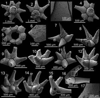
Chancelloriid sclerites, sample TE2F: (1–6) Chancelloria sp. indet.: (1) specimen USTL 3234-2, abaxial view; (2, 3) specimen USTL 3234-3: (2) abaxial view; (3) detail of boxed area (verruculose texture) in (2); (4) specimen USTL 3233-14, abaxial view; (5, 6) specimen USTL 3234-1: (5) abaxial view; (6) detail of boxed area in (5); (7–14) Allonnia tetrathallis (Jiang in Luo et al., Reference Luo, Jiang, Wu, Song and Ouyang1982): (7, 8) specimen USTL 3233-13, 4 + 0 type: (7) abaxial view; (8) oblique view; (9, 10) specimen USTL 3233-8, 5 + 0 type: (9) abaxial view; (10) oblique view; (11, 12) specimen USTL 3233-9, 6 + 0 type: (11) abaxial view; (12) oblique view; (13, 14) specimen USTL 3233-6: (13) abaxial view; (14) oblique view; (15–17) Archiasterella charma Moore, Li, and Porter, Reference Moore, Li and Porter2014, specimen USTL 3233-10: (15) basal view, (16) lateral view; (17) detail of boxed area (verruculose texture) in (16).

Description
The 7 + 1 sclerites consist of radiating lateral rays surrounding a larger, subvertical central ray in a star-like appearance (Fig. 20.1, 20.2, 20.4, 20.5). Lateral rays are mostly regularly oriented, straight, and parallel to the basal surface at least in their most proximal part (tips are not preserved). Rays are separated from one another by deep narrow grooves reflecting the original extent of now-dissolved, presumably calcitic walls. Grooves appear to be larger toward the sclerite center (Fig. 20.4, 20.7, 20.11), indicating larger wall thickness in this area. One specimen appears to preserve a mineral phase upon contact with the missing central ray (Fig. 20.5). However, closer inspection of this sclerite reveals the presence of a gap between radiating rays and central mineral inclusions, suggesting that the latter are likely remnants of the missing central ray. A verrucose texture produced by small, densely distributed bumps is visible on the external surface of some rays, tending to be better developed proximally (Fig. 20.3). Scale-like structures visible on the external surface of some rays can locally result in a fibrous appearance (Fig. 20.6). The maximum ray length is 1.03 mm (all fragmentary). The angle between rays varies 40–75° for irregular sclerites and is ~60° for regular ones. The diameter of the central ray ranges 352–755 μm.
Materials
A dozen slightly broken to complete internal molds of sclerites, including the figured specimens USTL 3233-14, 3234-1, 3234-2, and 3234-3.
Remarks
The specimens described above are assigned to Chancelloria because of their flattened basal surface consisting of seven regularly oriented lateral rays distributed over one plane, surrounding a larger, subvertical central ray (Fig. 20.1, 20.2, 20.4). Specimens from the Terrades inlier are comparable to Chancelloriella irregularis Qian, Reference Qian1989, for which 7 + 1 sclerites are common (Moore et al., Reference Moore, Li and Porter2014). However, the irregularity in orientation and shape of the lateral rays contrasts with the sclerites described above. Small cylindrical to conical bumps (Fig. 20.3) have already been reported among the Chancelloriidae and interpreted to represent infills of pores initially piercing the calcareous wall of the sclerite dissolved by acid treatment (Moore et al., Reference Moore, Li and Porter2014). Scale-like textures of internal molds (Fig. 20.6) are known from several chancelloriid genera and are interpreted to represent inclined projections of an underlying layer of aragonitic fibers (Porter, Reference Porter2008; Moore et al., Reference Moore, Li and Porter2014).
Genus Allonnia Doré and Reid, Reference Doré and Reid1965
Type species
Allonnia tripodophora Doré and Reid, Reference Doré and Reid1965; Carteret, Armorican Massif, France; Cambrian Age 2?.
Diagnosis
See Moore et al. (Reference Moore, Li and Porter2014).
Allonnia tetrathallis (Jiang in Luo et al., Reference Luo, Jiang, Wu, Song and Ouyang1982)
Figure 20.7–20.14
Holotype
[No catalog number] (Luo et al., Reference Luo, Jiang, Wu, Song and Ouyang1982, pl. 23, fig. 1); Chenggong pumping station, Kunming, Yunnan, China; upper part of Badaowan Member, Qiongzhusi Formation.
Occurrence
Sample TE2F.
Description
The 4 + 0, 5 + 0, and 6 + 0 sclerites exhibit lateral rays that bend steeply away from the basal surface; no central ray was observed. The maximum width of the rays is reached at its proximal end, tapering distally into a pointed tip (Fig. 20.8, 20.9, 20.11, 20.12). Deep and narrow grooves separate each lateral ray. In 4 + 0 sclerites, two rays bear three articulatory facets and therefore touch three other rays, whereas two rays bear two articulatory facets and are in contact with only two other rays (Fig. 20.7, 20.8). Rays bearing three articulatory facets are slightly longer than the others. The rays contact nearly at right angles to each other in abaxial view. The 5 + 0 sclerites have one larger ray in contact with the four others, two in contact with three other rays, and two in contact with two other rays (Fig. 20.9, 20.10). The 6 + 0 type sclerites are represented by two specimens with variable arrangement of the lateral rays (Fig. 20.11–20.14). The length of rays ranges 380–725 μm, the angles between rays are ~70° for 6 + 0-type sclerites and 90° for 4 + 0-type sclerites. The angle between lateral rays and the basal surface ranges 25–55° for 6 + 0 and 4 + 0 sclerite types, and is ~30° for the 5 + 0 sclerite type.
Materials
A dozen slightly broken to complete internal molds of sclerites, including the figured specimens USTL 3233-6, 3233-8, 3233-9, and 3233-13.
Remarks
The sclerites described above are assigned to the genus Allonnia because of the absence of a central ray and the strongly angled position of the lateral rays from the basal plane, extending out from the flattened basal surface. Within the genus, the Pyrenean sclerites are assigned to Allonnia tetrathallis due to similarities between the 4 + 0 and 5 + 0 forms already assigned to the species in the literature (see Moore et al., Reference Moore, Li and Porter2014). The 6 + 0 forms are also assigned to Allonnia tetrathallis owing to their co-occurrence and general resemblance with 4 + 0 and 5 + 0 forms. Similar 6 + 0 forms were also found in the Cambrian Puerto Blanco Formation of NW Mexico and tentatively assigned to Allonnia tetrathallis (see Devaere et al., Reference Devaere, Clausen, Sosa-León, Palafox-Reyes, Buitrón-Sánchez and Vachard2019). Nevertheless, in the absence of known scleritomes combining 4 + 0, 5 + 0, and 6 + 0 sclerites, the possibility that these sclerite types belong to different species cannot be ruled out, not least because of the similarity between 4–6 + 0 sclerites from Chancelloria and Allonnia scleritomes (see Moore et al., Reference Moore, Li and Porter2014).
Genus Archiasterella Sdzuy, Reference Sdzuy1969
Type species
Archiasterella pentactina Sdzuy, Reference Sdzuy1969; Guadalcanal basin, Cazalla de la Sierra, Sierra Morena, Spain; Cambrian Age 3?.
Diagnosis
See Moore et al. (Reference Moore, Li and Porter2014).
Archiasterella charma Moore, Li, and Porter, Reference Moore, Li and Porter2014
Figure 20.15–20.17
Holotype
NIGP 159158 (Moore et al., Reference Moore, Li and Porter2014, fig. 11H); Xiaotan, Yongshan, Yunnan, China; Shiyantou Formation.
Occurrence
Sample TE2F.
Description
The 3 + 0 sclerites, without a central ray, consist of two horizontal rays oriented parallel to the basal surface and one principal ray recurved over the other two (Fig. 20.15–20.16). In its proximal region, the principal ray is larger than the two lateral rays (Fig. 20.16), but its basal facet is smaller (Fig. 20.15). The cross section of each ray is subcircular distally and oval proximally. It is pierced on its basal facet by a central, subcircular to oval foramen that is smaller in the principal ray (Fig. 20.15). A deep, narrow, Y-shaped groove is situated close to the foramina, separating each ray. Verrucose texture is visible on the external surface of the rays, but it is better expressed distally (Fig. 20.16–20.17). The principal ray curves at ~45° above the basal surface. The angle between lateral rays is ~145°. All rays are fragmented, with a maximum length of 515 μm. The major axis of the foramina is ~156 μm for lateral rays and 93 μm for the principal ray. The minor axis is ~99 μm for lateral rays and ~55 μm for the principal ray.
Materials
Two internal molds of sclerites, including the figured specimen USTL 3233-10.
Remarks
The specimens described above are assigned to the genus Archiasterella based on the absence of central rays and the coplanar position of the two lateral rays, with the principal ray recurved over the latter (Moore et al., Reference Moore, Li and Porter2014). In this genus, three-rayed sclerites have only been reported in the species Archiastrella charma.
Order Hyolithelminthida Fisher, Reference Fisher and Moore1962
Remarks
Due to the uncertain systematic position and paleobiology of hyolithelminth tubes, along with their conspicuously simple shape showing considerable variation, new genera and species within the order have proliferated. Discrimination among taxa relies on subtle differences related to general shape, outline of the cross section, angle of divergence, and the morphology, density, and prominence of the ornamentation. Numerous studies have shown high variability in these characters among single taxa and their probable relationship with preservational biases (e.g., Skovsted et al., Reference Skovsted, Brock and Paterson2006). Moreover, distinctions at the generic level have been questioned by the discovery of transitional forms (Paterson et al., Reference Paterson, Skovsted, Brock and Jago2007; Topper et al., Reference Topper, Brock, Skovsted and Paterson2009). Nonetheless, for reasons of clarity, traditional taxonomy is used here with open nomenclature for the following systematic analyses.
Family Hyolithellidae Walcott, Reference Walcott1886
Genus Hyolithellus Billings, Reference Billings1871
Type species
Hyolithes micans Billings, Reference Billings1871; Troy, New York, USA; Cambrian Age 4.
Hyolithellus sp. indet.
Figure 21.1–21.9
Hyolithelminth tubes: (1–9) Hyolithellus sp. indet.: (1–3) specimen USTL 3245-8, sample TE1E: (1) lateral view; (2) adapical view; (3) apertural view; (4, 6, 7) specimen USTL 3246-1, sample TE1E: (4) lateral oblique view; (6) detail of boxed area (transverse growth lines) in (4); (7) cross sectional view; (5, 8) specimen USTL 3246-14, sample TE1E: (5) lateral view showing interrupted transverse rib (arrow); (8) adapical view; (9) specimen USTL 3246-6, sample TE1E, lateral view, showing interrupted transverse ribs (arrows); (10–28) Torellella? sp. indet.: (10–13) specimen USTL 3251-8, sample TE1E: (10) lateral view; (11) apertural view; (12) apical view; (13) detail of boxed area in (11); (14, 15) specimen USTL 3240-10, sample TE2F: (14) apertural view; (15) lateral view; (16–18) specimen USTL 3245-3, sample TE1E: (16) lateral view; (17) apical view; (18) apertural view; (19, 21, 27) specimen USTL 3240-9, sample TE2F: (19) apertural view; (21) lateral view; (27) detail of boxed area (faint transverse ribs) in (19); (20, 22) specimen USTL 3240-1, sample TE2F: (20) lateral view; (22) apertural view; (23–25) specimen USTL 3252-1, sample TE1E: (23) apertural view; (24, 25) lateral views showing longitudinal ridge (arrows); (26, 28) specimen USTL 3241-1, sample TE2F: (26) lateral view; (28) detail of boxed area (transverse ribs) in (26).

Occurrence
Samples TE2F, TE1D, and TE1E.
Description
The subcylindrical tubes of phosphatic composition have a subcircular cross section at both opened ends (Fig. 21.2–21.4, 21.7, 21.8). All specimens are fragmentary. They are generally slightly tapering toward one end, and straight (Fig. 21.4, 21.5, 21.9) to curved (Fig. 21.1). The external surface of the tube is either smooth (Fig. 21.4) or with regularly spaced, faint (Fig. 21.6) or irregular, dense, transverse annulations that can be interrupted at midlength (Fig. 21.5, 21.9). The length ranges 571 μm−1.7 mm, maximum diameter of the cross section 136–670 μm, and the angle of divergence 1–8°. The space between ribs ranges 20–80 μm.
Materials
Approximately 50 fragmented phosphatic tubes, including figured specimens from sample TE1E (USTL 3245-8, 3246-1, 3246-6, and 3246-14).
Remarks
The specimens described above are assigned to the genus Hyolithellus based on their phosphatic composition, their slightly tapering cylindrical shapes, and their circular cross sections. Nearly straight tubes covered with weak and regularly spaced annulations are comparable to Hyolithellus micans, whereas tubes with more irregular, coarser ornamentation (Fig. 21.3, 21.4) are similar to Hyolithellus filiformis Bengtson in Bengtson et al., Reference Bengtson, Conway Morris, Cooper, Jell and Runnegar1990, although the available material is too fragmentary to preserve their complete curved shape (Demidenko in Gravestock et al., Reference Gravestock, Alexander, Demidenko, Esakova, Holmer, Jago, Lin, Melnikova, Parkhaev, Yu, Rozanov, Yu, Ushatinskaya, Zang, Zhegallo, Zhuravlev and Yu2001; Topper et al., Reference Topper, Brock, Skovsted and Paterson2009).
Family Torellellidae Holm, Reference Holm1893
Genus Torellella? Holm, Reference Holm1893
Type species
Hyolithes laevigatus Linnarsson, Reference Linnarsson1871; Sweden; Cambrian Age 3.
Remark
Phosphatic tubes with an elliptical cross section, a planar apertural margin, and transverse annulations are generally referred to Torellella (Skovsted et al., Reference Skovsted, Brock and Paterson2006). However, this genus shares an important set of features with Sphenothallus Hall, Reference Hall1847 and their distinction relies on highly subtle details, which can be strongly controlled by preservational biases (Yang, Reference Yang2014). They include ornamental features (Zhu et al., Reference Zhu, Van Iten, Cox, Zhao and Erdtmann2000), yet this criterion is delicate to take into account given the scarcity of well-preserved microstructures in the material. Another distinctive feature concerns longitudinal thickenings on the narrow sides of the tube (in transverse view). Due to the difficulties in identifying these characters, the present material is only tentatively assigned to Torellella.
Torellella? sp. indet.
Figure 21.10–21.28
Occurrence
Samples TE2F, TE1D, and TE1E.
Description
The straight (Fig. 21.20), curved (Fig. 21.14, 21.15, 21.25), or slightly undulating (Fig. 21.10, 21.19, 21.26) phosphatic tubes have an elliptical cross section (Fig. 21.11, 21.13, 21.14, 21.18, 21.19, 21.22, 21.23). The latter becomes circular adapically as the tube tapers (Fig. 21.12, 21.17). The angle of divergence (6–23°) generally increases toward the aperture (Fig. 21.15, 21.26). The apical end can bear an expanded margin separated from the rest of the tube by a low constriction (Fig. 21.15). In the apertural region, the broad sides are flat (Fig. 21.14, 21.19) to slightly convex (Fig. 21.13, 21.22). The external surface of the tubes is generally smooth but, when preserved, the external ornament consists of faint transverse ribs (Fig. 21.27, 21.28), separated by ~20 μm. Narrow sides are rounded and can exhibit fine, discrete longitudinal ridges (Fig. 21.24, 21.25). All specimens are fragmented, 938 μm−3 mm in length, and 140–813 μm in maximum diameter.
Materials
Approximately 100 phosphatic tubes, more or less fragmented, including figured specimens from sample TE1E (USTL 3245-3, 3251-8, 3252-1); and sample TE2F (USTL 3240-1, 3240-9, 3240-10, 3241-1).
Remarks
The conical structure at the apical end could act as an attachment mechanism to the substrate, although no attachment disks were recovered.
Family indet.
Genus Rhombocorniculum Walliser, Reference Walliser1958
Type species
Helenia cancellata Cobbold, Reference Cobbold1921; Comley, Shropshire, Britain; Cambrian Age 3, lower Cambrian.
Diagnosis
See Kouchinsky et al. (Reference Kouchinsky, Bengtson, Clausen and Vendrasco2015).
Rhombocorniculum cancellatum Cobbold, Reference Cobbold1921
Figure 22
Holotype
CAMSM A.420a, (Cobbold, Reference Cobbold1921, pl. 24, fig. 7A); Comley, Shropshire, Britain; Callavia Zone, lower Cambrian.
Rhombocorniculum cancellatum Cobbold, Reference Cobbold1921, sample TE2F: (1, 2, 7, 9) specimen USTL 3237-3: (1) lateral view; (2) apertural view; (7) detail of boxed area (cancellate ornamentation) in (1); (9) detail of boxed area (cancellate ornamentation) in (1); (3) specimen USTL 3237-1, apertural view; (4, 8) specimen USTL 3237-9: (4) apertural view; (8) detail of boxed area in (4); (5, 6) specimen USTL 3237-2: (5) apertural view; (6) detail of boxed area in (5).

Occurrence
Sample TE2F.
Description
The sclerites are elongate, variably curving to ~70°, and strongly twisting to ~90° clock- (Fig. 22.3, 22.4) or counterclockwise (Fig. 22.5). They consist of a slender type (Fig. 22.3–22.5) and a broad type, the latter represented by a single specimen (Fig. 22.1, 22.2). The slender sclerites exhibit two broad sides, one convex and the other slightly concave, and two thin margins, one thicker than the other, which can be blade-like thus producing an asymmetric drop-like cross section (Fig. 22.3–22.6, 22.8). The latter displays a small circular to oval opening, called a lumen, and is surrounded by a thick wall (Fig. 22.3, 22.4, 22.8). The lumen can also be a planar-convex, crescent-like opening paralleling the margins of the cross section, with a reduced wall (Fig. 22.5, 22.6). The external surface is covered by a cancellate ornamentation, consisting of a dense network of fine, regularly spaced tubercles (Fig. 22.1, 22.3, 22.7, 22.9). These tubercles, when well preserved, seem to be perpendicular to the surface. The broad type differs from the slender one in bearing two broad lateral surfaces, one flat and the other slightly convex, thus producing a thin, elongated, almost rectangular cross section (Fig. 22.2). In this morphotype, the lumen is small, oval in cross section, and central to the cross section of the sclerite. The sclerites are 1.09–1.74 mm in length (maximum distance between the apex and the base), 167–424 μm in width (maximum distance between the lateral sides of the basal cross section), and 47.5–120 μm in height (maximum distance between upper and lower sides of the basal cross section).
Materials
Five entire or partially broken sclerites, four figured herein (USTL 3237-1, 3237-2, 3237-3, and 3237-9).
Remarks
The recovered sclerites are assigned to the genus Rhombocorniculum on the basis of their typical cancellate ornamentation, elongated shape, and the presence of a central lumen (Kouchinsky et al., Reference Kouchinsky, Bengtson, Clausen and Vendrasco2015). The diagnostic microstructure of obliquely arranged tubules covering the wall is not preserved. The recovered sclerites show a twisted shape with tubercles perpendicular to their surface and a thick wall associated with a drop-shaped cross section, which enables the assignment to R. cancellatum. However, one specimen clearly shows a reduced wall and a strongly developed opening (Fig. 22.5, 22.6), which questions its affiliation to this species. The Pyrenean specimens differ from R. insolutum Missarzhevsky in Missarzhevsky and Mambetov, Reference Missarzhevsky and Mambetov1981, which shows bifurcated, spine-like, or very broad morphotypes.
Indeterminate subconical elements
Figure 23
Indeterminate subconical elements, sample TE2F: (1–3, 6, 9) specimen USTL 3241-25: (1) lateral view; (2) posterior view; (3) basal view; (6) detail of boxed area (lamellae) in (2); (9) detail of boxed area (polygonal network and row of pustules) in (1); (4, 5) specimen USTL 3241-27: (4) lateral view; (5) detail of boxed area (papillate structures) in (4); (7, 8, 13, 14) specimen USTL 3241-21: (7) detail of boxed area (longitudinal furrows and pits) in (13); (8) basal view; (13) oblique anterior view; (14) lateral view; (10, 22, 25, 27) specimen USTL 3241-5: (10) oblique view; (22) lateral view; (25) detail of boxed area (longitudinal rows of faint ridges) in (22); (27) transverse view; (11, 16) specimen USTL 3241-20: (11) lateral view; (16) basal view; (12, 15) specimen USTL 3241-19: (12) lateral view; (15) detail of boxed area (longitudinal furrows and pits) in (12); (17, 18, 20, 21) specimen USTL 3237-14: (17) posterior view; (18) lateral view; (20) detail of boxed area (rows of pustules) in (17); (21) detail of boxed area (spinose ornament) in (18); (19, 26) specimen USTL 3241-9: (19) lateral view; (26) detail of boxed area (pustulose ornament at apex) in (19); (23, 24, 29) specimen USTL 3241-4: (23) basal view; (24) lateral view; (29) detail of boxed area (polygonal structures) in (24); (28) specimen USTL 3241-8, basal view.

Occurrence
Sample TE2F.
Description
The subconical elements are straight (Fig. 23.19, 23.22, 23.24) to curved (Fig. 23.1, 23.4, 23.11–23.14, 23.18), 329 μm−2.16 mm in length, 128–417 μm in width, and 112–267 μm in height. The degree of curvature, when observable, gently increases toward the apex where it reaches its maximum. The apex is curved toward the arbitrarily defined posterior region. When preserved, the apical tip is sharply pointed (Fig. 23.4, 23.11, 23.12, 23.14, 23.18) to slightly rounded (Fig. 23.19, 23.22, 23.26). The abapical area is generally irregularly broken but one specimen has an inflated abapical end (Fig. 23.1–23.3). The elements are laterally flattened (Fig. 23.2, 23.13, 23.17). One specimen is strongly flattened to crinkled (Fig. 23.11, 23.16). One longitudinal furrow, situated slightly posterior to the midline of each lateral side, is variably expressed, from faint (Fig. 23.1, 23.4, 23.12) to strong (Fig. 23.11, 23.14, 23.18, 23.19, 23.22, 23.24). When present, furrows gradually faint toward the apex. The posterior and anterior sides are strongly convex, so the cross section of the sclerite is 8-shaped (Fig. 23.2, 23.3, 23.8, 23.10, 23.13, 23.16, 23.17, 23.23, 23.27, 23.28) with two laterally flattened lobes. The ornamentation of the internal molds consists of broadly and regularly spaced pustules, 10–15 μm in diameter, arranged in rows on the anterior and posterior margins of the element (Fig. 23.5, 23.19, 23.20); narrow longitudinal striae or furrows and elongated pits aligned in the same direction (Fig. 23.7, 23.15); and rows of faint ridges (Fig. 23.25). Spines are also visible on the posterior margin of one specimen (Fig. 23.18, 23.21). Polygonal structures can also be observed on internal molds, surrounding the pustules on the lateral surface (Fig. 23.29). One specimen exhibits a subconical element attached to a crinkled unidentified structure (Fig. 23.17, 23.18, 23.20, 23.21).
Materials
More than 50 broken internal molds, some with fragments of external coating, including the figured specimens USTL 3237-14, 3241-4, 3241-5, 3241-8, 3241-9, 3241-19 to 3241-21, 3241-25, and 3241-27.
Remarks
The crinkled specimens support the hypothesis that the shell/carapace was initially flexible. Such preservation is comparable to the bradoriids recovered from the same level. Moreover, the crinkled unidentified structure to which a conical element is connected broadly resembles that of the bradoriids. Finally, the recovered conical elements and bradoriids share similar ornamentation. Nevertheless, in the absence of articulated material, the phylogenetic affinity of these fragments remains problematic and is possibly varied. Ornamented spines comparable to the unidentified subconical elements described above have been interpreted to represent detached fragments of different panarthropod origins, including potential trilobites (Skovsted and Peel, Reference Skovsted and Peel2001; Dzik, Reference Dzik2003; Topper et al., Reference Topper, Skovsted, Brock and Paterson2007; Li et al., Reference Li, Steiner, Zhu and Zhao2012; Dzik, Reference Dzik2003; Caron et al., Reference Caron, Smith and Harvey2013; Zhang et al., Reference Zhang, Dong and Xiao2014; Kouchinsky et al., Reference Kouchinsky, Bengtson, Clausen and Vendrasco2015).
Biostratigraphic and biogeographic implications
The microfossil assemblage described above provides new data for potential international correlation of the Terrades inlier and the eastern Pyrenees. The previously recovered archeocyathan assemblage has recently been revised and assigned to Spanish Zone VI corresponding to the Botoman zone 1 (upper part of Cambrian Stage 3; upper Ovetian according to the regional Spanish chart; Menéndez et al., Reference Menéndez, Perejón and Moreno-Eiris2015). However, owing to their strong tendency to regional endemism, correlations based on archeocyaths are limited to a regional scale (Peng et al., Reference Peng, Babcock, Cooper, Gradstein, Ogg, Schmitz and Ogg2012). The microfossils formally identified in this study have wider geographic extensions and well-known stratigraphic ranges that enable us to constrain the former archeocyathan-based chronostratigraphic age. The tommotiid Kelanella altaica has been so far reported from Cambrian Stage 3 of South China (Yuan and Zhang, Reference Yuan and Zhang1983; Qian, Reference Qian1989; Yang et al., Reference Yang, Steiner and Keupp2015), Cambrian Stage 5 of Siberia (Rozanov and Missarzhevsky, Reference Rozanov and Missarzhevskiy [Missarzhevsky]1966; Missarzhevsky and Grigorieva, Reference Missarzhevsky and Grigorieva1981), and Cambrian stages 3–4 of Antarctica (Wrona, Reference Wrona1989, Reference Wrona2004) and France (Devaere et al., Reference Devaere, Clausen, Monceret, Vizcaïno, Vachard and Genge2014b). The chancelloriid Allonnia tetrathallis is known from Cambrian stages 2–3 of South China (Luo et al., Reference Luo, Jiang, Wu, Song and Ouyang1982; Qian, Reference Qian1989; Qian and Bengtson, Reference Qian and Bengtson1989; Li and Xiao, Reference Li and Xiao2004; Li et al., Reference Li, Zhu, Steiner and Qian2004; Steiner et al., Reference Steiner, Li, Qian, Zhu and Erdtmann2007; Moore et al., Reference Moore, Li and Porter2014; Yang et al., Reference Yang, Steiner and Keupp2015) and Mexico (Devaere et al., Reference Devaere, Clausen, Sosa-León, Palafox-Reyes, Buitrón-Sánchez and Vachard2019), Cambrian Stage 3 of Germany (Elicki, Reference Elicki1994), and possibly Cambrian Stage 4 of Antarctica (Wrona, Reference Wrona2004). Another chancelloriid, Archiasterella charma, has been recovered from Cambrian Stage 2 (possibly Cambrian Stage 3) of South China (Moore et al., Reference Moore, Li and Porter2014), and Cambrian stages 2–3 of Mexico (Devaere et al., Reference Devaere, Clausen, Sosa-León, Palafox-Reyes, Buitrón-Sánchez and Vachard2019). The most biostratigraphically significant microfossil recovered from the Terrades inlier is Rhombocorniculum cancellatum, which displays a worldwide distribution in Cambrian Stage 3 successions (equivalent to the upper Atdabanian–lower Botoman of the Russian chart; Kouchinsky et al., Reference Kouchinsky, Bengtson, Clausen and Vendrasco2015). Rhombocorniculum cancellatum occurs in the Rhombocorniculum cancellatum-Microcornus parvulus Assemblage Biozone of China (Qian and Zhang, Reference Qian and Zhang1983; Qian, Reference Qian1989; Yue and Gao, Reference Yue and Gao1992; Yang et al., Reference Yang, Steiner and Keupp2015); the Rhombocorniculum cancellatum Biozone of Kazakhstan (Missarzhevsky and Mambetov, Reference Missarzhevsky and Mambetov1981), Kyrgyzstan (Missarzhevsky and Mambetov, Reference Missarzhevsky and Mambetov1981), Mongolia (Missarzhevsky, Reference Missarzhevsky and Tatarinov1977; Brasier et al., Reference Brasier, Shields, Kuleshov and Zhegallo1996; Esakova and Zhegallo, Reference Esakova and Zhegallo1996), Germany (Elicki and Schneider, Reference Elicki and Schneider1992; Elicki, Reference Elicki1994, Reference Elicki2005; Elicki and Wotte, Reference Elicki and Wotte2003), and England (Cobbold, Reference Cobbold1921; Walliser, Reference Walliser1958; Brasier, Reference Brasier1986; Hinz, Reference Hinz1987; Brasier et al., Reference Brasier, Anderson and Corfield1992); the Judomia–Erbiella trilobite Zone of the Siberian Platform (Bengtson, Reference Bengtson1983; Vasilieva, Reference Vasilieva1998; Rozanov et al., Reference Rozanov, Parkhaev, Demidenko, Karlova, Korovnikov, Shabanov, Ivancov, Luchinina, Malakhovskaya, Melnikova, Naimark, Ponomarenko, Skorlotova, Sundukov, Tokarev, Ushatinskaya and Kipriyanova2010; Korovnikov and Novozhilova, Reference Korovnikov and Novozhilova2012; Kouchinsky et al., Reference Kouchinsky, Bengtson, Clausen and Vendrasco2015); the Callavia broeggeri trilobite Zone of Nova Scotia (Landing et al., Reference Landing, Nowlan and Fletcher1980; Landing, Reference Landing1995), eastern Massachusetts (Landing, Reference Landing1988), and Newfoundland (Bengtson and Fletcher, Reference Bengtson and Fletcher1983; Landing, Reference Landing1988; Brasier, Reference Brasier, Cowie and Brasier1989; Brasier et al., Reference Brasier, Anderson and Corfield1992; Landing, Reference Landing1995); the Pararaia tatei and Pararaia bunyerooensis trilobite Zones of Australia (Gravestock et al., Reference Gravestock, Alexander, Demidenko, Esakova, Holmer, Jago, Lin, Melnikova, Parkhaev, Yu, Rozanov, Yu, Ushatinskaya, Zang, Zhegallo, Zhuravlev and Yu2001); and the Holmia kjerulfi trilobite Zone of Sweden (Brasier, Reference Brasier1986, Reference Brasier, Cowie and Brasier1989). The concurrent range of other microfossils from the Terrades inlier is also restricted to Cambrian Stage 3, and as a result, the limestones from the Terrades inlier are attributed to this age, as confirmed regionally by the archeocyathan record.
The newly reported microfossils, along with previously reported archeocyaths further enable discussion of the paleobiogeographic affinities of the study area. According to recent Cambrian paleogeographic reconstructions (Álvaro et al., Reference Álvaro, Zamora, Clausen, Vizcaïno and Smith2013, Reference Álvaro, Casas, Quesada, Murphy, Strachan and Quesada2021; Pouclet et al., Reference Pouclet, Álvaro, Bardintzeff, Imaz, Monceret and Vizcaïno2016), the Pyrenean Domain was located along the western Gondwana margin, although its exact position regarding neighboring areas remains controversial. Together with the Montagne Noire (France) and Sardinia (Italy), West Gondwana comprised a mosaic of Cambrian platforms that were prolonged laterally through the Cantabrian, West Asturian-Leonese, and Central Iberian tectonostratigraphic zones of the Iberian Massif (Fig. 1). The Cantabro-Ebroan land area, which was one of the Cambrian source for this Gondwana margin, would lie at present below the Cenozoic Ebro Valley (Aramburu et al., Reference Aramburu, Méndez-Bedia, Arbizu, García-López and Vera2004; Romero and Schimmel, Reference Romero and Schimmel2018). Therefore, even if Cambrian Age 3 archeocyaths have been reported from the Variscan zones of the Iberian Massif, allowing direct biostratigraphic correlation with the Terrades inlier, these zones were originally situated laterally facing the same Gondwana margin. Similarly, Cambrian Age 3 archeocyathan-microbial reef complexes punctuated rifting branches of the so-called Atlas-Ossa Morena Rift, which fringed the above-reported Iberian zones (Álvaro et al., Reference Álvaro, Zamora, Clausen, Vizcaïno and Smith2013, Reference Álvaro, Bellido, Gasquet, Pereira, Quesada and Sánchez-García2014a, Reference Álvaro, Bauluz, Clausen, Devaere, Gil Imaz, Monceret and Vizcaïnob; Pouclet et al., Reference Pouclet, Álvaro, Bardintzeff, Imaz, Monceret and Vizcaïno2016). Although Epoch 2 archaeocyaths have been reported from the Ossa-Morena Zone, which show limited generic similarities with the Terrades assemblages, Cambrian paleobiogeographic affinities of the Terrades Inlier with the Occitan Domain (Montagne Noire) and Sardinia are more significant. The archaeocyathan assemblage from the Mattopa Formation (Nebida Group) of Sardinia is most similar to that reported from the Terrades bioconstructions (Abad, Reference Abad1988; Perejón et al., Reference Perejón, Moreno-Eiris and Abad1994; Laumonier et al., Reference Laumonier, Abad, Alonso, Baudelot, Bresiére, Besson, Centéne, Barnolas, Chiron and Guérangé1996). However, an Occitan affinity of the Terrades inlier is also supported by the microfossil assemblage described above. The Cambrian reefal and perireefal limestones of the Terrades show strong similarities to the Pardailhan Formation of the Montagne Noire (Fig. 4). The latter, which consists of alternations of reefal and perireefal limestones and green shales, with subsidiary sandstone and conglomerate interbeds (Debrenne et al., Reference Debrenne, Gandin and Courjault-Radé2002), is dated as Cambrian Age 3–4 based on archaeocyaths and microfossil occurrences (Álvaro et al., Reference Álvaro, Bauluz, Clausen, Devaere, Gil Imaz, Monceret and Vizcaïno2014b; Devaere et al., Reference Devaere, Clausen, Monceret, Tormo, Cohen and Vachard2014a, Reference Tateb). Its fossil assemblage includes hyoliths, chancelloriids, the brachiopod Eoobolus, the bradoriid Monceretia erisylvia Vannier and Williams in Vannier et al., Reference Vannier, Williams, Álvaro, Vizcaïno, Monceret and Monceret2005 (Cambridae), and the tommotiid Kelanella altaica. The occurrences of the latter in western Europe are restricted to the southern Montagne Noire and the Terrades inlier, which gives strong support to their close paleobiogeographic relationships.
The Terrades inlier exposes a small piece of the pre-Variscan basement in the South Pyrenean Zone, which represents a major nappe (the Cadí Nappe) overlapping the Ebro Foreland Basin to the south. However, the tectonic relationships between the basement exposed in Terrades and that of the Axial Pyrenean Zone have not yet been well established and different structural interpretations have been suggested (e.g., Martínez Catalán et al., Reference Martínez Catalán, Pérez-Estaùn, Bastida, Banda and Mendes Victor1988, Reference Martínez Catalán, Arenas, Díaz García and Abati1997; Pujadas et al., Reference Pujadas, Casas, Muñoz and Sabat1989; Baudin et al., Reference Baudin, Autran, Guitard and Laumonier2008; Laumonier, Reference Laumonier2015). However, no structural interpretations place the source of the Terrades inlier in a northern Pyrenean or even Occitan, pre-Alpine position, but instead all consider it as a part of or direct continuation of the Axial Pyrenean Zone or even of the South Pyrenean Zone. As a consequence, both fossil assemblages and lithostratigraphy of the Terrades succession support strong palaeo(bio)geographic affinities among the Montagne Noire, the Pyrenees, and Sardinia.
Conclusions
The reefal deposits of the Terrades inlier are the only known Cambrian carbonates from the eastern Pyrenees that have avoided the drastic tectonothermal effects of the Variscan orogeny. Unlike metamorphosed deposits from the coeval Valcebollère Formation, carbonates from the Terrades inlier preserve delicate thromboid textures, archeocyaths, and SSF. The latter, described for the first time herein, include hyoliths, hyolithelminths, chancelloriids, tommotiids, bradoriids, mollusks, and brachiopods. The systematic identification of many groups remains uncertain owing to their preservation as internal molds, their juvenile age and/or the need for taxonomic revision. Nevertheless, the recovered assemblage provides firm evidence for the Cambrian Age 3 (Epoch 2) of the succession, and confirms previous regional correlations based on archeocyaths. Moreover, the recovered fauna sheds light on the paleobiogeographical affinities of the Terrades inlier, showing striking similarities with the assemblage from the Pardailhan Formation of the southern Montagne Noire and subsidiarily with Sardinia (Italy). These results have implications on the structural interpretation of Terrades and the paleogeographic position of the Pyrenean domain as a whole, which might have formed the lateral prolongation of the Montagne Noire.
Acknowledgments
Financial support for this work was provided by the RGF program of the French Geological Survey (Bureau de Recherches Géologiques et Minières [BRGM]). JJA and SC were further supported by a project from the Spanish Ministry of Sciences and Innovation CGL2017-87631-P, and International Emerging Action ‘ECGO’ (PICS07982) cofunded by the Centre National de la Recherche Scientifique (CNRS; France) and the Consejo Superior de Investigaciones Cientificas (CSIC; Spain). LD benefited from a postdoctoral research fellowship of the Alexander von Humboldt Foundation. The authors thank the Région Hauts-de-France, and the Ministère de l'Enseignement Supérieur et de la Recherche (CPER Climibio; France), and the European Fund for Regional Economic Development for their financial support. Constructive reviews from Christian B. Skovsted and a second, anonymous reviewer are gratefully acknowledged.