Introduction
Cryopreservation is nowadays a crucial tool in reproduction as used for long-term storage of genetic material, not only in human but also in other animal species including farm animals. Cryopreservation enables obtaining higher numbers of offspring from a valuable individual using the preserved material even after several decades of storage (Makarevich et al., Reference Makarevich, Olexiková, Földešiová, Pivko and Kubovičová2018).
Although oocyte cryopreservation for many years appears to be an established method of assisted reproduction, there is still no relevant methodology that would avoid damage during freezing and thawing. Due to high variability in culture conditions, biological material, concentration of cryoprotectants (CPs) and physiological properties of oocytes across species, oocyte survival and further development are still low and variable among studies. Therefore, new methodological approaches are continuously being developed to improve the freezing and thawing process, including the choice of suitable cryoprotectants or the replenishment of the culture medium composition.
Cryopreservation is a term often used in reproduction. The possibility of cryopreservation in the field of reproduction was reported for first time by Polge et al. (Reference Polge, Smith and Parkes1949), who accidentally discovered the cryoprotective properties of glycerol (G) on human spermatozoa. This finding was a turning point in cryobiology and started a wave of studies exploring the possibility of cryopreservation.
Initially used slow freezing has now been replaced by vitrification, rapid freezing by direct immersion into liquid nitrogen. Vitrification, due to the high cooling rate, eliminates the problem of intracellular ice crystal formation. However, vitrification success is negatively influenced by the high concentration of cryoprotectants that may be toxic to cells during prolonged exposure. Nevertheless, vitrification increases the percentage of oocytes surviving after thawing as well as the percentage of morphologically normal cells (Valojerdi et al., Reference Valojerdi, Eftekhari-Yazdi, Karimian, Hassani and Movaghr2009).
Despite the fact that embryo cryopreservation has a relatively high success rate, oocytes are more difficult to vitrify, mainly because of chromosome abnormalities, microtubule depolymerization, changes in spindle structure and disruptions in the actin cytoskeleton structure (Saunders and Parks, Reference Saunders and Parks1999). Sprícigo et al. (Reference Sprícigo, Morais, Ferreira, Machado, Gomes, Rumpf, Franco and Dode2014) reported degeneration of the cytoplasm as the most serious damage. Oocytes of some animal species have high lipid content in the cytoplasm compared with human ones, making it difficult to detect the status of the meiotic spindle (Prates et al., Reference Prates, Nunes and Pereira2014). Bovine oocytes are more susceptible to cold temperatures when compared with oocytes of mice and rabbits that are more thermotolerant (Martino et al., Reference Martino, Pollard and Leibo1996a).
In our previous study (Makarevich et al., Reference Makarevich, Olexiková, Földešiová, Pivko and Kubovičová2018) we attempted to vitrify bovine ovarian fragments containing antral follicles as a possible alternative to oocytes. Unfortunately, the vitrified/warmed oocytes showed no further development to metaphase II (MII) phase due to extensive damage at the histological and ultrastructural level. From this point of view, cattle oocytes are more suitable to be vitrified because of their smaller size compared with ovarian fragments.
This review summarizes published techniques and methods of oocyte cryopreservation and their effect on the survival rate and post-thaw development of oocytes and embryos with a particular emphasis on bovine species. Moreover, the article discusses the efficiency of individual vitrification techniques for cryopreservation of bovine oocytes.
Oocyte stage suitable for vitrification
A fully grown Graafian follicle ready for ovulation with a developed antrum contains an oocyte surrounded by a layer of cumulus cells, the so-called cumulus oophorus. Under in vivo conditions the cumulus oophorus is present before fertilization (Van Soom et al., Reference Van Soom, Tanghe, De Pauw, Maes and Kruif2002). Different methodological approaches have to be taken into account under in vitro conditions, as an optimal environment for development must be created and, at the same time, appropriate stage for vitrification must be selected to minimize cell damage.
A summary of the bovine embryo development in relation to the presence/absence of cumulus cells or to the oocyte maturation status is shown in Table 1.
Development of embryos produced from vitrified bovine oocytes in relation to the presence/absence of cumulus cells or the maturation status

C-E, cumulus-enclosed oocytes; C-F, cumulus-free oocytes; P-D, partially denuded oocytes.
a Results obtained using electron microscopy grids for oocyte vitrification.
b Results obtained using open pulled straws for oocyte vitrification.
c Results obtained using the Cryotop system for oocyte vitrification.
d Values before and after the forward slash correspond to different concentrations of cryoprotectants used.
Effect of cumulus cells (cumulus oophorus)
Removal of the cumulus cells from cattle oocytes prior to in vitro fertilization (IVF) significantly reduces the per cent fertilization rate, suggesting the need for the presence of these cells during in vitro maturation (IVM; Tanghe et al., Reference Tanghe, Soom, Nauwynck, Coryn and Kruf2002). Ishii et al. (Reference Ishii, Tomita, Sakakibara and Ohkura2018) in their study also confirmed that the removal of cumulus cells from vitrified mature bovine oocytes before IVF reduced blastocyst formation (13.9%) compared with blastocysts obtained from oocytes with several layers of cumulus cells before IVF (23.3%). Miyake et al. (Reference Miyake, Kasai, Zh, Sakurai and Machida1993) reported that the presence of cumulus cells even prior to vitrification of mouse oocytes is important, as they prevent the penetration of a highly toxic cryoprotectant and, thereby, prevent the cell damage. This was also confirmed by Jin and Sun (Reference Jin and Sun2008), who showed that the presence of cumulus cells on human oocytes during vitrification provides better survival, cleavage and pregnancy rate compared with denuded oocytes.
Chian et al. (Reference Chian, Kuwayama, Tan, Tan, Kato and Nagai2004) found no difference in the development of bovine embryos obtained from the oocytes with or without cumulus cells (6.9% vs. 7.7%). In mice, the presence of cumulus cells during in vitro maturation improved the fertilization rate but not blastocyst formation (Zhou et al., Reference Zhou, Wu, Shen, Wang, Kong, Lu, Li, Zhou, Zhao and Liang2016). In cattle, complete removal of cumulus cells before IVM led to decrease in maturation, fertilization and developmental rates, although partial removal of cumulus cells significantly improved blastocyst rate. The presence of cumulus cells also improved penetration of sperm into bovine oocytes (Zhang et al., Reference Zhang, Jiang, Wozniak, Yang and Godke1995). Cumulus cells during fertilization capture and select the sperm, and are important for induction of capacitation, acrosomal reaction and penetration, while preventing zona pellucida hardening (Ortiz-Escribano et al., Reference Ortiz-Escribano, Smits, Piepers, Van den Abbeel, Woelderst and Van Soom2016).
As a consequence of the complexity of the vitrification process, partially denuded bovine oocytes better survived vitrification compared with cumulus–oocyte complexes (COCs) (Ortiz-Escribano et al., Reference Ortiz-Escribano, Smits, Piepers, Van den Abbeel, Woelderst and Van Soom2016). Dinnyés et al. (Reference Dinnyés, Dai, Jiang and Yang2000) suggested that the presence of several layers of cumulus cells before vitrification did not have a deleterious effect on bovine embryo division and development even after parthenogenetic activation. As referenced in Dhali et al. (Reference Dhali, Manik, Das, Singla and Palta2000), the presence of partially denuded cumulus cells in buffalo oocytes is the most suitable for vitrification and seems to be a solution to greater embryo survival and development. In contrast, Vajta et al. (Reference Vajta, Holm, Kuwayama, Booth, Jacobsen, Greve and Callesen1998) showed a higher percentage of bovine blastocysts (25%) in the denuded oocytes 6 h after maturation, compared with 11% of blastocysts obtained from the denuded oocytes up to 22 h after the onset of maturation.
Effect of oocyte maturation status
It has been documented that in vitro matured oocytes achieve a higher percentage cleavage rate after fertilization compared with immature oocytes (Chaves et al., Reference Chaves, Corbin, Almanana, Locatelli, Souza-Fabjan, Bhat, Freitas and Mermillod2017; Sprícigo et al., Reference Sprícigo, Morais, Ferreira, Machado, Gomes, Rumpf, Franco and Dode2014). Also, after vitrification with open pulled straw (OPS), mature oocytes showed a higher cleavage rate even if blastocyst rate was still low compared with the control group. By contrast, Chaves et al. (Reference Chaves, Corbin, Almanana, Locatelli, Souza-Fabjan, Bhat, Freitas and Mermillod2017) reported that bovine immature oocytes were more tolerant to cryoprotectant exposure compared with mature oocytes. Similarly, Magnusson et al. (Reference Magnusson, Feitosa, Goissis, Yamada, Tavares, D’Avila Assumpção and Visintin2008) reported lower tolerance of cattle mature oocytes to cryoprotectants compared with oocytes at the germinal vesicle (GV) stage.
Hajarian et al. (Reference Hajarian, Wahid, Rosnina, Daliri, Dashtizad, Karamishabankareh and Abas Manzi2011) obtained 3–5% bovine blastocysts from immature vitrified oocytes using various freezing methods compared with the control group, with 35.62% of blastocysts. Zhou et al. (Reference Zhou, Al Naib, Sun and Lonergan2010) observed significant differences between cumulus-enclosed and partially denuded bovine oocytes (GV stage) in cleavage (65.8% vs. 47.3%) and blastocyst rates (11.3% vs. 4%). At the same time, they obtained a higher percentage of blastocysts from cumulus-enclosed oocytes vitrified at the GV stage, compared with MII oocytes. Based on their results we can summarize that cumulus-enclosed oocytes vitrified at the GV stage developed more often to blastocysts (11.3%) compared with partially denuded oocytes at the GV stage (4%) and cumulus-enclosed or partially denuded oocytes at the MII stage (5% and 4.4%). By contrast, Men et al. (Reference Men, Monson and Rutledge2002) obtained 8.37% blastocysts from bovine oocytes vitrified at the MII stage vs. 1% at the germinal vesicle breakdown (GVBD) stage.
Matsumoto et al. (Reference Matsumoto, Jiang, Tanaka, Sasada and Sato2001) in their study failed to obtain blastocysts after vitrification of bovine immature oocytes. As referenced in Sprícigo et al. (Reference Sprícigo, Morais, Ferreira, Machado, Gomes, Rumpf, Franco and Dode2014), mature bovine oocytes should have a greater ability to resist vitrification as they have completed meiosis, avoiding problems with division of a first polar body, organelle distribution and removal of cumulus cells. However, they may show poor developmental competence due to meiotic spindle decomposition and subsequent chromosomal scattering. The authors found in their study different degrees of degeneration of oocytes vitrified at different stages of development (0, 8 and 22 h), when the lowest degree of degeneration was at 0 h (GV) and the highest was observed after 22 h (MII) of maturation. The cause may be the disorganization and disruption of the cytoskeleton, which is well organized during the MII stage. However, a higher blastocyst rate was obtained when oocytes were vitrified at the MII (4.1%) compared with GV (2.6%) or GVBD (1.8%) stages (Sprícigo et al., Reference Sprícigo, Morais, Ferreira, Machado, Gomes, Rumpf, Franco and Dode2014). Diez et al. (Reference Diez, Duque, Gómez, Hidalgo, Tamargo, Rodríguez, Fernández, Varga, Fernandéz, Facal and Carbajo2005) in their study suggested an association among the maturation stage of the cattle oocyte before vitrification, the incidence of oocyte degeneration and embryo development after IVF.
The above-mentioned reports demonstrated that there is no consistent and uniform opinion about which maturation stage (mature/immature) is more suitable for oocyte cryopreservation, because different authors used various maturation and/or vitrification schemes.
Effect of follicle size
Another important criterion for proper embryo development is a follicle size. Oocytes derived from cow ovarian follicles smaller than 4 mm showed a lower developmental competence compared with those from 4 to 10 mm, regardless of whether they had undergone vitrification or not (Anchamparuthy et al., Reference Anchamparuthy, Dhali, Lott, Pearson and Gwazdauskas2009). Lonergan et al. (Reference Lonergan, Monaghan, Rizos, Boland and Gordon1994) obtained many more blastocysts using oocytes from cow follicles that were greater than 6 mm in size vs. oocytes from follicles smaller than 6 mm (65.9% vs. 34.3%, respectively). Kahraman et al. (Reference Kahraman, Cetankaya, Cetinkaya, Tufekci, Ekmekci and Montag2018) obtained similar blastocyst yields from human oocytes, however the implantation rate of embryos derived from small and large human ovarian follicles did not differ significantly.
Culture systems and media for in vitro production (IVP) of embryos
In vitro maturation medium
Maturation medium plays a crucial role in the whole process of oocyte development to the blastocyst stage (Russel et al., Reference Russel, Baqir, Bordignon and Betts2006). Commercially produced medium M199 is the most commonly used maturation medium supplemented with serum, hormones, antibiotics and sodium pyruvate (Fuku et al., Reference Fuku, Xia and Downey1995; Agca et al., Reference Agca, Liu, Rutledge, Critser and Critser2000; Verma et al., Reference Verma, Pandey, Bhat, Mukesh, Anand, Chandra and Sharma2018; Makarevich et al., Reference Makarevich, Olexiková, Bedeová and Kubovičová2019a). Comparing compositions of different maturation media, Russel et al. (Reference Russel, Baqir, Bordignon and Betts2006) found that bovine COCs showed better cell expansion during maturation in M199 medium supplemented with 10% of fetal calf serum (FCS) when compared with bovine serum albumin (BSA).
Many authors tried out IVM medium enriched with various substances to improve in vitro maturation, resistance to cryopreservation, as well as development into the blastocysts. In particular, Wang et al. (Reference Wang, Hao, Li, Zhao, Wang, Yan, Du, Wang, Liu, Pang, Zhu and Zhao2017) obtained 30.1% of bovine blastocysts (versus 10.4% in the untreated group) after the addition of 10 μM BAPTA (calcium ion blocker) into IVM medium for the last 2 h after 20 h of IVM, and a combination of 10 μM BAPTA with 1 μM ruthenium red (RR; Ca2+ efflux inhibitor) both into the vitrification medium and after 0.5 h post-thaw incubation with 1 μM RR. This blastocyst rate (30.1%) was comparable with that of the control group (34.4%).
Zhao et al. (Reference Zhao, Hao, Du, Zhao, Wang, Wang, Wang, Liu, Qin and Zhu2015) improved bovine blastocyst development by supplementing IVM medium and vitrification medium with melatonin (10–9M). Phongnimitr et al. (Reference Phongnimitr, Liang, Srirattana, Panyawai, Sripunya, Treetampinich and Parnpai2013) described a positive effect of l-carnitine on the nuclear maturation of bovine oocytes that was, however, neutralized after vitrification. Later, Verma et al. (Reference Verma, Pandey, Bhat, Mukesh, Anand, Chandra and Sharma2018) obtained a higher percentage and post-thaw survival of buffalo blastocysts using 1.5 mM of l-carnitine in IVM medium compared with the control group and groups with 1 and 2 mM of l-carnitine. Oppositely, Knitlova et al. (Reference Knitlova, Hulinska, Jeseta, Hanzalova, Kempisty and Machatkova2017), when supplementing IVM medium with l-carnitine, did not obtain a significant difference in the blastocyst yield in meiotically more competent bovine oocytes, but observed a reduction in the number of lipid droplets and higher blastocyst rate during l-carnitine supplementation of less competent oocytes.
Rahim et al. (Reference Rahim, Jalal and Yosef2011) reported a higher percentage of mature bovine oocytes after cysteine supplementation (100 and 500 μM) of IVM medium. Rocha-Frigoni et al. (Reference Rocha-Frigoni, Leão, Nuqueira, Accorsi and Mingoti2013) confirmed that the addition of cysteine during IVM and first 72 h of in vitro culture (IVC) reduced reactive oxygen species (ROS) level and apoptotic rate in the bovine blastocysts, but did not affect embryo development after vitrification. However, to date there has been no report on if it had effect on embryo development after oocyte vitrification. The addition of coenzyme Q10 (50 μM) to IVM medium increased the survival rate of bovine oocytes after vitrification from 57.9 to 77.2% (Ruiz-Conca et al., Reference Ruiz-Conca, Vendrell, Sabés-Alsina, Mogas and López-Bejar2017).
Bernal-Ulloa et al. (Reference Bernal-Ulloa, Lucas-Hahn, Herrmann, Hadeler, Aldag, Baulain and Niemann2016) tested the effect of caffeine, known as an inhibitor of meiotic resumption. Addition of 10 mM caffeine for 2 h to pre-IVM improved the survival of IVP bovine blastocysts after vitrification (blastocoel re-expansion), but not blastocyst yield before vitrification.
Glutathione added to IVM medium (5 mM) significantly increased bovine blastocyst total cell number (n = 133.00) compared with the control group (n = 98.63) without affecting the blastocyst rate (Curnow et al., Reference Curnow, Ryan, Saunders and Hayes2010). Trapphoff et al. (Reference Trapphoff, Heiligentag, Simon, Staubach, Seidel, Otte, Frohlich, Arnold and Eichenlaub-Ritter2016) described the decrease in a ROS level as well as rapid recovery of meiotic spindle in mouse vitrified–thawed oocytes after addition of 1 mM glutathione ethyl ester during IVM.
Some authors have reported positive effects of resveratrol on bovine oocyte maturation (Wang et al., Reference Wang, Tian, Zhang, He, Ji, Li, Tan and Liu2014), the development of IVP embryos up to hatching (Salzano et al., Reference Salzano, Albero, Zullo, Neglia, Abdel-Wahab, Bifulco, Zicarelli and Gasparrini2014) as well as embryo survival and lipid content after cryopreservation (Abe et al., Reference Abe, Kawahara-Miki, Hara, Noguchi, Hayashi, Shirasuna, Kuwayama and Iwata2017). By contrast, Sprícigo et al. (Reference Sprícigo, Morató, Arcarons, Yeste, Dode, López-Bejar and Mogas2017) did not achieve better embryo development by addition of resveratrol or l-carnitine into the IVM medium after vitrification–thawing of the oocytes from prepubertal calf.
In vitro culture systems
There are several approaches for embryo culture. One of them is co-culture of embryos with a monolayer of somatic cells, such as cumulus cells derived from oocytes after maturation, buffalo rat liver (BRL) cells or other (Vero cells; STO fibroblasts) in the incubator under 5% CO2 in air conditions. An alternative to co-culture with somatic cells is a commercially provided synthetic culture medium, such as synthetic oviduct fluid (SOF), KSOM, CR2 and others. Some of these media required an incubation system with a reduced oxygen concentration from 20% down to 5%.
Blastocyst rate under different oxygen content and culture conditions
Carolan et al. (Reference Carolan, Lonergan, Van Langendonckt and Mermillod1995) in their study obtained 33% of bovine blastocysts at 5% O2 during incubation, compared with 9% of blastocysts at 20% O2. As only 2–9% of oxygen is present in the female´s reproductive tract, an oxygen reduction incubation system is often more preferable compared with a CO2-supplemented (5%) system. In addition to the air composition in the incubator, other factors as temperature, osmolality and pH of the culture medium are important.
To maintain an optimal pH value, the medium must be buffered; this is ensured by adding 4(2-hydroxyethyl)-1piperazine ethanesulfonic acid (HEPES) to the culture medium, especially during handling outside the incubator. Presence of 5% CO2 keeps the pH of bicarbonate buffers in the medium at the physiological level (Carter and Shieh, Reference Carter and Shieh2015).
Higdon et al. (Reference Higdon, Blackhurst and Boone2008) in their study with bovine oocytes using a dual-gas incubator (5% CO2 and 95% air) described a higher fertilization rate (95% vs. 84%) compared with a triple-gas incubator (7% O2, 5% CO2, 88% N2). However, blastocyst yield was significantly lower when using a dual-gas incubator for embryo culture (34% vs. 49% in a triple-gas incubator). Authors pointed out that pH was related indirectly to CO2 concentration in the bicarbonate-based medium. Therefore, it is preferable to fertilize an egg at a higher pH and then to reduce the pH during zygote cleavage.
Carolan et al. (Reference Carolan, Lonergan, Van Langendonckt and Mermillod1995) reported a significant difference in the bovine blastocyst rate (18%) on day 7 obtained using SOF culture medium without BSA in contrast with the presence of BSA (28%). However, there was no difference between groups with or without addition of FCS, when the SOF medium was supplemented with BSA. Lane et al. (Reference Lane, Maybach, Hooper, Hasler and Gardner2003) reported a significantly higher percentage of hatched blastocysts post-thawing under defined conditions following addition of hyaluronan to the culture medium together with recombinant albumin instead of serum.
Farin et al. (Reference Farin, Hasler, Martus and Stokes1997) observed that Menezo B2 medium was more suitable for co-culture of bovine embryos with BRL cells than M199. Olexiková et al. (Reference Olexiková, Kubovičová, Makarevič, Popelková and Pivko2009) obtained a higher blastocyst rate using Menezo B2 medium in combination with BRL cells (28.3%) compared with SOF medium without co-culture (6.1%).
Blastocyst cell number under different oxygen content and culture conditions
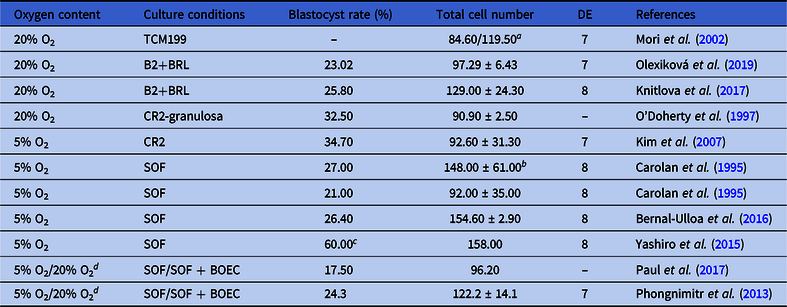
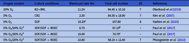
Tables 2 and 3 summarize the data on the total cell numbers of blastocysts cultured under different culture conditions, obtained from fresh (Table 2) and vitrified (Table 3) bovine oocytes. The published data on blastocyst cell numbers in different articles are often inconsistent. In particular, some authors report average cell number counted for all blastocysts, including all, expanded, normal and, sometimes, hatching blastocysts, while other authors represent the numbers of expanded and normal blastocysts separately. This fact may cause high variability in embryo cell counts among studies.
Difference in total cell number of blastocysts obtained from fresh oocytes using different oxygen content and culture conditions

B2+BRL, B2 medium + buffalo rat liver cell monolayer; BOEC, oviduct epithelial cells; DE, day of evaluation; SOF, synthetic oviduct fluid.
a Value before the forward slash indicates the cell number in blastocysts and the value after the forward slash indicates the number of cells in expanded blastocysts evaluated on day 7.
b Improved total cell number using addition of 10% FCS to SOF after 20–24 h of in vitro culture.
c Blastocysts harvested on days 7+8.
d Oxygen content (from 5% to 20%) and IVC medium (from SOF to SOF + OEC) were changed when embryos reached 8-cell stage.
Difference in total cell number of blastocysts obtained from vitrified–thawed oocytes using different oxygen content and culture conditions

B2+BRL, B2 medium + buffalo rat liver cell monolayer; BOEC, bovine oviduct epithelial cells; DE, day of evaluation; SOF, synthetic oviduct fluid.
a Blastocysts harvested on days 7+8.
b The blastocyst rate and cell number obtained using two-step vitrification.
c The blastocyst rate and cell number obtained using three-step vitrification.
d Oxygen content (from 5% to 20%) and IVC medium (from SOF to SOF + OEC) were changed when embryos reached the 8-cell stage.
Some studies on cattle embryos (Carolan et al., Reference Carolan, Lonergan, Van Langendonckt and Mermillod1995; Curnow et al., Reference Curnow, Ryan, Saunders and Hayes2010; Yashiro et al., Reference Yashiro, Tagiri, Ogawa, Tashima, Takashima, Hara, Hirabayashi and Hichi2015; Bernal-Ulloa et al., Reference Bernal-Ulloa, Lucas-Hahn, Herrmann, Hadeler, Aldag, Baulain and Niemann2016) documented a significantly higher number of cells in embryos incubated at reduced oxygen content (5% O2) compared with the classical system (5% CO2 and 20% O2). The number of cells in blastocysts can also be influenced by maturation time. For example, Park et al. (Reference Park, Kim, Kim and Park2005) in their study described 130-cell (average value) bovine blastocysts developed after an 18 h maturation compared with 112-cell blastocysts at 24 h maturation. Mori et al. (Reference Mori, Otoi and Suzuki2002) counted 84.6 cells in day 7 bovine blastocysts, while expanded blastocysts contained up to 119.5 cells. In our previous study (Olexiková et al., Reference Olexiková, Makarevich, Bedeová and Kubovičová2019), blastocysts derived from vitrified bovine oocytes contained on average 84.45 cells vs. 97.29 cells in the blastocysts of the control fresh group. A higher blastocyst cell number (n = 148) was achieved by Carolan et al. (Reference Carolan, Lonergan, Van Langendonckt and Mermillod1995) after 20–24 h culture in SOF medium supplemented with serum at 5% O2, compared with 92 cells without serum addition.
Yashiro et al. (Reference Yashiro, Tagiri, Ogawa, Tashima, Takashima, Hara, Hirabayashi and Hichi2015) reported that the addition of α-tocopherol to the recovery medium improved the survival of bovine oocytes after thawing, the blastocyst rate as well as the total cell number (143.4) compared with the untreated group (107.8). Irrespectively of the addition of tocopherol, the number of cells in blastocysts from vitrified oocytes was still higher when the culture was performed under conditions of 5% O2 using SOF culture medium.
Type and concentration of cryoprotectants
During vitrification, an intracellular ice is formed that can damage the cell. This damage can be reduced or eliminated using cryoprotectants (Kasai, Reference Kasai2002; Chian et al., Reference Chian, Wang and Li2014); the type and concentration of cryoprotectants are important.
Before placing the oocytes into freezing medium with a cryoprotectant, most authors (Martins et al., Reference Martins, Costa, Chagas, Ignácio, Torres and McManus2005; Ortiz-Escribano et al., Reference Ortiz-Escribano, Smits, Piepers, Van den Abbeel, Woelderst and Van Soom2016; Makarevich et al., Reference Makarevich, Olexiková, Bedeová and Kubovičová2019b) used pre-incubation of bovine oocytes in the equilibration medium with a cryoprotectant beginning at concentrations lower (3–40%) than would be used in the vitrification medium (30–40%). However, vitrification of murine oocytes was performed without equilibration with almost a 50% successful birth rate after the transfer of two-cell embryos (Nakagata, Reference Nakagata1989). Martins et al. (Reference Martins, Costa, Chagas, Ignácio, Torres and McManus2005) did not observe any differences in the survivability of vitrified bovine oocytes, which were pre-equilibrated for 0.5, 5 or 15 min in different concentrations of CPs. However, different concentrations and time of equilibration produced significant differences in the ability of oocytes to reach the MII stage after vitrification. The highest concentration of ethylene glycol (EG; 40%) in equilibration solution was the most harmful after 5 and 15 min of equilibration. The medium EG concentration (20%) and 5 min of equilibration showed the highest (44.5%) MII rate after vitrification–thawing of immature oocytes. Moreover, the lowest tested concentration (3%) of EG showed better results (MII rate: 18.1–19.4%) compared with the 40% of EG during 5 and 15 min of equilibration.
During vitrification, the cryoprotectant penetrates the oocyte and causes replacement of intracellular water, its dehydration and subsequent rehydration after thawing. Therefore, different membrane permeabilities of the oocyte at different stages of maturation should be considered. In particular, the movement of a cryoprotectant and water is slower in bovine oocytes at the MII stage, as the plasma membrane has a low permeability coefficient (Mogas, Reference Mogas2019).
The most commonly used penetrating CPs used for bovine oocytes are EG and dimethylsulfoxide (DMSO). Some authors have also reported the effects of other cryoprotectants, such as 1,2-propanediol (PROH), glycerol (cattle: Chian et al., Reference Chian, Kuwayama, Tan, Tan, Kato and Nagai2004; buffalo: El-Shahat and Hammam, Reference El-Shahat and Hammam2014) or polymer Ficoll 70 (Abdel-Gawad et al., Reference Abdel-Gawad, Abdel-Halim, Helmy and Badr2016). The latter authors tested the effects of different CP combinations on vitrification of bovine oocytes at different volume ratios: 20% DMSO + 20% EG + 0.3M trehalose vs. 40 % EG + 18% Ficoll 70. Although they did not find any significant differences in blastocyst rate (blastocysts from vitrified oocytes) between the tested concentrations, significant difference was noted in the recovery rate of frozen–thawed blastocysts between the DMSO group (45%) and the Ficoll 70 group (79.28%). Anchamparuthy et al. (Reference Anchamparuthy, Dhali, Lott, Pearson and Gwazdauskas2009) obtained a lower blastocyst rate from vitrified oocytes using Ficoll in combination with EG and sucrose in vitrification solution compared with Abdel-Gawad et al. (Reference Abdel-Gawad, Abdel-Halim, Helmy and Badr2016).
Sucrose as a non-penetrating cryoprotectant is often added to the vitrification medium together with EG, as it acts as a membrane stabilizer, minimizing the harmful effects of EG on membrane integrity of mouse oocytes (Hotamisligil et al., Reference Hotamisligil, Toner and Powers1996). The beneficial effects of sucrose added at the 0.4–0.8 M range have also been reported for the vitrification of mouse ovarian tissues (Zhang et al., Reference Zhang, Li, Liu, Yang and Wan2009). Martins et al. (Reference Martins, Costa, Chagas, Ignácio, Torres and McManus2005) also achieved a better percentage of bovine oocyte maturation using sucrose compared with trehalose.
Several authors have described the positive effects of antifreeze proteins (AFPs) on cryopreservation of mouse oocytes (Jo et al., Reference Jo, Jee, Lee and Suh2011), as well as rabbit (Nishijima et al., Reference Nishijima, Tanaka, Sakai, Koshimoto, Morimoto, Watanabe, Fan and Kitajima2014) and cattle (Liang et al., Reference Liang, Yuan, Jin, Zhang, Bang and Kim2017) embryos. AFPs are able to bind to ice during vitrification and therefore prevent crystallization. However, Chaves et al. (Reference Chaves, Campelo, Silva, Bhat, Teixeira, Melo, Souza-Fabjan, Mermillod and Freitas2016) did not note any cryoprotective effects of these proteins during vitrification of bovine oocytes/embryos. Supplementation of AFPs improved embryo development only up to day 5, while none of the developing embryos reached the blastocyst stage on day 7 when using 500 ng/ml AFPs compared with 6.3% of blastocysts using 1000 ng/ml of AFPs (Chaves et al., Reference Chaves, Campelo, Silva, Bhat, Teixeira, Melo, Souza-Fabjan, Mermillod and Freitas2016).
A combination of multiple CPs is used mostly at lower concentrations of the individual components to reduce the toxic effect of a high total concentration of CPs. The combination of DMSO and EG with the addition of a non-penetrating CP such as sucrose is used more frequently. Chian et al. (Reference Chian, Kuwayama, Tan, Tan, Kato and Nagai2004) obtained a higher percentage of bovine blastocysts (7.4%) using the combination of EG with PROH compared with 1.7% blastocysts using EG with DMSO. Therefore, DMSO appears to be less suitable for cryopreservation of bovine oocytes compared with EG, corresponding to the study by Cetin and Bastan (Reference Cetin and Bastan2006) who found that EG was less harmful to bovine oocytes compared with DMSO. Despite this, DMSO is still a commonly used cryoprotectant, because it is a smaller molecule and, therefore, more permeable to membranes compared with glycerol (Yamada et al., Reference Yamada, Caetano, Simoes, Nicacio, Feitosa, Assumpção and Visintin2007).
An important factor for successful cryopreservation is the CP concentration. At vitrification after 18 h of maturation, less oocytes developed to the blastocyst stage compared with immature bovine oocytes vitrified using 40% EG (7.17 M) (Magnusson et al., Reference Magnusson, Feitosa, Goissis, Yamada, Tavares, D’Avila Assumpção and Visintin2008). The authors further reported a higher cleavage (19.39% vs. 7.29%) and blastocysts rate (8.16% vs. 4.17%) at a higher concentration of EG (7.17 M) compared with a lower concentration (5.37 M) at the MII stage. Papis et al. (Reference Papis, Shimizu and Izaike2000) obtained the highest blastocyst rate (29.6%) using 3% of EG in the equilibration medium for 12 min followed by 30 s in vitrification medium with 5.5 M EG and 1 M sucrose.
Yamada et al. (Reference Yamada, Caetano, Simoes, Nicacio, Feitosa, Assumpção and Visintin2007) reported a higher percentage of mature bovine oocytes (29.2%) after vitrification with a higher concentration of CPs (25% EG + 25% DMSO + 0.5 M trehalose) compared with a lower concentration of CPs (20% EG + 20% DMSO + 0.5 M trehalose; 11.7% of mature oocytes). The lowest maturation rate (4.3% of mature oocytes) was reached using a combination of EG and glycerol (25% EG + 25% G + 0.5 M trehalose). However, the differences were not only in concentration, but also in length of CP exposure but without vitrification. The exposure of oocytes to low CP concentrations in a vitrification medium for 30 s did not show any significant difference compared with the fresh control. Nevertheless, higher concentrations of CPs seemed to be more toxic especially at 60 s of exposure compared with the control fresh group.
El-Shahat and Hammam (Reference El-Shahat and Hammam2014) compared the effects of EG (7 M), DMSO (7 M) and G (4 M) on embryo development from vitrified immature buffalo oocytes. Developmental capacity of embryos derived from vitrified thawed oocytes using glycerol as the cryoprotectant was lower compared with EG and DMSO. Lim et al. (Reference Lim, Ko, Hwang, Chung and Niwa1999) reported similar results, i.e. a reduced proportion of morphologically normal bovine oocytes after vitrification using G versus DMSO and propylene glycol. In immature buffalo oocytes, Wani et al. (Reference Wani, Misra and Maurya2004) obtained a lower oocyte maturation rate after the exposure to 7 M of G compared with DMSO, EG and PROH.
As referenced in Paynter (Reference Paynter2005), glycerol appears to be unsuitable as a cryoprotectant, particularly for mature mouse oocytes, as it has low permeability and causes extreme cell shrinkage compared with other CPs. This author also mentioned the temperature as an important factor for cell shrinkage and swelling. At higher temperatures, oocyte shrinkage decreases, reducing osmotic stress, but the potential toxicity of CPs increases. For example, at 30°C, oocytes shrank more in the presence of EG compared with DMSO, while at 10°C the volume change was almost the same. Temperature range may vary with type of cryoprotectant, so it is important to adapt the protocol for each cryoprotectant to minimize cell damage.
Permeability of EG and DMSO is different for GV or MII stages of bovine oocytes (Agca et al., Reference Agca, Liu, Peter, Critser and Critse1998). Therefore, the developmental stage of the oocyte should be considered when determining the CP concentration.
Cooling/warming rate and post-thaw oocyte development
Cooling rate (CR) and warming rate (WR) can also be key factors causing differences in bovine oocyte or embryo survival after vitrification. At slow freezing using programmable devices, the CR at the beginning of the procedure is around 0.3–2°C/min, compared with more than 20,000°C/min CR during vitrification using carriers developed for direct contact with LN2 (Chian et al., Reference Chian, Wang and Li2014).
Arav et al. (Reference Arav, Natan, Kalo, Komsky-Elbaz, Roth, Levi-Setti and Leong2018) in their study using a Sarah® vitrification device with a CR of 18,000°C/min, obtained 7% blastocysts from vitrified bovine oocytes. Kuwayama (Reference Kuwayama2005) in their study using human oocytes obtained a higher blastocyst rate (22.9%) using a Cryotop system with the highest CR of 22,800°C/min, compared with OPS (7.4% blastocysts with 16 340°C/min CR) and in-straw dilution (5.3% blastocyst with CR 4460°C/min). Zhou et al. (Reference Zhou, Al Naib, Sun and Lonergan2010) in their study using a Cryotop system with a CR of 23,000°C/min obtained 4.0–11.3% bovine blastocysts.
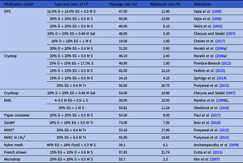
Misumi et al. (Reference Misumi, Hirayama, Egawa, Yamashita, Hoshi and Imai2013) found that the CR value using the microvolume air cooling (MVAC) technique (860°C/min) was approximately 20 times slower compared with CR using the Cryotop system (17,250°C/min). However, they did not note differences in survival and hatching rates after IVC of vitrified porcine embryos using these two CR rates. Later, Punyawai et al. (Reference Punyawai, Anakkul, Srirattana, Aikawa, Sangsritavong, Nagai, Imai and Parnpai2015), using the above-mentioned vitrification carriers (MVAC and Cryotop) for bovine mature oocytes and embryos, observed no differences in the hatched blastocyst rates after vitrification of bovine blastocysts. For vitrified oocytes, there was only a slight insignificant increase in blastocyst rate using MVAC compared with the Cryotop system (Table 4).
Development of embryos from vitrified oocytes using various vitrification carriers

D, DMSO; EG, ethylene glycol; EMG, electron microscopy grids; Gal, galactose; MVAC, micro volume air cooling; OPS, open pulled straw; S, sucrose; Tr, trehalose.
a IVM oocytes vitrified using an MVAC device without direct contact with LN2.
b IVM oocytes vitrified using an MVAC device directly plunged into LN2.
Hochi et al. (Reference Hochi, Akiyama, Minagawa, Kimura and Hanada2001), using vitrification of bovine oocytes in capillaries of 1400 µm with CR 3000°C/min and WR 8000°C/min, obtained the highest fertilization rate compared with other tested values of CR and WR. However, they also concluded that survival of in vitro matured COCs vitrified using OPS was not affected by WR. Conversely, Mazur and Seki (Reference Mazur and Seki2011) reported that mouse oocyte survival after vitrification using the Cryotop system depended on the WR rather than the previous CR. In particular, up to 70–80% oocytes survived using the highest WR (117,000°C/min), while only 0–25% of oocytes survived using the lowest WR (600°C/min) regardless of the CR (95 to 69,000°C/min). These authors also noted that the higher the CR used, the higher was the WR required to achieve better oocyte survival.
Although most authors in their reports specified only CR values, the above cited studies suggested that ‘WR’ is also an important parameter of successful vitrification (blastocyst rate). Therefore, WR should be taken into consideration when evaluating vitrification success.
Vitrification carriers and post-thaw embryo development
Vitrification carriers are increasingly used to provide a minimum sample volume prior to freezing. A minimum volume of vitrification medium allows a sufficiently high CR for successful vitrification. This method is suitable for oocytes, because they are vitrified in small numbers compared with other cells or tissues, which have larger volumes. The survival and quality of oocytes undergoing cryopreservation has improved using a minimal volume technique. The only difficulty of this approach is the need for experience in handling oocytes and small volumes of medium (Zhang et al., Reference Zhang, Catalano, Gurkan, Khimji and Demirci2011). A summary of blastocyst production from vitrified oocytes using various vitrification carriers is given in Table 4.
The most commonly used commercial carriers are the OPS system, Cryotop and Cryoloop. Another alternative is electron microscope grids (EM grids), which are mainly used to provide volumes less than 1 μl (Zhang et al., Reference Zhang, Catalano, Gurkan, Khimji and Demirci2011). Morató et al. (Reference Morató, Izquierdo, Paramio and Mogas2008a) described Cryotop as a more effective vitrification carrier compared with OPS in cattle, because the percentage of blastocyst development after in vitro fertilization was 0% with OPS, while with Cryotop it was 2.4%. Similar results (1% of blastocysts) using the OPS method have been reported by Chaves et al. (Reference Chaves, Corbin, Almanana, Locatelli, Souza-Fabjan, Bhat, Freitas and Mermillod2017). Slightly better results (6.1% of blastocysts on day 8) were reported by Vieira et al. (Reference Vieira, Mezzalira, Barbieri, Lehmkuhl, Rubin and Vajta2002) using immature bovine oocytes.
Zhou et al. (Reference Zhou, Al Naib, Sun and Lonergan2010) in their study, using the Cryotop method, obtained 5% bovine blastocysts from oocytes at the MII stage after vitrification vs. 35.6% in the control group. A similar blastocyst rate in the control group (34.4%) was described by Prentice-Biensch et al. (Reference Prentice-Biensch, Singh, Mapletoft and Anzar2012), whereas they obtained only a 1.6% rate for blastocysts after oocyte vitrification. Sprícigo et al. (Reference Sprícigo, Diógenes, Leme, Guimaräes, Muterlle, Silva, Sola-Oriol, Pivato, Silva and Dode2015) also obtained a low blastocyst rate (2.8%) compared with the control fresh group (37.9%).
Cryoloops are devices that use a small Nylon loop attached to a lid by a small metal tube, allowing easier handling (Mukaida et al., Reference Mukaida, Takahashi and Kasai2003).
Dinnyés et al. (Reference Dinnyés, Dai, Jiang and Yang2000) used solid surface vitrification of mature bovine oocytes. Interestingly, they obtained a higher percentage of blastocysts (15%) at immediate freezing and thawing procedures compared with 6% blastocysts, when thawing was performed after 2–3 weeks. Checura and Seidel (Reference Checura and Seidel2007) did not find any significant differences in blastocyst rate between the groups of vitrified/immediately thawed and long-term stored/thawed bovine oocytes. Using the Cryoloop technique, they observed a greater percentage of blastocysts (12.9%) compared with OPS (5.4%).
Anchamparuthy et al. (Reference Anchamparuthy, Dhali, Lott, Pearson and Gwazdauskas2009) produced 5.1% bovine blastocysts using a Nylon mesh as the vitrification carrier, compared with 22.9% in the non-vitrified group.
EM grids are another carrier useful for oocyte vitrification, due to the ability to aspirate the maximum excess freezing medium from the mesh. Using EM grids, Martino et al. (Reference Martino, Songsasen and Leibo1996b) obtained 15% bovine blastocysts, although this was lower compared with the control group (41%). In our previous study (Olexiková et al., Reference Olexiková, Makarevich, Bedeová and Kubovičová2019), we approached the results of Martino et al. (Reference Martino, Songsasen and Leibo1996b), achieving 11% blastocysts derived from vitrified mature oocytes. However, Kim et al. (Reference Kim, Kim, Yi, Yoon, Park, Chung and Lim1998) produced 20% blastocysts from immature vitrified bovine oocytes using EM grids.
Dutta et al. (Reference Dutta, Dev and Raj2013) obtained a high cleavage rate (97.8%) and blastocyst rate (21.7%) after vitrification of immature bovine oocytes in 0.25 ml French straws.
Among other various alternative carriers, a paper container was used, which was advantageous for immediate aspiration of cryoprotectant (Kim et al., Reference Kim, Uhm, Gupta, Yang, Lim, Das, Heo, Chung, Kong, Kim, Lee and Ko2012; Paul et al., Reference Paul, Liang, Srirattana, Nagai and Parnpai2017). However, to date, a methodological approach for separating oocytes or embryos from a paper container has not been described and, therefore, difficult to reproduce. Another option is the use of automatic vitrification devices, such as Sarah®, which produced 7% bovine blastocysts compared with the control group (11%; Arav et al., Reference Arav, Natan, Kalo, Komsky-Elbaz, Roth, Levi-Setti and Leong2018). Punyawai et al. (Reference Punyawai, Anakkul, Srirattana, Aikawa, Sangsritavong, Nagai, Imai and Parnpai2015) tested the MVAC technique, which prevents the contact of cryopreserved cells with liquid nitrogen, on bovine oocytes. They did not find any significant difference in the blastocyst rate compared with the Cryotop technique (17.9% vs. 20.7%). Papis et al. (Reference Papis, Shimizu and Izaike2000) combined the straw freezing system with this popular technique using a minimum volume of medium in microdroplets without carrier and obtained 6.1–29.6% bovine blastocysts when using different concentrations of equilibration medium, compared with the control group (41.5%). Kim et al. (Reference Kim, Park, Kim, Hwang, Yang, Im, Chung, Seong, Moon and Yang2007) yielded 2.3% bovine blastocysts after freezing in microdroplets using a glass micropipette.
Effect of age of oocyte donors
The age of oocyte donors (cows or heifers) can also affect blastocyst yield after oocyte vitrification. In particular, Albarracín et al. (Reference Albarracín, Morató, Rojas and Mogas2005) in their study reported a large difference in blastocyst yield between cows and heifers (33% vs. 7.8%) in the control group, with only 2.5% blastocysts from cow oocytes and none from heifer oocytes after vitrification with OPS. Morató et al. (Reference Morató, Izquierdo, Albaracín, Anguita, Palomo, Jiménez-Macedo, Paramio and Mogas2008b) did not obtain any blastocysts after vitrification using the OPS method either in cows or heifers, but the embryo cleavage rate in heifers was significantly lower compared with that in cows (23.5% vs. 34%). These authors obtained 3.2% blastocysts from cow oocytes after vitrification in the presence of 1 M taxol, an actin cytoskeleton stabilizer. This effect of taxol was not observed in heifers. It appears that oocytes from heifers are more sensitive to cytoskeletal damage by vitrification compared with oocytes from other cows.
Conclusion
Vitrification of oocytes is still challenging not only in cattle but also in other animal species. The optimization of the methodology is demanding due to some factors that affect the vitrification process, in vitro fertilization and subsequent embryo development. Even using the same protocols and culture systems, the results of embryo development after oocyte vitrification are influenced, in addition to operator skill and experience, by the geographical location of experimental institutions or research centres, local climate conditions, animal breeds used as an oocyte donor and quality of biological material delivered to the laboratory.
These factors cause relatively high variability among results from individual studies. The selection of the appropriate maturation stage of an oocyte before vitrification, as well as presence or absence of cumulus cells, are the basic factors that ensure the success of the whole process. Based on the results summarized in this review, it is difficult to suggest unambiguously which is the proper maturation stage of oocytes that is most suitable for vitrification, as well as to certainly confirm if the presence or absence of cumulus cells is beneficial. This task hinges on individual experimental design, research conditions in the laboratory and probably the personal experience of the researcher.
Culture conditions and medium composition for IVP of embryos is another criterion required for better embryo development after cryopreservation. The addition of different substances such as antioxidants, hormones and growth factors to maturation or culture media is a sufficient way to increase oocyte cryotolerance and ensure lower cell damage and higher blastocyst rate. To confirm the influence of tested substances on embryos, completely defined culture conditions are required.
From the results of different studies that tested different types of CPs, it was obvious that glycerol was not adequate as a cryoprotectant for bovine oocytes, compared with EG or DMSO. For sugars, the use of trehalose, as a non-penetrating cryoprotectant, produced worse results compared with sucrose. Usually, the combination of several CPs in vitrification medium was used to minimize toxicity as opposed to the use of one CP alone at high concentrations. The concentration and exposure time of CPs also played a crucial role in the whole vitrification process. Higher concentrations and longer exposure times could cause extensive damage during vitrification.
The results of studies focussed on measurement of cooling and warming rates suggested the importance of these parameters (in their mutual interrelation) for successful oocyte survival after vitrification.
The specimen carrier chosen for vitrification also played an important role in the process of vitrification and storage of oocytes in liquid nitrogen. In this regard, minimum volume size carriers represented a better choice, as they ensure faster freezing of the oocyte, however final success depended on the proper combination, type and, especially, concentration of cryoprotectants.
Age of oocyte donors appeared to be a factor contributing to the variability in vitrification survival results. As reviewed here, heifer oocytes were more vulnerable to vitrification compared with cow oocytes.
This review presents a summary of the positive and adverse factors influencing cryotolerance and development of bovine oocytes following cryopreservation using different vitrification protocols. At present, no consistent approach for successful oocyte vitrification has been published, this review may generate possible ideas for further modification of cryopreservation and IVP protocols, adjusted to the conditions of a specific laboratory, to ensure both better oocyte cryotolerance and higher blastocyst rate.
Acknowledgements
The authors thank Andrea Kunova (Università degli Studi di Milano) for critical editing and language correction.
Financial support
This study was realized with funding from the Agency for the Support of Science and Technology in the Slovak Republic (grant APVV-15–0196).
Conflict of interest
Hereby, I declare that there are no financial or personal relationships between all co-authors and other people or organizations that could inappropriately influence their work.
Ethical standards
With reference to this type of article (review), no animals were involved.