Introduction
Oocyte cryopreservation is an important practice in assisted reproductive technology. Despite the first pregnancies obtained with transfers of embryos derived from frozen oocytes reported in the 1980s (Chen, Reference Chen1986), it was only with the advent of vitrification that oocyte cryopreservation was considered safe and efficient to achieve successful in vitro fertilization (IVF) outcomes (Coello et al., Reference Coello, Pellicer and Cobo2018). Vitrification is now considered the gold standard in oocyte freezing (Iussig et al., Reference Iussig, Maggiulli, Fabozzi, Bertelle, Vaiarelli, Cimadomo, Ubaldi and Rienzi2019).
Many indications exist for oocyte cryopreservation, such as fertility preservation in oncological patients and in those wishing to delay motherhood for personal reasons. In addition, oocyte cryopreservation can be used for oocyte donation treatment, which is a well established treatment for female infertility that allows oocyte recipients to become mothers.
Oocyte donation practice possesses some intrinsic difficulties related to synchronization between the donor cycle and the recipient endometrium (Trokoudes et al., Reference Trokoudes, Pavlides and Zhang2011), therefore the use of cryopreserved donor oocytes is often seen in donation cycles. Moreover, egg-cryobanking provides recipients with more options in oocyte selection, and higher flexibility in pregnancy scheduling (Solé et al., Reference Solé, Santaló, Boada, Clua, Rodríguez, Martínez, Coroleu, Barri and Veiga2013).
Brazilian IVF centres may not conduct oocyte donation cycles for commercial purposes. Therefore, young patients undergoing IVF treatment who have surplus oocytes make oocyte cryobanking and donation accessible in this country. In this way, oocytes available for donation are obtained from infertile couples, therefore creating an interesting scenario in which the competence and development potential of vitrified and fresh oocytes from the same donor can be compared.
At present, there is no evidence that ensures that cryopreservation of shared eggs is not detrimental to recipient treatment outcomes. The objective of this study was to investigate the influence of cryopreservation on donated eggs in terms of laboratory outcomes of intracytoplasmic sperm injection (ICSI) cycles.
Materials and Methods
Patients and experimental design
This retrospective cohort study was performed in a private university-affiliated IVF centre, between January 2015 and January 2018. Data were obtained via chart review of 267 vitrified oocyte donor ICSI cycles, and 320 oocyte recipients who were undergoing 307 vitrified and 119 fresh oocyte recipient ICSI cycles, and participating in an egg-sharing donation programme.
Oocyte donors were between the age of 19 and 34 years, and recipients were between the age of 26 and 48 years. The effect of cryopreservation on recipient ICSI outcome was investigated.
All patients signed a written informed consent form in which they agreed to share their treatment outcomes for research purposes; the study was approved by the local Institutional Review Board.
Controlled ovarian stimulation and laboratory procedures
Controlled ovarian stimulation was performed using recombinant follicle-stimulating hormone (r-FSH; Gonal-F®, Serono, Geneva, Switzerland) and pituitary blockage using a gonadotropin-releasing hormone (GnRH) antagonist, cetrorelix acetate (Cetrotide®, Merck KGaA, Serono, Geneva, Switzerland).
Leuprolide acetate (Lupron; TAP Pharmaceuticals, North Chicago, IL, USA) was administered to trigger the final follicular maturation. Oocytes were collected 35 h later through transvaginal ultrasound ovum pickup.
The recovered oocytes were assessed to determine their nuclear status, and those in metaphase II were vitrified, if destined for the oocyte recipients.
Oocyte vitrification and warming
Both vitrification and the warming procedures were performed using the Cryotop method (Kuwayama et al., Reference Kuwayama, Vajta, Kato and Leibo2005). Briefly, within 3 h after ovum pickup, vitrification was achieved by exposure of oocytes initially to the equilibration solution, followed by a 30-second exposure to the vitrification solution.
Individual oocytes were then picked up in an extremely small volume (<0.1 ml) of vitrification solution, to facilitate rapid cooling, and placed on top of a very fine polypropylene strip attached to a hard-plastic handle. As soon as the oocyte was placed onto the thin polypropylene strip of the Cryotop, it was immediately submerged vertically into liquid nitrogen. Then the thin strip was covered with a hard-plastic cover on top of the Cryotop sheet.
For warming, the protective cover was removed from the Cryotop while it was still submerged in liquid nitrogen, and the polypropylene strip of the Cryotop was immersed directly into the thawing solution at 37 °C for 1 min. Oocytes were retrieved and transferred into dilution solution for 3 min and then washed twice in the washing solution for 5 min each. The tools and solutions required for the vitrification and warming processes were obtained from Kitazato® (Tokyo, Japan). Within 3 h after warming, oocytes were submitted to ICSI.
Embryo culture and morphology evaluation
Approximately 16 h after ICSI, fertilization was confirmed by the presence of two pronuclei and the extrusion of the second polar body. The embryos were maintained in a 50-µl drop of culture medium (Global®; LifeGlobal, CT, USA) covered with paraffin oil in a humidified atmosphere under 6% CO2 in air at 37°C for 5 days.
Embryo morphology was assessed 16–18 h post-ICSI and on the mornings of days 2, 3, and 5 of embryo development using an inverted Nikon Diaphot microscope (Eclipse TE 300; Nikon, Tokyo, Japan) with a Hoffmann modulation contrast system under ×400 magnification.
High-quality cleavage-stage embryos were defined as those with all of the following characteristics: 4 cells on day 2 or 8−10 cells on day 3, <15% fragmentation, symmetrical blastomeres, and absence of multinucleation.
Blastocyst morphology was evaluated according to the degree of expansion and hatching status. Full blastocysts onwards, presenting morphologically normal inner cell mass and trophectoderm (TE) were defined as high-quality blastocysts.
Endometrial preparation
For recipients, after menses, endometrial development was followed by ultrasound examination, and the patients received 200 µg of transdermal 17β estradiol every 3 days (Estradot®, Noven Pharmaceuticals, Miami, USA).
Approximately 7 days after initiation of 17β estradiol administration, serum E2 levels and endometrial thickness were determined. When the endometrium showed proliferative morphology and thickness of at least 7.5 mm, 600 mg of progesterone (Utrogestan®, Farmoquímica, Rio de Janeiro, Brazil) was vaginally administered per day.
Both 17β estradiol and progesterone were administered concomitantly after embryo transfer and were suspended following a negative βhCG test. After a positive βhCG test, 17β estradiol and progesterone treatments were maintained until weeks 6 and 12 of gestation, respectively.
Embryo transfer and clinical follow-up
Embryo transfers were performed on day 5 of embryo development and one or two embryos were transferred per patient.
A clinical pregnancy was diagnosed when the fetal heartbeat was detected at 6 weeks’ pregnancy. Pregnancy rates were calculated per transfer. The implantation rate was calculated by dividing the number of gestational sacs with fetal heartbeats by the number of transferred embryos. Miscarriage was defined as clinical pregnancy loss before 20 weeks.
Data analysis and statistics
The sample size calculation suggested that 199 cycles would be enough to demonstrate a 20% effect with 80% power and 5% significance level, considering a primary outcome blastocyst development rate.
The effect of oocyte cryopreservation on recipient ICSI outcome was investigated using a general mixed models fit by restricted maximum likelihood, followed by Bonferroni post hoc test to compare the means between fresh and warm oocyte donation groups. Linear mixed effect models were generated using covariates as fixed effects and egg donors and egg recipients as random effects, with unstructured covariance structure, adjusted for oocyte dimorphisms. A Gaussian distribution was assumed and the normal distribution of model residuals was checked to confirm goodness of fit. Final model selection was decided using the Akaike information criterion and Schwarz’s Bayesian criterion. Results were expressed as means ± standard error and P-value. A P-value < 0.05 was considered statistically significant. Data analyses were conducted using SPSS Statistics 21 (IBM, New York, NY, USA).
Results
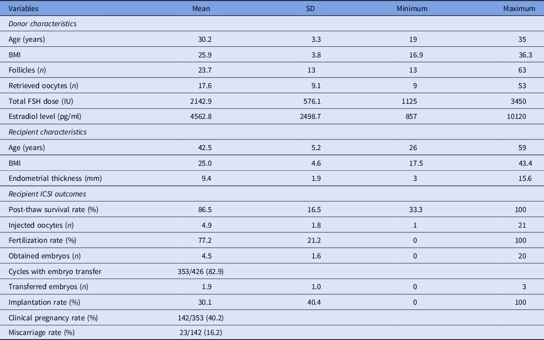
Demographic data concerning donors and recipients, as well as general characteristics of recipient ICSI cycles are shown in Table 1.
Donors and recipients demographic data and recipients’ ICSI cycles outcomes (n = 426)

Note: BMI, body mass index; ICSI, intracytoplasmic sperm injection; IU, international unit; SD, standard deviation. Values are mean ± SD, unless otherwise noted.
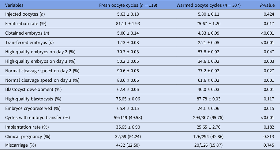
Results from generalized method of moments (GMM) analysis are shown in Table 2. The fertilization rate (81.1% vs. 75.7%, P = 0.017), high-quality embryos rate on days 2 (70.3% vs. 57.8%, P = 0.047) and 3 (50.2% vs. 34.6%, P = 0.003), normal cleavage speed rate on days 2 (90.6% vs. 77.2%, P = 0.027) and 3 (83.6% vs. 61.6%, P = 0.001), and blastocyst development rate (62.4% vs. 40.0%, P = 0.001) were significantly higher for fresh oocyte donation cycles compared with warmed oocyte donation cycles. There were no statistically significant differences between fresh and warmed oocyte donation cycles in terms of high-quality blastocyst rate, implantation rate, clinical pregnancy rate, and miscarriage rate. The surplus embryos cryopreservation rate was significantly higher for fresh cycles compared with warmed cycles (65.4% vs. 24.1%, P = 0.015).
Descriptive analysis of recipients’ ICSI cycles outcomes using fresh (n = 119) or warmed oocytes (n = 307)

Note: ICSI, intracytoplasmic sperm injection. Values are mean ± standard error, unless otherwise noted.
Discussion
The present study compared development and morphology of embryos generated in fresh and frozen donated oocytes cycles, with the aim of detecting whether the performance after thawing was similar to that observed in the corresponding fresh cycle.
The first significant difference was a reduced fertilization rate in thawed oocytes, representing their decreased viability irrespective of survival to cryopreservation. It was also noted that embryo development and morphology were reduced in thawed cycles compared with their fresh counterparts. This was evident immediately from day 2 of embryo development to blastocyst development. Cleavage defects after oocyte cryopreservation are a consequence of cryogenic damage that may ultimately affect pregnancy outcomes.
Although evaluation of clinical outcomes was beyond the objective of this study, there were no statistically significant differences between fresh and warmed oocyte donation cycles in terms of pregnancy outcomes. We wonder if there would be a significant difference in implantation and pregnancy outcomes if a higher proportion of embryo transfers had been performed in fresh oocyte cycles, therefore allowing an appropriate comparison with the outcomes of thawed oocytes. As expected, a higher rate of cryopreserved embryos was observed in fresh oocyte donation cycles compared with thawed cycles, mainly because fresh embryo transfer in fresh oocyte donation cycles are hindered by a lack of synchronization between donors and recipients.
Our results were in accordance with those from previous studies that have shown lower fertilization and embryo quality in thawed oocytes compared with fresh oocytes (Chen et al., Reference Chen, Lien, Chang, Tsai, Ho and Yang2004; Chamayou et al., Reference Chamayou, Alecci, Ragolia, Storaci, Maglia, Russo and Guglielmino2006; Magli et al., Reference Magli, Lappi, Ferraretti, Capoti, Ruberti and Gianaroli2010; Forman et al., Reference Forman, Li, Ferry, Scott, Treff and Scott2012; Siano et al., Reference Siano, Engmann, Nulsen and Benadiva2013). A previous study from our group showed that reduced rates of fertilization, high-quality embryos on days 2 and 3, and blastocyst development were obtained when warmed oocytes were compared with their fresh counterparts (Braga et al., Reference Braga, Setti, Figueira, Azevedo Mde, Iaconelli, Lo Turco and Borges2016). In addition, morphokinetic differences were observed from time to 2-cell to blastocyst development (Cobo et al., Reference Cobo, Coello, Remohí, Serrano, de Los Santos and Meseguer2017), disfavouring vitrified oocytes when compared with fresh oocytes. Conversely, comparable success rates to those achieved with fresh oocytes in terms of clinical outcomes, blastocyst formation, embryo cleavage and fertilization have been observed by other groups (Cobo et al., Reference Cobo, Kuwayama, Pérez, Ruiz, Pellicer and Remohí2008; Rienzi et al., Reference Rienzi, Romano, Albricci, Maggiulli, Capalbo, Baroni, Colamaria, Sapienza and Ubaldi2010; García et al., Reference García, Noriega-Portella and Noriega-Hoces2011; Parmegiani et al., Reference Parmegiani, Cognigni, Bernardi, Cuomo, Ciampaglia, Infante, Tabarelli de Fatis, Arnone, Maccarini and Filicori2011; Trokoudes et al., Reference Trokoudes, Pavlides and Zhang2011; Goldman et al., Reference Goldman, Noyes, Knopman, McCaffrey and Grifo2013). The discrepancy observed between those studies and the present one might be explained by our cohort of oocytes, which was derived from infertile couples, and could be responsible for the reduced outcomes.
The results of this study suggested that low temperatures may alter the regulation of oocyte activation and embryo cleavage. The oocyte is the largest cell in the mammalian body and it is well known that, in cryobiology, the smaller the size of the cell, the better the cryopreservation results.
Several types of injury may occur to human oocytes during cryopreservation. There is a close relationship between the incidence of cryoinjury and the oocyte cryobiological properties, such as sensitivity to chilling and to toxicity of the cryoprotectant, permeability of the plasma membrane to cryoprotectant and water, and tolerance for osmotic swelling and shrinkage (Moussa et al., Reference Moussa, Shu, Zhang and Zeng2014). Some mechanisms of cryoinjury, such as chilling injury, ice crystal formation, and osmotic stress are avoided by vitrification, by virtue of high concentrations of cryoprotectants and high cooling rates associated with the technique.
However, another form of cryoinjury, named multiple aster formation, may occur, especially during vitrification (Hara et al., Reference Hara, Hwang, Kagawa, Kuwayama, Hirabayashi and Hochi2012). During vitrification, oocyte exposure to cryoprotectant and high cooling rate induces multiple aster formation close to male pronucleus, which in turn causes delayed first cleavage, and reduced blastocyst development potential (Hara et al., Reference Hara, Hwang, Kagawa, Kuwayama, Hirabayashi and Hochi2012).
In addition, thawed oocytes showed severe alteration of spindles (Rienzi et al., Reference Rienzi, Martínez, Ubaldi, Minasi, Iacobelli, Tesarik and Greco2004) followed both slow freezing and vitrification methods, suggesting that meiotic and mitotic spindles are especially sensitive to temperature oscillations (Pickering et al., Reference Pickering, Braude, Johnson, Cant and Currie1990; Boiso et al., Reference Boiso, Veiga and Edwards2002). Maternal genes and oocyte proteins (Boiso et al., Reference Boiso, Veiga and Edwards2002), and epigenetic factors such as mitochondrial distribution (Van Blerkom et al., Reference Van Blerkom, Davis and Alexander2000) support the first embryo cleavage. It has been hypothesized that mitochondrial alteration in thawed oocytes could induce flaws in intracellular calcium signalling post sperm injection, which in turn could harm normal embryogenesis (Jones et al., Reference Jones, Van Blerkom, Davis and Toledo2004). More recently, oocyte vitrification was proven to shift the intracellular redox potential towards oxidation (Nohales-Córcoles et al., Reference Nohales-Córcoles, Sevillano-Almerich, Di Emidio, Tatone, Cobo, Dumollard and De Los Santos Molina2016), and the authors suggested that the addition of protective antioxidant molecules to the vitrification and post-warming culture medium to improve embryo cleavage deserved further research.
The strength of this study was the use of a nested statistical analysis that allowed affirmation of the reduced embryo development observed in thawed oocytes compared with fresh oocytes to be independent from oocyte donor characteristics such as age and body mass index as the statistical analysis auto-adjusts the results for potential differences. However, this study also has limitations, such as its retrospective design, and that oocytes included in this study originated from infertile couples undergoing IVF. Nevertheless, oocyte donors were young, and showed adequate ovarian reserve and response to ovarian stimulation. The importance of this study relied on the fact that, in general, the literature reported comparable development in embryos derived from fresh and warmed oocytes but, in the egg-sharing programme scenario, warmed oocytes appeared to behave differently, yielding poorer embryo quality.
In conclusion, in an egg-sharing donation programme, fertilization and embryo developmental competence are reduced when vitrified oocytes are used for ICSI, compared with fresh oocytes. Although no statistical significant differences were observed in terms of pregnancy outcomes, efforts must be made to make donor–recipient matching possible when receiving fresh eggs.
Ethical standards
All patients signed a written informed consent form in which they agreed to share their treatment outcomes for research purposes, and the study was approved by the local Institutional Review Board.
Funding
This research did not receive any specific funding.
Conflicts of interest
The authors declare no conflicts of interest.