Case report
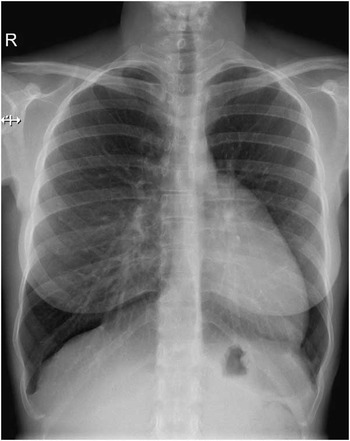
A 33-year-old asymptomatic woman was referred to the cardiology clinic after the sudden death of her brother, whose autopsy revealed a previously undiagnosed hypertrophic cardiomyopathy. Physical examination was unremarkable, except for leftward displacement of the apical impulse. Her 12-lead electrocardiogram showed incomplete right bundle branch block and poor R-wave progression. Chest radiography suggested levoposition of the heart, with no projection of the cardiac silhouette on the right side of the spine, and a band of lucency between the heart and the diaphragm (Fig 1). The transthoracic echocardiogram required the use of atypical lateral views but was considered normal except for the unusual cardiac orientation. Finally, cardiac MRI was performed to confirm the absence of phenotypic signs of hypertrophic cardiomyopathy and ascertain the reason for the other uncommon findings.
Chest radiography showing leftward displacement of the heart (with no projection of the cardiac silhouette on the right side of the spine) and a band of lucency between the heart and the diaphragm.

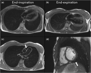
Cardiac MRI confirmed the leftward cardiac displacement and showed marked mobility of the heart with respiratory movements, with the cardiac apex pointing laterally and anteriorly in end-inspiration and slightly posteriorly in end-expiration (Fig 2a and b). The parietal pericardium could not be visualised over the left side of the heart. Cardiac MRI depicted the interposition of lung parenchyma between the aorta and the pulmonary artery and between the inferior surface of the heart and the diaphragm (Fig 2c and d), thus establishing the diagnosis of congenital absence of the pericardium. There were no signs of herniation or incarceration of cardiac structures, and no other developmental anomalies were identified. There were no phenotypic signs of hypertrophic cardiomyopathy either. Given the low risk of complications and as the patient was asymptomatic, a conservative approach was adopted.
( a and b ) Axial T2-weighted half-Fourier acquisition single-shot turbo spin-echo (HASTE) in end-inspiration and expiration, respectively, showing the marked cardiac mobility with respiratory movements; ( c ) axial segmented turbo spin-echo image showing the interposition of lung tissue between the aorta and the main pulmonary artery; ( d ) balanced steady-state free precession (b-SSFP) in left ventricular short-axis view, depicting the presence of lung tissue between the diaphragm and the inferior surface of the heart.

Discussion
Congenital absence of the pericardium is a rare cardiac anomaly. It was first described in the 16th century and is thought to result from premature atrophy of the left common cardiac vein with insufficient blood supply to the pleuropericardium, leading to its agenesis in the early embryonic stage.Reference Faridah and Julsrud 1 Patients are usually asymptomatic, but may sometimes present with positional chest pain, syncope, or even sudden death as a result of cardiac herniation through partial pericardial defects.Reference Gatzoulis, Munk, Merchant, Van Arsdell, McCrindle and Webb 2 The chest radiograph and echocardiogram are very useful in suggesting the diagnosis, which is usually confirmed with cardiac MRI or computed tomography. Rather than documenting the absence of the pericardium itself (which can be very challenging in the absence of adjacent fat), these techniques establish the diagnosis by showing indirect signs that are considered pathognomonic of this condition, such as the interposition of lung tissue between the aorta and the pulmonary artery and between the diaphragm and the inferior surface of the heart.Reference Hutchison 3 Prognosis is usually benign, and most cases are managed conservatively. Surgical pericardioplasty can be considered in highly symptomatic patients or when partial absence of the pericardium leads to herniation or strangulation of the cardiac or vascular structures.Reference Gatzoulis, Munk, Merchant, Van Arsdell, McCrindle and Webb 2 , Reference Abbas, Appleton, Liu and Sweeney 4
Acknowledgements
None.
Financial Support
This research received no specific grant from any funding agency, commercial, or not-for-profit sectors.
Conflicts of Interest
None.