Introduction
The interaction between nanoparticles (NPs) and proteins has long been an active area of study for a wide range of nanotechnology applications.Reference Niemeyer1 For example, NPs have been used as a vehicle to deliver proteins into the cytosol of cells,Reference Tang, Kim, Solfiell, Rana, Mout, Velázquez-Delgado, Chompoosor, Jeong, Yan, Zhu, Kim, Hardy and Rotello2 taking advantage of the enhanced results obtained by NP–protein conjugates compared to proteins alone.Reference Mcnaughton, Cronican, Thompson and Liu3 Similarly, controlling the interaction of NPs with receptors and other biomacromolecules has served as the basis for diagnosticReference Jain4 and sensingReference Saha, Agasti, Kim, Li and Rotello5 applications, where specific and/or selective interactions between NPs and proteins play a defining role. In addition, new hybrid materials can be assembled using NPs and proteins as unit cells, yielding properties unique to these kinds of nanocomposites.Reference Rana, Yeh and Rotello6 Understanding the nature of the interaction between NPs and proteins is of critical importance to achieving tunability of these systems and improving their functional capabilities.
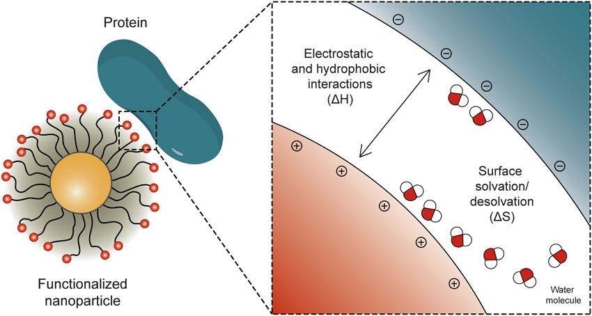
The interaction between NPs and proteins is multiparametric in nature; both NPs and proteins come in a variety of sizes and shapes, each with different charges, acidities, and chemical functionalities. Traditionally, the nature of NP–protein complexes has been assessed using techniques such as dynamic light scattering (DLS), size-exclusion chromatography (SEC), sodium dodecyl sulfate polyacrylamide gel electrophoresis (SDS-PAGE), mass spectrometry (MS), and surface plasmon resonance (SPR).Reference Walkey and Chan7 These studies showed that electrostatic attraction is the most significant noncovalent force acting at the NP–protein interface (Figure 1).Reference You, De and Rotello8 The pH of the solution generally dictates the charge states of proteins and NPs and, hence, the electrostatic force between them.
(a) Schematic of the binding between nanoparticles (NPs) and proteins, illustrating the effects of electrostatic and hydrophobic interactions, as well as the changes in the hydration shells. By controlling the contribution of either of these components (i.e., by engineering the surface chemistry of the NPs), the NP–protein interaction can be modulated or eliminated as desired.

Isothermal titration calorimetry (ITC) studies have shed more light on the fundamental thermodynamics behind the NP–protein interaction.Reference De, Miranda, Rana and Rotello9 These studies have shown that water molecules surrounding both the NP and the protein surface play a significant role in the binding (Figure 1). A model based on the ITC results describes the binding of NPs and proteinsReference De, Miranda, Rana and Rotello9 by the following equations:
 $${\rm{Protein}}\,{\rm{ + }}\,\rightleftharpoons\,{\rm{NP}}{\rm{Protein}} - {\rm{NP}}$$
 $$x{\rm{(H}}_{\rm{2}} {\rm{O)}}_p \,{\rm{ + }}\,y{\rm{(H}}_{\rm{2}} {\rm{O)}}_n \,\rightleftharpoons\,{\rm{(}}x{\rm{\ + \ }}y - z{\rm{)(H}}_{\rm{2}} {\rm{O)}}_{p.n} {\rm{\ + \ }}z{\rm{(H}}_{\rm{2}} {\rm{O)}}$$
 $${\rm{Protein\cdot}}x{\rm{(H}}_{\rm{2}} {\rm{O)}}_p {\rm{\ + \ NP\cdot}}y{\rm{(H}}_{\rm{2}} {\rm{O)}}_n \,\rightleftharpoons\,{\rm{Protein}} - {\rm{NP\cdot(}}x{\rm{\ + \ }}y - z{\rm{)(H}}_{\rm{2}} {\rm{O)}}_{p.n} {\rm{\ + \ }}z{\rm{(H}}_{\rm{2}} {\rm{O),}}$$
where (H2O)p, (H2O)n, and (H2O)p.n represent water molecules associated with proteins, NPs, and NP–protein complexes, respectively. Equation 1 describes the binding between the NP and the protein, where the change in enthalpy (formation of new bonds) and the change in entropy (two entities come together, losing degrees of freedom) are negative. This equation describes a simple interaction, whose favorability would depend only on the release of energy during bond formation.
However, the solvent reorganization (Equation 2) has to be taken into account. The release of water from the solvation shells from both NP and protein implies an increase in both entropy (ΔS > 0, because of the gain of free molecules) and enthalpy (ΔH > 0, because of the rupture of bonds) (Figure 1). Adding the contribution of each of these components to give the overall reaction (Equation 3) strongly modifies the thermodynamic forces behind the NP–protein interaction. If the “bare” interaction between NPs and proteins is predominant, the process will be exothermic in nature (vide supra). However, if the desolvation is the principal component, the overall reaction will be entropically driven and most probably endothermic.
These results imply that even if particles do not have a large charge complementarity or strong noncovalent interaction, they can still form complexes if the release of water molecules overcomes the energetic needs of the interaction. Similarly, these results suggest that if NP–protein interaction is not desired, the properties of the system can be changed to a point where the process is not energetically favorable (i.e., ΔG > 0). In the following sections, we discuss how the chemical functionality at the NP surface can be tuned to control the interaction with proteins and how the promotion or the inhibition of these interactions improves the use of nanomaterials for a variety of applications.
Controlling nanoparticle–protein interactions
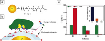
The logical way of understanding protein–NP interactions is to functionalize NPs with specific chemical groups to quantify their interactions with proteins.Reference Moyano and Rotello10 Researchers first explored the role of the net charge on the nanoparticle surface using neutral, cationic, and anionic functionalities to study its interaction with proteins. For example, carboxylic acid functionalized nanoparticles (Figure 2a) have been shown to interact with α-chymotrypsin (ChT, a model protein), through the complementary surface charge at the active pocket of the protein (Figure 2b).Reference You, Agasti, De, Knapp and Rotello11 This supramolecular association modulates the enzymatic activity of the protein toward cationic, neutral, and anionic derivatives of the substrate (N-succinyl-l-phenylalanine p-nitroanilide, SPNA).
(a) Chemical structure of anionic amino-acid-functionalized gold nanoparticles. (b) Schematic representation of the interaction between negatively charged nanoparticles (NPs) and chymotrypsin (ChT), illustrating the effect of the charge not only on the binding but also on the regulation of the enzymatic activity. (c) Generation rates for each substrate (S1, cationic; S2, neutral; and S3, anionic) evidencing the binding selectivity due to the electrostatic interactions. The inset shows the normalized activity of the ChT-NP complex to ChT toward different substrates. Reproduced with permission from Reference 11. © 2006 American Chemical Society.
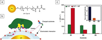
For instance, association of ChT with negatively charged particles leads to a threefold enhancement in the catalytic activity for the cationic derivative of SPNA, while reducing the activity by 50% and 95% for the neutral and anionic derivatives, respectively, of SPNA (Figure 2c). This phenomenon of substrate selectivity can be attributed to the electrostatic interaction and steric hindrance of the NP surface with the substrate. Another case was presented in an analysis of NPs binding to proteins with overall positive charges, albeit different surface properties.Reference De, You, Srivastava and Rotello12 Whereas ChT with 17 positively charged residues (isoelectric point, pI ≈ 8) binds to these anionic NPs principally because of a large change in enthalpy (ΔH), the binding with histone (62 positively charged residues) and cytochrome C (20 positively charged residues, pI ≈ 11) is entropically driven; these results showed that charge is not the only component of the binding, and other characteristics of the protein may be involved in the process.
Following this idea, a series of cationic NPs were used against a panel of proteins with different sizes and geometries to analyze the thermodynamic forces behind the binding.Reference De, Miranda, Rana and Rotello9 One would expect that, because of charge complementarity (all of the proteins were partially negative at the pH of the study), the principal component of the binding would be ΔH. However, this factor was dominant only when the proteins were large enough to accommodate several NPs. In the case of smaller proteins, when one NP bound to several proteins, entropy was the driving force of the process, presumably because more water molecules per NP were released in the process. It is important to note that, during these binding events, entropy–enthalpy compensation was observed, suggesting significant conformational changes for both the proteins and the NPs. This compensation can be attributed to the structural features of the functionalized NPs, which are expected to reorganize to attain maximum stability of the complexes.Reference Houk, Leach, Kim and Zhang13
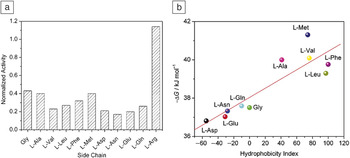
Charge is not the only variable of the NP surface that can be tailored. Other chemical motifs, such as polar, hydrophobic, and aromatic groups, have been studied for recognition of protein surfaces. As an example, amino-acid-terminated ligands of different structures have been fabricated to probe the effect of the NP surface on ChT binding (Figure 2a).Reference You, De, Han and Rotello14 Most of these amino-acid-functionalized NPs showed an inhibitory effect on the activity of ChT, with the only exception being l-arginine-functionalized NPs (positively charged, with reduced interaction with the positively charged ChT). NPs with polar side chains such as aspartic acid (Asp), glutamic acid (Glu), and asparagine induced ∼80% inhibition of the ChT activity, whereas NPs with hydrophobic side chains (for example alanine and methionine) showed an inhibition of ∼60% (Figure 3a). This observation can be attributed to the difference in hydrophobicity of the ChT active site.
(a) Inhibition of chymotrypsin (ChT) activity by different amino-acid-functionalized nanoparticles (NPs) (scheme in Figure 2b). (b) Correlation of the Gibbs free energy of binding between NP and ChT and the hydrophobicity index of the amino acids at the NP ligand termini. Reproduced with permission from Reference 14. © 2005 American Chemical Society.
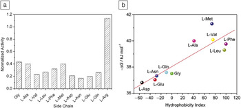
Presumably, the hydrophobic active site of ChT is more accessible to SPNA when hydrophobic amino acids are present than when surrounded by NPs bearing hydrophilic chains. This is because the hydrophobic phenylalanine residue in the substrate first needs to be bound to the active site during the enzyme-promoted cleavage of peptide bonds. As a result, more residual ChT activity is observed for NPs with hydrophobic amino acids. Moreover, the binding constants between ChT and amino-acid-functionalized NPs show a similar trend that is more evident when the Gibbs free energy changes (ΔG) for ChT–NP complexation are plotted against the hydrophobicity indexes of the amino acids (Figure 3b).
Finally, the denaturation kinetics of ChT interactions with NPs were measured by fluorescence studies. A plot of the logarithm of the ChT concentration against time showed a first-order denaturation profile for the protein, demonstrating hydrophobic interactions between hydrophobic patches on the protein and the NP surface, enhancing NP–protein complex stability. The secondary structure of ChT might be stabilized by the hydrophobicity of the NPs, whereas the hydrophilic side chains might destabilize the protein structure through either competitive hydrogen bonding or breakage of salt bridges. For instance, NPs with di-anionic moieties such as Asp and Glu induce fast denaturation of ChT during complexation.
The effects of NP surface hydrophobicity on the binding of NPs and proteins seem to be an underlying and key characteristic of this phenomenon. For example, Moyano et al. screened a series of cationic NPs with different chemical moieties, such as H-bond donors and acceptors, aromatic and hydrophobic groups, and observed their interactions with proteins by a displacement assay.Reference Moyano, Rana, Bunz and Rotello15 The results showed that, when hydrophobic moieties were introduced onto the NP surface, proteins were differentially adsorbed. In addition, by using only hydrophobic motifs at the NP surface, the same researchers were able to selectively differentiate protein isomers that differed only in the hydrophobicity of two amino acids, confirming the importance of hydrophobicity in the binding of NPs and proteins.Reference Chen, Rana, Moyano, Xu, Guo and Rotello16
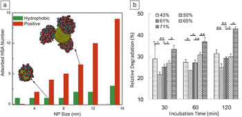
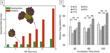
Gessner et al. presented another example of this phenomenon when they analyzed the adsorption of proteins on NPs of different hydrophobicity.Reference Gessner, Waicz, Lieske, Paulke, Mäder and Müller17 They found not only a quantitative increase in adsorbed proteins when the NP surface was more hydrophobic, but also a change in the nature of the adsorbed proteins. In fact, theoretical studies suggested that hydrophobicity could be as relevant as charge for the adsorption of proteins on NPs of certain sizes (Figure 4a).Reference Ding and Ma18 Finally, the increased protein adsorption due to enhanced NP surface hydrophobicity altered the cellular uptake of cationic NPsReference Zhu, Posati, Moyano, Tang, Yan, Vachet and Rotello19 and induced increased degradability of encapsulated proteins in anionic systems upon contact with cells (Figure 4b).Reference Shima, Akagi and Akashi20 These results become logical from the perspective of the hydration shells: hydrophobic structures significantly change the organization of water molecules surrounding them, with this change directly affecting the binding with proteins.
(a) Adsorbed proteins (human serum albumin, HSA) as a function of the size of charged and hydrophobic nanoparticles (NPs). (b) Increasing the hydrophobicity of NPs (represented with an increase in the percentage % value) increases the degradability of proteins upon incubation with cells. Note. n.s., no significant differences. (a) Reproduced with permission from Reference 18. © 2014 Elsevier. (b) Reproduced with permission from Reference 20. © 2014 Royal Society of Chemistry.
Avoiding protein interactions
Whereas NP–protein interactions are desirable for sensing and protein delivery applications, “stealthy” particles that avoid protein interactions are quite useful for other delivery applications. For example, proteins adsorbed on the NP surface can mask the targeting capabilities of nanoprobes.Reference Salvati, Pitek, Monopoli, Prapainop, Bombelli, Hristov, Kelly, Aberg, Mahon and Dawson21 This undesirable adsorption of proteins, commonly known as the protein corona formation, is one of the most critical challenges in nanomedicine today. A protein corona not only reduces the targeting capabilities of the NPs but also promotes elimination of nanomaterials from the body.Reference Aggarwal, Hall, McLeland, Dobrovolskaia and McNeil22 Given the significance of this phenomenon, different approaches have been proposed to reduce the favorability of the interaction between NPs and proteins, thus conferring nonfouling characteristics to these systems (low adsorption of proteins).
The conventional approach involves the use of poly(ethylene glycol) (PEG), a polymer with the capacity of increasing the blood circulation time of nanomaterials by reducing the nonspecific adsorption of proteins.Reference Jokerst, Lobovkina, Zare and Gambhir23 For example, research has shown that by adjusting oligo(ethylene glycol) (OEG) linkers, interactions between the NP interior and ChT can be decreased with increasing PEG length, that is, 1EG > 2EG > 3EG > 4EG.Reference You, De and Rotello24 It has been proposed that the rationale behind this reduced protein adsorption is the wettability (water solvation) of the polymer chains.Reference Gombotz, Guanghui, Horbett and Hoffman25 The interaction of PEG ligands with numerous water molecules (primarily through H-bonds) might be energetically more favored than the interaction with proteins. In practical terms, the interaction with water molecules creates a “barrier” of solvent between the NP surface and proteins, impeding their interaction.
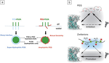
The interaction between PEG and water molecules is relatively weak, so there are proteins for which PEG does not prevent adsorption. For example, C3b, a protein found in human serum, has been shown to interact strongly with the hydroxyl moieties of PEG, taking advantage of their nucleophilic properties.Reference Reddy, van der Vlies, Simeoni, Angeli, Randolph, O’Neil, Lee, Swartz and Hubbell26 Similarly, specific antibodies can be generated against this molecule, and these antibodies are able to recognize and opsonize PEG-functionalized NPs. These observations demonstrate that PEG structures at the NP surface are more exposed than originally thought, and that proteins can reach beyond the “water wall” surrounding them. These results are not unexpected given the fact that PEG is not purely hydrophilic in nature, but rather is amphiphilic. Some reports even suggest that PEG nanostructures composed of oil–water suspensions might not have the stability required for clinical purposes, as PEG is soluble in both phases (Figure 5a).Reference Cao and Jiang27 In addition, research has been presented showing that, although PEG can stabilize the structure of enzymes, it might also decrease their activity through interactions with the substrate, even for substrates with a hydrophobic nature (Figure 5b).Reference Keefe and Jiang28
(a) Schematic representation of nanostructures featuring zwitterionic poly(carboxybetaine) (PCB) and poly(ethylene glycol) (PEG) ligands on a poly(lactic-co-glycolic acid) (PLGA) core, illustrating the lack of a well-defined hydration shell for PEG nanoparticles due to the amphiphilic nature of this molecule. (b) Influence of PEG and zwitterions on the interactions of enzymes and their substrates. Whereas PEG competes in the binding because of its amphiphilic nature, zwitterions promote the enzyme–substrate binding. (a) Reproduced with permission from Reference 27. © 2012 Elsevier. (b) Reproduced with permission from Reference 28. © 2011 Nature Publishing Group.

Zwitterions (overall neutral molecules with both cationic and anionic charges) have been used recently as an alternative to PEG NPs. Their utility is based on the fact that the hydration process of zwitterions is electrostatic in nature, making their binding with water molecules stronger and increasing the energy barrier required for protein adsorption.Reference Chen, Zheng, Li and Jiang29 This ability to interact strongly with water molecules might also explain the resistance of zwitterionic NPs to freezing damage during freeze-drying cycles in contrast to PEG systems, which often require cryoprotectants. In addition, zwitterions create a purely hydrophilic environment at the NP surface, allowing the presentation of the functional groups exclusive to the aqueous environment (Figure 5a). Comparative studies have been performed between PEG and zwitterionic materials to observe the interactions with proteins from human serum, revealing that, whereas PEG NPs adsorb a layer of proteins, zwitterionic NPs do not form a protein corona.Reference Moyano, Saha, Prakash, Yan, Kong, Yazdani and Rotello30 Other reports have shown that zwitterionic-functionalized NPs do not trigger antibody generation, in contrast to the case of PEG, further supporting their enhanced stealth properties.Reference Yang, Liu, Bai, Keefe, Zhang, Ella-Menye, Li and Jiang31 In practical terms, these results imply that zwitterionic nanomaterials exhibit longer circulation times and larger tumor-site accumulation, potentially increasing the therapeutic capabilities of these systems.Reference Arvizo, Miranda, Moyano, Walden, Giri, Bhattacharya, Robertson, Rotello, Reid and Mukherjee32
Given the proven advantages of zwitterions, the modification of their chemical properties to modulate their activity is the next step and represents the current forefront of research in this area. For example, using different classes of carboxybetaine moieties, Leng et al. showed that the surface hydration of ligands can be modified by changes in pH and salt concentration, whereas sulfobetaine analogues are unaffected by such changes.Reference Leng, Han, Shao, Zhu, Li, Jiang and Chen33 This research also used computational studies to better understand the hydration process around the zwitterionic moieties, providing further information on their nonfouling properties.Reference Shao and Jiang34 Similarly, Pillai et al. opted for the use of a mixed monolayer of ligands to modify the zwitterion character and control not only the stability of NPs in solution (NP–NP interactions), but also their cellular uptake.Reference Pillai, Huda, Kowalczyk and Grzybowski35
Finally, Moyano et al. modified the hydrophobicity of NPs, similarly to their previously cited work, but in this case maintaining the nonfouling properties through the use of a zwitterionic backbone.Reference Moyano, Saha, Prakash, Yan, Kong, Yazdani and Rotello30 This approach allowed the direct presentation of specific hydrophobic motifs at the surface of the NPs (due to the absence of a protein corona), residues that change the biological response toward these particles, presumably as a result of the hydration changes imposed by the hydrophobic portions.
Conclusions
In summary, the NP surface structure determines hydration, which then dictates protein-binding properties. We have discussed the multidimensionality of the interaction between NPs and proteins. The nature of the hydration shell can be controlled by modifying the chemistry at the surface of the NPs, for example, by the inclusion of hydrophobic portions and/or changes in the net charge. As a result, the interactions between NPs and proteins can be tuned, achieving strong binding events or completely avoiding binding when desired. These results not only offer new insights into the fundamentals of NP–protein interactions, but also provide design principles for achieving a wide range of desired capabilities from improvement of delivery vehicles to the creation of nanocomposites.
Acknowledgment
V.M.R. acknowledges the NIH (EB014277 and GM077173).