In 1979, exercise-induced abnormalities of ventricular mural motion were identified using cross-sectional echocardiography in patients with ischaemic heart disease.Reference Wann, Faris, Childress, Dillon, Weyman and Feigenbaum1 From then on, assessment of ventricular function with stress imaging has been widely applied in patients with coronary arterial disease.Reference Wahl, Paetsch and Gollesch2 In these patients, stress imaging is able to demonstrate abnormalities of ventricular mural motion and coronary arterial perfusion not apparent at rest, and can provide information on risk factors and prognostic factors.Reference Wahl, Paetsch and Gollesch2–Reference Kuijpers, Janssen, van Dijkman and Oudkerk4 The use of stress imaging has subsequently been extended to patients with dilated or hypertrophic cardiomyopathy, valvar heart disease, and those with congenitally malformed hearts. In patients with dilated or hypertrophic cardiomyopathy, this form of imaging is used to assess, in addition to abnormalities of mural motion, gradients across the outflow tracts and the contractile reserve, that is the change in ventricular ejection fraction induced by stress.Reference Wu, Bhavsar, Aziz and Sadaniantz5 In patients with congenitally malformed hearts, stress imaging is used to determine all these factors, but also to assess diastolic and vascular function. In this review, we will discuss the use, limitations, and future applications of stress imaging in patients with congenitally malformed hearts, placing our emphasis on echocardiography and cardiovascular magnetic resonance imaging.
Stressors
Physical exercise
Physical exercise is the ideal stressor. It results in the combined activation of cardiac, pulmonary, vascular, neurohormonal, muscular, and metabolic systems involved in the adaptations to stress. Common tools to perform physical exercise during imaging procedures are the treadmill, bicycle ergometer, or handgrip. Specific magnetic resonance-compatible supine or upright bicycle ergometers have been used for exercise testing in combination with magnetic resonance imaging. In this respect, the posture of the patient is important, since this has a significant effect on central haemodynamics, with lower cardiac output, higher stroke volumes, and lower heart rates in those tested in supine as opposed to upright positions.Reference Cheng, Herfkens, Lightner, Taylor and Feinstein6
Different protocols have been described for study of patients with congenitally malformed hearts, including
• exercise to a certain percentage of their maximal exercise capacity or oxygen consumptionReference Alpert, Bloom and Olley7–Reference Roest, Helbing and Kunz9
• symptom-limited exerciseReference Hauser, Bengel and Kuhn10–Reference Cyran, Grzeszczak and Kaufman12
• exercise at 0.5 and 2.5 W/kg.Reference Oyen, Ingerfeld, Ignatzy and Brode13–Reference Pedersen, Stenbog and Frund16
Physical exercise has several limitations. Maximal physical stress is dependent on the motivation and cooperation of the patient, and cannot be attained in patients with certain co-existing neurologic or orthopaedic diseases. Hyperventilation during physical exercise hinders breath-holding during the acquisition of images. In combination with echocardiography, it is difficult to acquire images during physical exercise. Hence, images are usually obtained directly after exercise. This is also applicable to magnetic resonance imaging,Reference Roest, Kunz and Helbing8, Reference Roest, Helbing and Kunz9, Reference Pedersen, Stenbog and Frund16–Reference Roest, Kunz, Lamb, Helbing, van der Wall and de Roos18 although with real-time imaging this limitation has been overcome, and scanning during rapid breathing is feasible, even in patients with complex lesions.Reference Hjortdal, Christensen, Larsen, Emmertsen and Pedersen14, Reference Hjortdal, Emmertsen and Stenbog15
Pharmacological stress
During the 1980s, pharmacological agents were introduced as stressors to overcome the limitations of physical exercise.Reference Berthe, Pierard and Hiernaux19, Reference Picano, Lattanzi, Masini, Distante and L’Abbate20 Use of these agents has extended the use of stress imaging to patients with constraints that preclude adequate exercise. The most commonly used drugs are adenosine, dipyridamole, and dobutamine.
Adenosine and dipyridamole are coronary vasodilators that generate a reduced supply of oxygen in myocardial areas supplied by stenotic coronary arteries, and are used for coronary perfusion imaging. Adenosine is a naturally occurring substance in the body that causes coronary vasodilation through activation of A2-receptors. This results in increased flow in normal coronary arteries compared to stenotic ones, a steal phenomenon which results in a perfusion mismatch. It does not necessarily cause ischaemia. Adenosine has a very short half life of only 2 seconds, and therefore needs to be administered through continuous intravenous infusion. The dose used for stress-testing is 0.14 mg/kg/min over 6 minutes, or until the patient suffers significant discomfort, whichever comes first. Common side effects are flushing, dyspnoea, chest pain, gastro-intestinal discomfort, headache, and light-headedness. Most side effects disappear shortly after discontinuation of administration of the agent, and do not require medical treatment. Adenosine is contra-indicated in patients with active restrictive airway disease, 2nd or 3rd degree atrioventricular block, and in patients taking dipyridamole. Since theophylline and caffeine are adenosine receptor antagonists, abstention from use of these substances is required for 1 to 2 days before stress testing with adenosine or dipyridamole. In patients with congenitally malformed hearts, adenosine has been rarely used for stress imaging, since coronary arterial abnormalities are not common.
Dipyridamole inhibits reuptake of adenosine by vascular endothelial cells, and indirectly causes coronary vasodilation. It has a considerably longer half-life than adenosine, and its haemodynamic effects can persist up to 30 minutes. The agent is administered at a rate of 0.56 mg/kg over 4 minutes. Side effects and contraindications for the use of dipyridamole are similar to those of adenosine.
Dobutamine increases the myocardial demand for oxygen similar to physical exercise, and is used to study contractile reserve and abnormalities of ventricular mural motion. It is a synthetic catecholamine with positive inotropic and, to a lesser extent, chronotropic effects. In healthy children, positive inotropic effects occur from 1 to 2 μg/kg/min, with dose-dependent increases in measures of systolic ventricular function,Reference Berg, Padbury, Donnerstein, Klewer and Hutter21 while chronotropic effects are seen from 5 to10 μg/kg/min.Reference Michelfelder, Witt, Khoury and Kimball22 The agent is also known to enhance diastolic function, and to decrease preload and afterload.Reference Harada, Tamura, Ito, Suzuki and Takada23 In normal subjects, during its infusion, stroke volume increases, end-diastolic volume does not change, end-systolic volume decreases, and ejection fraction increases.Reference De Wolf, Suys, Verhaaren, Matthys and Taeymans24 The half-life is only 2 minutes, and therefore it should be given through continuous intravenous infusion. In patients with congenitally malformed hearts, different dosages are used to answer different research questions. When used at low or moderate doses, dobutamine is used to assess cardiac contractile reserve. The infusion is started at 2.5 or 5 μg/kg/min, and increased every 3 to 5 minutes with 2.5 or 5 μg/kg/min to 5 to 20 μg/kg/min. At high doses, dobutamine is used to detect mural abnormalities in patients with coronary arterial abnormalities, as in patients with Kawasaki disease or after the arterial switch operation. Infusion usually starts at 10 μg/kg/min, and the rate is increased every 3 to 5 minutes with 10 μg/kg/min, until the heart rate is 85% of the maximal predicted heart rate for age, or to 40 μg/kg/min maximum. If 85% of the maximal predicted heart rate for age is not reached at 40 μg/kg/min, atropine can be administered in conjunction with dobutamine to reach the target heart rate. Heart rate, blood pressure, and cardiac rhythm if possible, are continuously monitored during dobutamine stress-testing. Common minor side effects, such as headache, nausea, hypertension, hypotension, and haemodynamically insignificant arrhythmias, can occur in up to one-fifth of children.Reference Noto, Ayusawa and Karasawa25 Major side effects have not been reported in patients with congenitally malformed hearts. Side effects of atropine are dry mouth, tachycardia, and hallucinations. Side effects of both dobutamine and atropine can be treated with β-blockers or calcium antagonists, albeit the latter preferably not in children, and these agents should be readily available during stress-testing. Administration of dobutamine is contra-indicated in patients with a mechanical obstruction of systemic ventricular filling or ejection, in patients with 2nd or 3rd degree atrioventricular block, and in patients with a history of sustained ventricular tachycardia.
The majority of stress imaging studies in patients with congenitally malformed hearts has been performed with pharmacological stress, almost exclusively using dobutamine. Only one study reported on the safety and feasibility of adenosine-stress cardiovascular magnetic resonance imaging in children with congenital aortic stenosis or after an arterial switch operation.Reference Taylor, Dymarkowski and De Meerleer26 Stress imaging has only been performed in a limited number of studies using high doses.Reference Li, Hornung and Francis27–Reference Strigl, Beroukhim and Valente30 In studies performing stress imaging at low doses, the doses have ranged from 5.0 to 20 μg/kg/min. In our experience, a dosage of 7.5 μg/kg/min is safe, with a low incidence of only minor adverse effects. Furthermore, it is sufficiently high to elicit a significant cardiovascular stress response.Reference van den Berg, Strengers and Wielopolski31–Reference Robbers-Visser, Ten Harkel and Kapusta34 Arrhythmias are side effects of special concern in this group of patients. Although the incidence of arrhythmias is low in the studies performed so far, most occurred with a dosage of at least 10 μg/kg/min.Reference Hui, Chau, Leung, Chiu and Cheung35–Reference Tulevski, van der Wall and Groenink38 In patients who had undergone a Fontan operation for functionally univentricular hearts, administration at 7.5 μg/kg/min provoked an increase in heart rate of more than 150% from baseline in 10 of 37 patients.Reference Robbers-Visser, Ten Harkel and Kapusta34 This was well tolerated, and lowering the dosage to 5 μg/kg/min was sufficient to decrease the heart rate and successfully complete the study protocol, producing images adequate for analysis.
Imaging modalities
Radionuclide myocardial perfusion imaging
Positron emission tomography and single-photon emission computed tomography allow evaluation of myocardial perfusion and the effects of myocardial hypoperfusion on metabolic activity and myocardial contractility.Reference Vesely and Dilsizian39 Positron emission tomography and single-photon emission computed tomography cameras capture the photons emitted by radiopharmaceuticals, such as nitrogen-13 labeled ammonia, and technetium-99m tetrophosmin or sestamibi, and translate the information into digital data representing the magnitude and location of the emission in the heart. Positron emission tomography has a better performance on spatial and temporal resolution, with less attenuation of the emitted signal than single-photon emission computed tomography. In children and patients with congenitally malformed hearts, positron emission tomography and single-photon emission computed tomography are rarely used, predominantly in patients with coronary arterial disease.Reference Fukuda, Ishibashi, Shinohara, Miyake, Kudoh and Saga40–Reference Hauser, Bengel and Kuhn45 These studies assessed myocardial perfusion and coronary flow reserve at rest and during adenosine or dipyridamole infusion. Although showing signs of coronary arterial damage or decreased coronary vasoreactivity, use of these imaging modalities is limited because of concerns on radiation exposure in young children. Furthermore, coronary arterial abnormalities can also be assessed with radiation-free imaging modalities, such as echocardiography and cardiovascular magnetic resonance imaging.
Echocardiography
Conventional echocardiography, M-mode, Doppler and tissue Doppler imaging have all been performed in combination with stress in patients with various forms of congenitally malformed hearts.Reference Alpert, Bloom and Olley7, Reference Hauser, Bengel and Kuhn10–Reference Oyen, Ingerfeld, Ignatzy and Brode13, Reference Li, Hornung and Francis27–Reference Brili, Alexopoulos and Barberis29, Reference Hui, Chau, Leung, Chiu and Cheung35, Reference Brili, Stamatopoulos and Barbetseas36 Although the first report on stress imaging in these patients involved echocardiography, its implementation has been largely hampered by the limitations of the acoustic window. Suboptimal acquisition of images, producing images of suboptimal image, is common in patients with complex cardiac anatomy. Combined with an abnormal orientation of the heart and great vessels in the chest, and the problems encountered in imaging the functionally univentricular arrangements as well as the morphologically right ventricle,Reference Helbing, Bosch and Maliepaard46, Reference Hirsch, Kilner, Connelly, Redington, St John Sutton and Somerville47 this has led to limited use of stress echocardiography in the setting of congenital cardiac disease.
Magnetic resonance imaging
In patients with coronary arterial disease, echocardiography was the modality of choice for stress imaging up until the early 1990s. In 1992, the use of dobutamine stress cardiovascular magnetic resonance imaging was introduced,Reference Pennell, Underwood and Manzara48 and has proven to be a valuable tool to detect ischaemia in patients with known or suspected coronary arterial disease.Reference Jahnke, Nagel and Gebker3, Reference Paetsch, Jahnke and Wahl49 Dobutamine stress cardiovascular magnetic resonance imaging can assess abnormalities of mural motion, contractile reserve, abnormalities of diastolic function, and vascular function. It has a sensitivity and specificity comparable to dobutamine stress echocardiography for identifying coronary arterial disease.Reference Paetsch, Jahnke and Wahl49 The technique overcomes the limitations of echocardiography in selected patients with congenitally malformed hearts.Reference Tulevski, Lee and Groenink50 A few groups have reported on the use of this modality to evaluate the reaction of the cardiovascular system in such patients when stressed.Reference Roest, Kunz and Helbing8, Reference Roest, Helbing and Kunz9, Reference Hjortdal, Christensen, Larsen, Emmertsen and Pedersen14–Reference Pedersen, Stenbog and Frund16, Reference Roest, Kunz, Lamb, Helbing, van der Wall and de Roos18, Reference Taylor, Dymarkowski and De Meerleer26, Reference Strigl, Beroukhim and Valente30–Reference Robbers-Visser, Ten Harkel and Kapusta34, Reference Fratz, Hager and Busch37, Reference Tulevski, van der Wall and Groenink38, Reference Tulevski, Lee and Groenink50–Reference Tulevski, Hirsch, Dodge-Khatami, Stoker, van der Wall and Mulder54 These studies have shown abnormal systolic, and diastolic function, and abnormal vascular responses which help elucidate the pathophysiological processes. An important advantage of dobutamine stress cardiovascular magnetic resonance imaging is that all these parameters can be obtained in one single study. Disadvantages are the exclusion of patients with claustrophobia, severe obesity, and magnetic resonance-incompatible devices, and the need for sedation or general anaesthesia in young children, usually those under the age of 5 to 6 years.
Stress imaging in children and patients with congenital cardiac disease
Stress echocardiography
The first report of stress-testing by supine exercise combined with echocardiography in children with left-sided cardiac disease was published in 1980.Reference Alpert, Bloom and Olley7 Thereafter, stress echocardiography combined with dobutamine was used to assess the contractile reserve in children with thalassemia major, children with Kawasaki disease, those having chemotherapy for childhood cancer, and those with transplanted hearts.Reference De Wolf, Suys, Verhaaren, Matthys and Taeymans24, Reference Noto, Ayusawa and Karasawa25, Reference Lanzarini, Bossi, Laudisa, Klersy and Arico55–Reference Larsen, Applegate and Dyar58 These studies showed that the technique could provide important information on subclinical global ventricular dysfunction or coronary arterial disease.
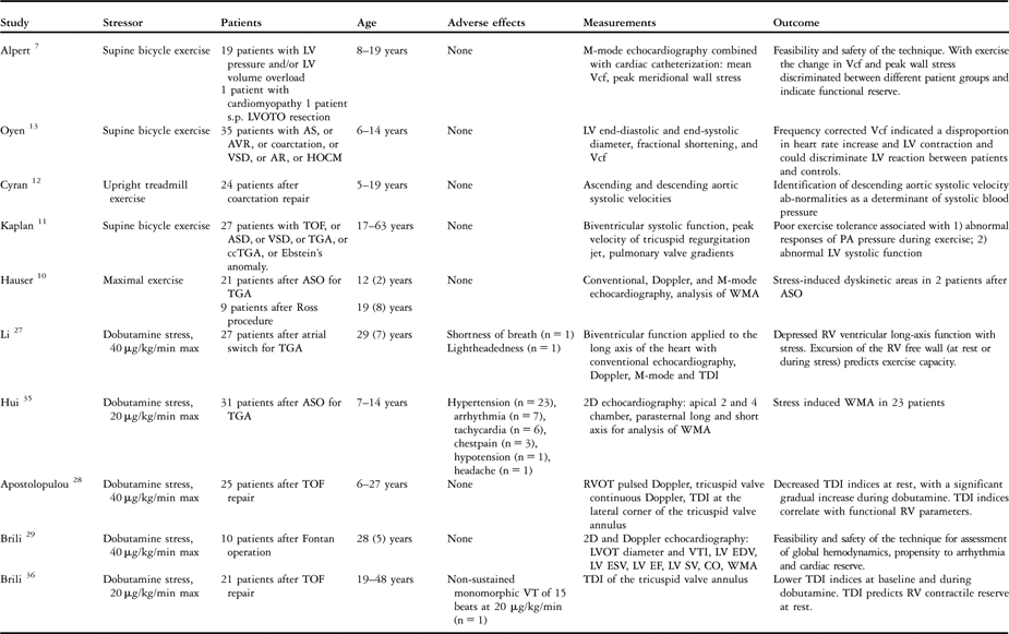
There are, however, relatively few studies reported on use of stress echocardiography in patients with congenital cardiac disease, albeit that the technique has been used in patients with complex lesions, such as patients after atrial or arterial corrections for transposition, those with the Fontan circulation, and patients with tetralogy of Fallot.Reference Hauser, Bengel and Kuhn10–Reference Oyen, Ingerfeld, Ignatzy and Brode13, Reference Li, Hornung and Francis27–Reference Brili, Alexopoulos and Barberis29, Reference Hui, Chau, Leung, Chiu and Cheung35, Reference Brili, Stamatopoulos and Barbetseas36 The results of these studies are summarized in Table 1. Half of these studies have been performed in combination with physical exercise. The other half has been performed using dobutamine stress-testing, with dosages ranging from 20 to 40 μg/kg/min. With infusions of at least 20 μg/kg/min, no major side effects have been reported, albeit that one study reported hypertension in two-thirds of the studied patients.Reference Hui, Chau, Leung, Chiu and Cheung35 The definition of hypertension used by these investigators, however, was an increase in systolic blood pressure of greater than 30% from baseline, while others have defined hypertension as an increase in systolic blood pressure of greater than half from baseline.Reference De Wolf, Suys, Verhaaren, Matthys and Taeymans24
Stress echocardiography in congenital heart disease.
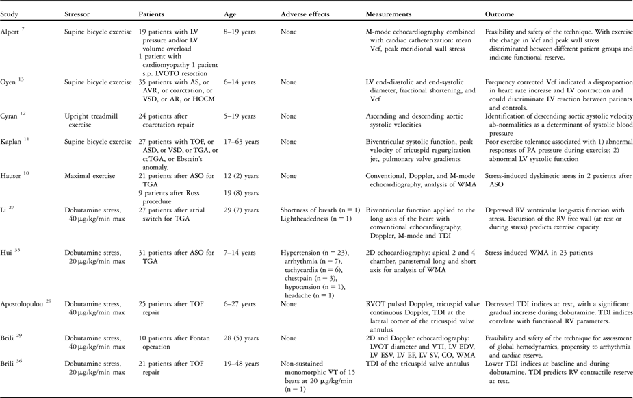
Ages are given as ranges, or mean (standard deviation). Abbreviations: AR = aortic regurgitation, AS = aortic stenosis, ASD = atrial septal defect, ASO = arterial switch operation, AVR = aortic valve replacement, ccTGA = congenitally corrected transposition of the great arteries, CO = cardiac output, EDV = end-diastolic volume, EF = ejection fraction, ESV = end-systolic volume, HOCM = hypertrophic obstructive cardiomyopathy, LV = left ventricle, LVOT = left ventricular outflow tract, LVOTO = LVOT obstruction, PA = pulmonary artery, RV = right ventricle, RVOT = right ventricular outflow tract, SV = stroke volume, TDI = tissue Doppler imaging, TGA = transposition of the great arteries, TOF = tetralogy of Fallot, VSD = ventricular septal defect, VT = ventricular tachycardia, VTI = velocity time integral, WMA = wall motion abnormalities.
Outcomes of these studies cannot be easily compared. There is a wide variety in the age and diagnoses of the patients, the type of measurements with echocardiography, and the type of stressor. All studies demonstrated abnormal ventricular and/or vascular responses with stress-testing. Some studies identified these abnormal responses as predictors of exercise capacity.Reference Kaplan, Foster, Redberg and Schiller11, Reference Li, Hornung and Francis27 In general, the clinical relevance of these abnormal findings with stress-testing is unknown. Standardized studies using identical stressors and identical methods of measurement are needed to determine their clinical relevance. Furthermore, the combination of new techniques, such as 3-dimensional echocardiography, with its capability for assessment of ventricular volumes and dyssynchronicity, and of tissue Doppler and speckle tracking techniques with stress imaging need further exploration.
Stress cardiovascular magnetic resonance imaging
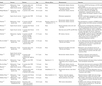
Only a few groups have performed stress cardiovascular magnetic resonance imaging in patients with congenital cardiac disease (Table 2). In more than 200 patients, these studies showed the feasibility and safety of the technique. Unfortunately, as for those using stress echocardiography, differences in diagnoses, ages, measurements, and protocols preclude direct comparison of the reported outcomes.
Stress cardiovascular magnetic resonance imaging in congenital heart disease.

Ages are given as ranges, or mean (standard deviation). Abbreviations: AS = aortic stenosis, ASO = arterial switch operation, CMR = cardiovascular magnetic resonance, ccTGA = congenitally corrected transposition of the great arteries, DCMR = dobutamine CMR; EDV = end-diastolic volume, EF = ejection fraction, ESV = end-systolic volume, IVC = inferior caval vein, LV = left ventricle, RV = right ventricle, SV = stroke volume, SVC = superior caval vein, TGA = transposition of the great arteries, TOF = tetralogy of Fallot.
Studies in have been performed predominantly in
• patients with a pressure overloaded right ventricle, such as in congenitally corrected transposition, pulmonary arterial stenosis, or the Eisenmenger syndrome;Reference Roest, Kunz and Helbing8, Reference Fratz, Hager and Busch37, Reference Tulevski, van der Wall and Groenink38, Reference Tulevski, Lee and Groenink50–Reference Tulevski, Hirsch, Dodge-Khatami, Stoker, van der Wall and Mulder54
• patients with a volume overloaded right ventricle, as after correction for tetralogy of Fallot;Reference Roest, Helbing and Kunz9, Reference van den Berg, Strengers and Wielopolski31, Reference van den Berg, Wielopolski and Meijboom32, Reference Tulevski, Hirsch, Dodge-Khatami, Stoker, van der Wall and Mulder54 and
• patients with a potentially impaired ventricular preload as a result of atrial baffle reconstruction after creation of the Fontan circulation or atrial correction for transposition.Reference Hjortdal, Christensen, Larsen, Emmertsen and Pedersen14–Reference Pedersen, Stenbog and Frund16, Reference Robbers-Visser, Helderman and Strengers33, Reference Robbers-Visser, Ten Harkel and Kapusta34, Reference Tulevski, van der Wall and Groenink38, Reference Tulevski, Lee and Groenink50, Reference Oosterhof, Tulevski and Roest51, Reference van der Zedde, Oosterhof, Tulevski, Vliegen and Mulder53
In patients with a pressure overloaded right ventricle, the technique demonstrated abnormal responses of the systemic right ventricle. In patients with congenitally corrected transposition, different responses of the systemic right ventricle have been reported, according to the type of repair. SomeReference Tulevski, van der Wall and Groenink38, Reference Dodge-Khatami, Tulevski and Bennink52 reported that stroke volume did not increase, with the latter studyReference Dodge-Khatami, Tulevski and Bennink52 reporting a normal response to stress, with an increase in stroke volume, in patients with congenitally corrected transposition who had not undergone repair compared to patients after physiologic repair. OthersReference Fratz, Hager and Busch37 also demonstrated an increase in stroke volume with dobutamine stress cardiovascular magnetic resonance imaging in unoperated patients with congenitally corrected transposition. The first groupReference Dodge-Khatami, Tulevski and Bennink52 suggested that unoperated patients, with a favourable anatomy, may never require an operation, and that dobutamine stress cardiovascular magnetic resonance imaging might be helpful in identifying the patients who will need anatomic correction. In patients with pulmonary arterial stenosis, and in those with Eisenmenger syndrome, end-diastolic volume was significantly larger compared to controls, but ejection fraction was normal at rest. With stress-testing, end-diastolic volume, end-systolic volume, and stroke volume all decreased, and ejection fraction did not change, clearly demonstrating the highly abnormal response of these morphologically right ventricles to stress.Reference Tulevski, van der Wall and Groenink38
Several investigators have reported on the biventricular stress response in patients with a volume overloaded right ventricle. In one study,Reference Roest, Helbing and Kunz9 supine bicycle exercise demonstrated a decrease in pulmonary regurgitation with stress-testing, and a normal left ventricular stress response in patients with tetralogy of Fallot. Others reported on the use of low-dose dobutamine stress cardiovascular magnetic resonance imaging in patients after contemporary repair of tetralogy of Fallot.Reference van den Berg, Strengers and Wielopolski31, Reference van den Berg, Wielopolski and Meijboom32 They showed well preserved functional reserve in all patients despite important pulmonary regurgitation and right ventricular dilation, as well as an abnormal relaxation with stress-testing in patients with end-diastolic forward flow, which was not appreciated at rest. Many of these patients with normal contractile reserve had right ventricular volumes that would have made them candidates for replacement of the pulmonary valve according to criterions based on right ventricular volumes.Reference van den Berg, Strengers and Wielopolski31, Reference Valsangiacomo Buechel, Dave and Kellenberger59 This demonstrates the potential added value of stress imaging in clinical decision making.
In patients with an impaired ventricular preload as a result of atrial baffle reconstruction, several groups have performed various stress cardiovascular magnetic resonance studies, studying the systemic venous return, pulmonary arterial circulation, and systemic ventricular function.Reference Hjortdal, Christensen, Larsen, Emmertsen and Pedersen14–Reference Pedersen, Stenbog and Frund16, Reference Robbers-Visser, Helderman and Strengers33, Reference Robbers-Visser, Ten Harkel and Kapusta34 Measurements were made by some in both pulmonary arteries, along with flow in the caval veins, immediately after exercise or during exercise using real-time cardiovascular magnetic resonance imaging.Reference Hjortdal, Christensen, Larsen, Emmertsen and Pedersen14–Reference Pedersen, Stenbog and Frund16 All the children, with mean ages between 9 and 12 years, were able to perform and complete the protocol. They demonstrated that an increase in cardiac output with exercise is predominantly caused by an increase in heart rate, and that the influence of inspiration on systemic venous return is less pronounced during exercise.
Using pharmacological stress with dobutamine at 7.5 μg/kg/min, weReference Robbers-Visser, Ten Harkel and Kapusta34 demonstrated an abnormal decrease in end-diastolic volumes and no increase in stroke volumes with stress-testing in patients having conversion to the Fontan circulation at a young age. We alsop showed abnormal reactions of flow variables, distensibility, and wall shear stress with stress-testing in the pulmonary arteries.Reference Robbers-Visser, Helderman and Strengers33 These results demonstrate that low-dose dobutamine stress cardiovascular magnetic resonance imaging revealed abnormal findings that were not apparent at rest, and helped in understanding the pathophysiology of this group of patients, notably the progression of dysfunction during their follow-up.
In patients undergoing the atrial switch operation, stroke volume did not increase with pharmacological stress-testing.Reference Fratz, Hager and Busch37, Reference Tulevski, van der Wall and Groenink38, Reference Tulevski, Lee and Groenink50, Reference Oosterhof, Tulevski and Roest51 A smaller increase in stroke volume was noted in patients after the atrial switch when compared to healthy subjects, and a prolonged stroke volume recovery using supine bicycle exercise testing.Reference Roest, Kunz and Helbing8 In patients after intra-atrial correction for transposition, heart rate increase is significantly higher when compared to patients with congenitally corrected transposition or healthy subjects, probably as a compensatory mechanism for the lack of increase in stroke volume with stress-testing.Reference Fratz, Hager and Busch37, Reference Tulevski, van der Wall and Groenink38
Recently, data has been published on the feasibility of high-dose dobutamine stress cardiovascular magnetic resonance imaging in children and young adults with suspected coronary arterial abnormalities by examination of abnormalities of mural motion.Reference Strigl, Beroukhim and Valente30 No major adverse effects were found, and high-quality imaging was obtained in all ventricular mural segments in patients from less than 1 year of age. In only 1 patient was there an inducible abnormality, with the group recommending further exploration of high-dose dobutamine stress cardiovascular magnetic resonance in children.
The type of stressor should be taken into account when comparing the results of stress studies. One groupReference Coma-Canella, Garcia Velloso and Maceira60 showed a different response in healthy volunteers to physical exercise and to dobutamine stress. Global and regional ejection fraction increased more with dobutamine than with physical exercise. Another groupReference Oosterhof, Tulevski and Roest51 demonstrated a discrepancy in response to physical exercise and low-dose dobutamine stress in patients after intra-atrial correction for transposition, whilst in controls the response to both stressors was comparable. They suggested that this disagreement was the result of differences in preload and afterload. Both dobutamine and physical exercise, however, increase contractility, and decrease preload and afterload.Reference Michelfelder, Witt, Khoury and Kimball22, Reference Kimball, Mays, Khoury, Mallie and Claytor61 There are also some differences in acquisition between the studies depending on dobutamine as opposed to physical exercise studies that might explain the discrepancies. Additional studies are needed to clarify this phenomenon.
Although the studies cited this far showed pathophysiologically relevant observations in patients with various types of congenital cardiac disease, the clinical significance of these abnormal responses still has to be assessed. In congenital cardiology, surrogate endpoints for clinical trials are necessary. Primary endpoints, such as death and major complications, are rare, requiring large study populations or prolonged study length to acquire adequate statistical power, while the population is relatively small.Reference Mahony, Sleeper and Anderson62 Surrogate endpoints are expected to predict a primary endpoint, and have a higher incidence in the population under investigation. Thus, surrogate endpoints facilitate clinical trials in congenital cardiology by reducing the numbers of subjects needed and by reducing the length of the study. The abnormal stress responses found in patients with congenital cardiac disease are potential surrogate endpoints for this purpose and their clinical usefulness has to be addressed in follow-up studies.
Future applications
As for echocardiography, standardized dobutamine stress cardiovascular magnetic resonance studies using identical stressors and identical methods of measurement are needed to determine the clinical relevance of the abnormal stress responses in patients with congenital cardiac disease. Such future applications might involve the assessment of pressure-volume loops. Pressure-volume loops give information on myocardial contractility, ventricular pump function, and ventriculo-arterial coupling, which might help explain the pathophysiological processes in cardiac function in patients with congenital cardiac disease.Reference Derrick, Narang and White63 The feasibility of magnetic resonance-derived pressure volume loops in the right ventricle of patients with chronic pressure overload has already been demonstrated.Reference Kuehne, Yilmaz and Steendijk64
Magnetic resonance tissue-tagging with radiofrequency signals destroys the spins in a selected plane, resulting in a line or grid of signal void on the image.Reference Fogel65 These lines or grids deform with ventricular contraction and relaxation and this allows quantification of strain, that is the deformation of an object normalized to its original shape, as a measure of regional myocardial function. In combination with stress-testing, this technique could reveal important regional functional information in patients with complex lesions. In patients with coronary arterial disease, the response of systolic strain to low-dose dobutamine has a significant promise in discriminating between viable and non-viable myocardium.Reference Bree, Wollmuth and Cupps66
Lastly, in pathways with complex patterns of flow, such as the Fontan circulation, or intra-atrial baffles, 3-directional flow measurements can give important additional information on shear stress and energy dissipation and the influence of increased flow during stress on these variables.
Conclusions
Stress imaging is a tool that has been used in limited fashion in patients with congenitally malformed hearts when compared to its implementation in patients with coronary arterial disease. Several studies, nonetheless, have shown its feasibility and safety. Stress imaging is able to assess systolic, diastolic, and vascular responses in patients with various types of complex lesions. At present, magnetic resonance imaging has been used more often for this purpose than echocardiography. The clinical relevance of these findings needs to be investigated in standardized protocols. The abnormal stress responses can then potentially be used for risk assessment in the follow-up of patients with congenital cardiac disease.