
Introduction
In the ordered mesoporous silica field, SBA-15 materials with cylindrical pore structure, controllable morphology and pore diameter have become one kind of the most attractive materials due to their wide potential applications in catalysis, sensors, adsorption, separation, drug delivery, and nanotechnology.[ Reference Zhao, Sun, Li and Stucky 1 , Reference Zhao, Feng, Huo, Melosh, Fredrickson, Chmelka and Stucky 2 ] As a desired carrier material, the morphology and pore structure of SBA-15 were the critical factors in its applications.[ Reference Yu, Fan, Tian, Zhao and Stucky 3 – Reference Fan, Lei, Wang, Yu, Tu and Zhao 5 ] In previous studies, many attentions have been paid on the morphology control and the pore regulation, and great progresses have been achieved. For example, the SBA-15 with various shapes, such as sphere, rod, fiber, plate, sheet, etc.[ Reference Zhao, Sun, Li and Stucky 1 , Reference Johansson, Córdoba and Odén 6 – Reference Chen, Tang, Chuang, Lee, Tsai, Chan, Lin, Liu and Cheng 9 ] have been synthesized by means of adjusting synthesis conditions, such as temperature, reactant concentration, agitating time, and the amount of the organic or inorganic additives.[ Reference Zhao, Sun, Li and Stucky 1 , Reference Chen, Wang and He 10 – Reference Linton, Wennerstrom and Alfredsson 12 ] The reported works demonstrated that adjusting the reactant concentration was one of the effective methods for the morphology control.[ Reference Naik, Elangovan, Okubo and Sokolov 13 , Reference Ding, Yin, Liao, Huang, Chen, Yao and Li 14 ] In general, the reactant and the template would simultaneously affect the formation of SBA-15 in the growth process, and their synergistic effects would especially play important roles in the morphology control. However, to the best of our knowledge, most researches focused on the single-factor effect of the reactant or the template, and few analyses on the growth process of SBA-15, which could provide a lot of important information about the morphology control, were reported.[ Reference Ding, Yin, Liao, Huang, Chen, Yao and Li 14 – Reference Moulin, Schmitt, Lecchi, Degrouard and Impéror-Clerc 19 ] The synergistic effects of the reactant and the template on the SBA-15 morphology have not been fully understood.
In this work, hexagonal plate-like SBA-15 particles were prepared in aqueous acid condition using amphiphilic triblock copolymer (Pluronic-123 (P123)] as the template and tetraethoxysilane (TEOS) as the silica source. Through only adjusting the initial solution volume with the same amounts of silica source and template, a novel gear-like SBA-15 was synthesized. According to the morphology transformations, the growth process of the gear-like SBA-15 particles and the synergistic effects of the reactants and the template have been investigated.
Experimental details
SBA-15 materials were synthesized according to the procedure of the reported literature.[ Reference Zhao, Feng, Huo, Melosh, Fredrickson, Chmelka and Stucky 2 ] In a typical synthesis, 1.4 g triblock copolymer P123 (Sigma-Aldrich Company) was dissolved in 1.5 M HCl solution with water bath stirring at 32 °C. After 3 h, 3.2 mL TEOS (Chengdu Kelon Chemical Reagent Company) was added dropwise, and the stirring was stopped after another 5 min. The P123 concentration was decreased from 2.28% to 1.83%, 1.53% and 1.32% by adjusting the solution volume from 60 to 75, 90, and 105 mL, respectively (concentration calculation example: when the solution volume was controlled at 60 mL, roughly 1.4/61.4 g × 100% = 2.28 wt.% of P123), and the TEOS concentration decreased from 4.75% to 3.83%, 3.22%, and 2.77%, respectively. Then the mixture was kept under static condition for 20 h and subsequently the resultant mixture was transferred into a Teflon bottle for hydrothermal treatment at 100 °C for 24 h. After filtered and washed by distilled water, the white precipitation was dried in vacuum oven at 100 °C for 2 h and then calcined in air at 550 °C for 5 h to remove the template. The sample was denoted as SiO2-1, SiO2-2, SiO2-3, and SiO2-4 corresponding to the P123 concentration of 2.28%, 1.83%, 1.53%, and 1.32%, respectively.
To study the morphology transformations of the sample SiO2-3 in different growth stages, the samples were taken out directly from the reacting solution using a dropper and diluted about 120 times immediately. After centrifuged and washed six times by double-distilled water, the sample was diluted again and dropped on silicon wafer. At last, the sample was dried under the room temperature. The sampling time began to count when the agitation was stopped.
Small-angle x-ray scattering (SAXS) patterns of the calcined samples were obtained on a Bruker D8 diffractometer using a Cu Kα source. Wide-angle x-ray diffraction (XRD) patterns were obtained on a DX-1000 diffractometer using a Cu Kα source. Surface morphologies were observed by scanning electron microscope (SEM, Hitachi, S-4800). Microstructure images were recorded by transmission electron microscopy (TEM, TECNAI G2 F20). Nitrogen adsorption–desorption isotherms were obtained with a Gemini VII instrument. The pore size distribution was analyzed by the adsorption isotherm, using the Barrett–Joyner–Halenda (BJH) method. The diameter and thickness were analyzed by Nano Measure 1.2.5 Software (Fudan University).
Results and discussion
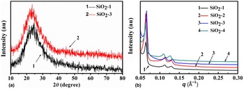
XRD patterns of the calcined samples [Fig. 1(a)] were consistent with the characteristic peaks of the orthorhombic SiO2 (JCPDF 78-1423). SAXS patterns of the calcined samples [Fig. 1(b)] exhibited three well-resolved diffraction peaks, which were indexed to the (100), (110), and (200) reflections of a two-dimensional (2D) hexagonal symmetry lattice (p6 mm). XRD and SAXS results demonstrated that the calcined samples were the typical SBA-15 mesoporous material.
(a) XRD and (b) SAXS patterns of calcined samples at different P123 concentration: 2.28% (SiO2-1), 1.83% (SiO2-2), 1.53% (SiO2-3), and 1.32% (SiO2-4).

Nitrogen adsorption–desorption isotherms of all calcined samples corresponded to type-IV isotherms with an H1-type hysteresis loop [Fig. 2(a)], which were the characteristics of hexagonal mesoporous materials with cylindrical channels.[ Reference Liu, Yang, Zhao and Zhang 20 ] The calcined samples possessed narrow pore distribution with an average diameter of approximate 5.95 nm [Fig. 2(b)]. Uniform pore channels perpendicular to the end face of the plate-like particle were clearly observed in Figs. 2(c)–2(h). For the calcined sample of SiO2-1 and SiO2-3, the well-ordered pore channels without apparent defections could be seen from the end view [Figs. 2(e) and 2(h)].
(a) Nitrogen adsorption–desorption isotherms, (b) pore diameter distribution curves and TEM images of the samples: (c–e) SiO2-3 and (f–h) SiO2-1.

SEM observations [Figs. 3(a)–3(d)] displayed that the morphologies of the calcined samples varied with the change of the P123 and TEOS concentration. Figure 3(a) showed that SiO2-1 possessed a hexagonal plate-like morphology with the diameter of 1.2–1.7 µm and the average thickness of about 0.65 µm. (Particle diameter was measured along the diagonal of hexagonal particle and particle thickness was measured at the edge of the particle along the hexagonal axis.) With the P123 concentration decreasing to 1.83%, 1.53%, and 1.32%, respectively, the particle morphology gradually changed from the hexagonal plate-like shape to the gear-like shape, which consisted of a hexagonal rod-like shaft at the end of the particle and a toothed ring-like plate, and the grooves between the adjacent teeth became deeper and deeper [Figs. 3(b)–3(d)]. Simultaneously, the diameter of the plate increased and the thickness decreased. The sample SiO2-4 had the diameter of 1.6–2.1 µm and the average thickness of about 0.25 µm [Fig. 3(d)]. There were no apparent morphology and dimension variations of the hexagonal rod-like shaft with the decrease of the P123 and TEOS concentration. Figure 2(d) demonstrated that the hexagonal rod-like shaft possessed the same pore structure than the rest of the particle.
SEM images of the samples synthesized at different P123 concentration: (a) 2.28% (SiO2-1), (b) 1.83% (SiO2-2), (c) 1.53% (SiO2-3), (d) 1.32% (SiO2-4) and particle morphologies of SiO2-3 in different growth stages: (e) 45 min, (f) 60 min, (g) 75 min, (h) 90 min, (i) 105 min, (j) 120 min, (k) 150 min, (l) 180 min.

Figures 3(e)–3(l) showed the morphology transformations of SiO2-3 in different growth stages. In the early growth, a blurry particle morphology transformed into agglomerated rod-like morphology [Figs. 3(e) and 3(f)]. After that, plate-like particle gradually appeared and some of the particles had obvious seriated morphology [Fig. 3(g)]. With the prolongation of the sampling time, the gear-shaped morphology was clearer [Figs. 3(h)–3(l)]. Moreover, the particle size changed correspondingly. The average particle diameter increased from 1.37 to 1.68 µm and the average thickness increased from 0.17 to 0.26 µm [Figs. 3(g)–3(l)].
The formation of mesoporous silica was initiated when the silica source was added into the micelle solution. Previous studies of the formation of SBA-15 demonstrated that initial spherical micelles evolved to threadlike micelles as the silica species associated with the ethyleneoxide chains.[ Reference Ruthstein, Schmidt, Kesselman, Talmon and Goldfarb 21 , Reference Manet, Schmitt, Imperor-Clerc, Zholobenko, Durand, Oliveira, Pedersen, Gervais, Baccile, Babonneau, Grillo, Meneau and Rochas 22 ] These threadlike micelles aggregated as the silica polymerization progressed, leading to a subsequent development to a 2D hexagonal mesostructure.[ Reference Ruthstein, Schmidt, Kesselman, Talmon and Goldfarb 21 ] Here we focused on the growth mechanism of the gear-like particles with well-defined morphologies. The particle generally can be known as a result of the nucleation and growth processes, which was explained by four sequential steps described below, and the schematic representation of the growth process for the gear-like SBA-15 particle was displayed in Figure 4.
Schematic representation of the growth process for the gear-like SBA-15 particle (shaft in purple was just for easy observation).

Aggression of threadlike micelles and nucleation
The aggression of threadlike micelles was a consequence of micellar collisions and led to the formation of micelle groups. It was known that micelle groups developed to nuclei must overcome a nucleation barrier. As the micelle saturation point was reached and the silica polymerization proceeded,[ Reference Flodström, Wennerström and Alfredsson 23 ] the nucleation barrier would be passed eventually and then the nucleation process was triggered [Fig. 4(b)]. The nucleation step occurred in a single short burst[ Reference Privman, Goia, Park and Matijević 24 ] and generated a large number of nuclei, which would determine the particle number (the primary particle number).[ Reference Linton and Alfredsson 15 ]
Growth of primary particles
After nucleation, the nuclei grew through accreting threadlike micelles under the silica polymerization process, which led to larger primary particles with an internal 2D hexagonal mesophase [Fig. 4(c)].[ Reference Ruthstein, Schmidt, Kesselman, Talmon and Goldfarb 21 ] The accretion occurred both in the side and the end.[ Reference Naik, Elangovan, Okubo and Sokolov 13 , Reference Park, Lee, Jeon, Jo and Park 25 ] The favor accreting direction was strongly affected by synthesis conditions specially the temperature and resulted in different dimension hexagonal rod-like primary particles.[ Reference Linton and Alfredsson 15 , Reference Linton, Rennie, Zackrisson and Alfredsson 16 ]
Association of primary particles
The primary particles could also associate due to the silica polymerization.[ Reference Linton, Wennerstrom and Alfredsson 12 , Reference Linton and Alfredsson 15 ] The temperature strongly affected the aggregation manner and resulted in the particle morphology differences.[ Reference Linton, Rennie, Zackrisson and Alfredsson 16 , Reference Schmitt, Kjellman, Kwasniewski, Meneau, Pedersen, Edler, Rennie, Alfredsson and Imperor-Clerc 26 , Reference Lee, Kim, Stucky, Shi, Pak and Kim 27 ] The side-to-side association occurred under relative low temperature and led to the plate-like morphology,[ Reference Linton and Alfredsson 15 , Reference Linton, Rennie, Zackrisson and Alfredsson 16 ] while in higher temperature the end-to-end association was favored and resulted in the rod-like morphology.[ Reference Schmitt, Kjellman, Kwasniewski, Meneau, Pedersen, Edler, Rennie, Alfredsson and Imperor-Clerc 26 ] The formation of plate-like particles in Fig. 3 indicated that the side-to-side association occurred at our synthesis temperature. In the association process, the aggregation of several primary particles in the side direction resulted in the forming of initial hexagonal plate-like particles [Fig. 4(d)].
Growth of initial plate-like particles
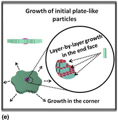
Initial plate-like particles would continue to grow by accreting rest micelles toward the particle under the silica polymerization process until the micelles consumed out. This growth occurred both in the side face direction and the end face direction [Fig. 4(e)], which was demonstrated by the both increase of the particle width and thickness [Figs. 3(g)–3(l)].
Reducing the P123 and TEOS concentration through increasing the initial solution volume to 75, 90, and 105 mL, respectively, a series of gear-like particle were obtained [Figs. 3(b)–3(d)]. The particle teeth grew from the corner of the hexagonal plate-like particle was clearly observed in Fig. 3(b), which was attributed to the preferential growth at the corner of the initial hexagonal plate-like particle under low concentration of the rest micelle. Decreasing the P123 and TEOS concentration by increasing the initial solution volume would result in more micelles remaining in the solution as the micelle concentration decreased under the micelle saturation point and the nucleation stopped. Furthermore, the reactant concentration decrease would lead to the decrease of the nucleation.[ Reference Turkevich, Stevenson and Hillier 28 ] Thus, the lower of the P123 and TEOS concentration, the more micelles were supplied for the initial plate-like particle growing, which was demonstrated by the deeper grooves and bigger diameter of the gear-like particle [Figs. 3(b)–3(d)]. While the P123 concentration was controlled at 2.28%, the final particle displayed a hexagonal plate-like morphology [Fig. 3(a)], which might attribute to the higher nucleation under the relative higher reactants concentration[ Reference Turkevich, Stevenson and Hillier 28 ] and the micelles almost consumed at the primary particle growth stage.[ Reference Linton and Alfredsson 15 ]
The gradual increase of the particle thickness in the growth process [Figs. 3(g)–3(l)] and the layer-like stack in the particle end face [Figs. 3(a)–3(d)] indicated that there was a layer-by-layer growth in the end face. The stack of a certain number of the not fully growing layer resulted in the generation of the shaft of the gear-like particle [Fig. 4(e)].
Conclusions
In this study, it was found that the synergistic effect of P123 and TEOS concentrations played an important role in controlling the particle morphology of SBA-15 mesoporous material. With the decrease of the P123 and TEOS concentration (i.e., the increase of the initial solution volume), the morphology of the plate-like SBA-15 particles changed gradually from hexagonal shape to the gear-like shape. Based on the morphology changes, a four-step growth process with aggression of threadlike micelles and nucleation, growth of primary particles, association of primary particles forming the initial plate-like particle and growth of initial plate-like particles was proposed.
Acknowledgment
This work was supported by the National Science Foundation of China (grant number 51372157)