Implications
Reducing dietary CP level is an effective way to improve the efficiency of N utilization, while maintaining normal growth performance of pigs once the indispensable amino acids (IAA) requirements are met. The results of this study suggest that not only IAA, but also the intact protein, dispensable amino acids and/or nitrogen should be taken into consideration in extremely low-CP diet to maintain normal growth and health of pigs.
Introduction
Reducing the CP level in diets has been recognized as an effective strategy to reduce the incidence of diarrhea in piglets (Yue and Qiao, Reference Yue and Qiao2008; Heo et al., Reference Heo, Kim, Hansen, Mullan, Hampson and Pluske2010). It is well documented that reducing dietary CP level from 16% to 12%, supplemented with indispensable amino acids (IAA), could maintain similar growth performance as pigs fed control diet (Figueroa et al., Reference Figueroa, Lewis, Miller, Fischer, Gómez and Diedrichsen2002 and Reference Figueroa, Lewis, Miller, Fischer and Diedrichsen2003; Kerr et al., Reference Kerr, Yen, Nienaber and Easter2003). In addition, a moderate reduction of dietary CP level has been shown to decrease the harmful microbial metabolites in the digesta such as ammonia N (Nyachoti et al., Reference Nyachoti, Omogbenigun, Rademacher and Blank2006; Opapeju et al., Reference Opapeju, Krause, Payne, Rademacher and Nyachoti2008), as well as improving the villous morphology of weaned piglets (Gu and Li, Reference Gu and Li2004; Opapeju et al., Reference Opapeju, Krause, Payne, Rademacher and Nyachoti2008). In contrast, several studies have shown that pig growth performance can decrease when the dietary CP level is reduced by more than 4% , even though supplemented with IAA (Powell et al., Reference Powell, Bidner, Payne and Southern2011; Gloaguen et al., Reference Gloaguen, Le Floc’h, Corrent, Primot and van Milgen2014), moreover, poor intestinal growth and morphology, reduced disaccharidase activities in small intestine were observed in pigs receiving extremely low-CP diets (Guay et al., Reference Guay, Donovan and Trottier2006; Yue and Qiao, Reference Yue and Qiao2008). However, there is little information whether there are alterations on immunological traits of pigs receiving extremely low-CP diet supplemented with IAA.
We hypothesized that feeding a moderate reduction of dietary CP level supplemented with IAA would maintain normal growth and immune function of pigs, while a high reduction of dietary CP level have a negative effect on growth performance and immune function. Therefore, the objective of the current study was to evaluate the effects of both a moderate and a high reduction of dietary CP level, supplemented with IAA, on growth performance and immune function of pigs.
Material and methods
All experimental protocols for the present study were approved by the Animal Care and Use Committee of Sichuan Agricultural University.
Experimental diets
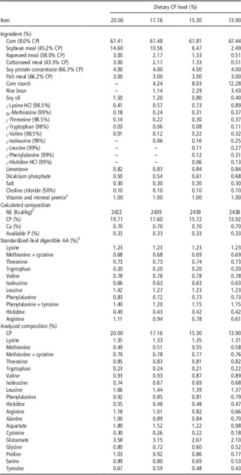
Four diets based on corn, soybean meal, rapeseed meal and cottonseed meal were used in the current study. Diets differed in CP level but contained the same amount of net energy and other nutrients which met or exceeded the requirements recommended by National Research Council (2012) for 11- and 25-kg pigs, industrial AA were added to the four diets to cover the requirements of standardized ileal digestible (SID) AA. Low-CP diets were formulated by mainly reducing the soybean meal but increasing the cornstarch contents. Feed ingredients contributing to AA were analyzed for CP and AA contents, the analyzed values were used in dietary formulation. Dietary composition and analysis is presented in Table 1. In the 20.00% CP diet, lysine, methionine, threonine, tryptophan and valine were limiting; thus, industrial l-lysine.HCl, dl-methionine, l-threonine, l-tryptophan and l-valine were included. In the 17.16% CP diet, additional isoleucine was limiting; thus, industrial l-isoleucine was included. In the 15.30% and 13.90% CP diets, nine IAA with the exception of arginine were limiting and they were included in their industrial form.
Ingredients composition and nutrient levels of the experimental diets (as-fed basis)

NE=net energy; SID=standardized ileal digestible.
1 Supplied the following per kg of diet: 8000 IU, vitamin A; 2400 IU, vitamin D3; 20 mg, vitamin E; 15 mg, pantothenic acid; 5 mg, vitamin B6; 0.3 mg, biotin; 3 mg, folic acid; 0.03 mg, vitamin B12; 40 mg, ascorbic acid; 120 mg, Fe; 25 mg, Cu; 20 mg, Mn; 20 mg, Zn; 0.5 mg, I; 0.30 mg, Se.
2 Values for net energy were calculated according to Noblet et al. (Reference Noblet, Fortune, Shi and Dubois1994), the contents of ether extract, starch, CP and ADF were analyzed.
3 Values for standardized ileal digestible (SID) concentrations of amino acids for the diets were estimated using SID coefficients for the various ingredients provided by NRC (2012).
Pigs and housing
A total of 40 crossbred barrows (Duroc×(Landrace×Yorkshire)), with an initial BW of 13.50±0.50 (mean±SEM) kg and aged at 45±2 days, were randomly assigned into four groups in a randomized complete block design. Pigs were individually housed in metabolic cages (1.5×0.7×1.0 m). Each cage was equipped with a feeder and a nipple drinker. Pigs had free access to feed and water over the 4-week study. The room temperature was maintained at 22–24°C. BW and feed intake were monitored weekly, average daily feed intake (ADFI), average daily gain (ADG) and feed conversion ratio (FCR) were calculated.
Blood sampling
On day 28, blood samples were collected by jugular venipuncture in the morning (0800 h) after an overnight fast according to our previous study (Hu et al., Reference Hu, Liu, Yan, Peng, Xu, Xuan, Han, Tian, Fang, Lin, Xu, Zhang, Chen, Wu and Che2015). Approximately 1 ml of the blood was injected into ethylene–diamine–tetraacetic acid-coated tubes for the determination of peripheral blood T-cells subsets. The rest were contained in heparinized tubes, followed by 3000×g at 4°C for 15 min, plasma was separated and then immediately stored at −80°C for later analysis.
Chemical analysis
The dry matter, ether extract and crude fiber were determined according to AOAC (2003) procedures. Nitrogen content (CP=6.25×N) was determined using the Kjeldahl methodology. The ADF content was determined following the procedures of Van Soest et al. (Reference Van Soest, Robertson and Lewis1991). The starch of ingredients was analyzed using the method of McCleary et al. (Reference McCleary, Solah and Gibson1994). The AA contents in ingredients and diets were determined by ion exchange chromatography using an auto amino acid analyzer (L-8800; Hitachi, Tokyo, Japan) after hydrolysis of the samples in 6 N HCl for 24 h at 110°C under a nitrogen atmosphere, whereas methionine and cystine were measured after performic acid oxidation, and the tryptophan content was determined after alkaline hydrolysis (AOAC, 2003).
Tissue sample collection and histological assessment
At the end of week 4, eight pigs with similar BW were selected from each treatment and sedated with an intravenous injection of sodium pentobarbital (80 mg/kg of BW) before jugular exsanguination. After the abdomen was exposed, heart, spleen, pancreas, kidney and liver were quickly removed and weighed. All tissue samples were rinsed by saline water (0.9% NaCl), pieces of spleen and intestinal ileum tissues were immediately frozen in liquid nitrogen and stored at −80°C until analysis. Another portion of intestinal tissue in duodenum, jejunum and ileum (~2 cm long, respectively) was collected and stored in 4% methanol solution for histological measurement.
The duodenal, jejunal and ileal samples were embedded in paraffin. The villous height and crypt depth were determined according to our previous study (Hu et al., Reference Hu, Liu, Yan, Peng, Xu, Xuan, Han, Tian, Fang, Lin, Xu, Zhang, Chen, Wu and Che2015). Briefly, each tissue sample was used to prepare five slides and each slide had three sections (5 µm thick), which were stained with hematoxylin and eosin for intestinal morphology analysis of 20 intact well-oriented crypt–villus units each section (Scion Image software, Version 4.02, 2004).
Plasma urea nitrogen, total protein and AA analysis
Plasma urea nitrogen (PUN) and total protein (TP) were measured using a biochemistry analyzer (Beckman CX4, Beckman Coulter Inc., Fullerton, CA, USA) and commercial kits (Sino-German Beijing Leadman Biotech Ltd, Beijing, China) according to the manufacturer’s instructions. Plasma AA contents were determined as previously described (Kong et al., Reference Kong, Yin, He, Yin, Liu, Li, Huang, Geng, Ruan, Deng, Xie and Wu2009). Briefly, 1 ml of plasma and 2.5 ml of 7.5% trichloroacetic acid were mixed thoroughly and centrifuged at 12 000×g at 4°C for 15 min. The supernatant was analyzed for amino acids using an auto amino acid analyzer (L-8800; Hitachi, Tokyo, Japan).
Immunogloblins and peripheral blood T-cells subsets analysis
Plasma concentrations of IgA, IgG and IgM were measured using the commercially available ELISA kits from R&D system (Minneapolis, MN, USA). The methods were referred to the manufacturer’s instructions. Each plasma sample was analyzed in duplicates. The plate wells were added with 50 µl of standard and plasma samples. Each well was then added with 50 µL of horseradish peroxidase, sealed and mixed prior to incubation at 37°C for 60 min and then completely washed 5 times using a washing buffer. Afterwards, 50 µl of Chromogen Solution A and 50 µl of Chromogen Solution B were added to each well and incubated at 37°C in the dark for 15 min, subsequently added 50 µl of stop solution. After completely mixed, the optical density (OD) values of standards and plasma samples were determined at 450 nm within 15 min (MuLtisKan MK3; Thermo Labsystems, Milford, MA, USA). The concentration of the plasma sample was quantified against the standard curve which was drawn from the concentration and absorbance of standards. The minimal detection limit was 3.12 μg/ml for IgA, 15.6 μg/ml for IgG and 2.5 μg/ml for IgM, respectively.
The peripheral blood of eight pigs each group were used to determine the percentages of CD3+, CD3+CD4+, CD3+CD8+T cells and CD4+/CD8+ratio by the flow cytometry method, as described by our previous study (Hu et al., Reference Hu, Liu, Yan, Peng, Xu, Xuan, Han, Tian, Fang, Lin, Xu, Zhang, Chen, Wu and Che2015). Total peripheral blood lymphocytes were separated from anti-clotting peripheral blood by separation medium. The cells were stained with mouse anti-porcine CD3 (Cat. NO. 561485; BD Pharmingen, Franklin Lakes, NJ, USA), mouse anti-porcine CD4 (Cat. NO. 561472; BD Pharmingen) and mouse anti-porcine CD8 (Cat. NO. 551303; BD Pharmingen). PBS (1×; Gibco, Carlsbad, CA, USA) and 1.0% bovine serum albumin (ICN Biomedicals, Aurora, OH, USA) were used as diluent and washing buffer. Flow cytometry analysis was performed on a FACS Calibur flow cytometer (Becton Dickinson, San Jose, CA, USA).
Total RNA extraction and RT reaction
Total RNA was isolated from tissue samples of spleen and ileum using TRIzol Reagent (TaKaRa Biotechnology Dalian Co., Ltd, Dalian, China) according to the manufacturer’s instructions. The quality and purity of RNA samples were verified by both agarose gel (1.0%) electrophoresis and nucleic acid analyzer (Beckman DU-800; Beckman Coulter Inc.). Both genomic DNA elimination and RT were performed using PrimeScript RT reagent kit with gDNA eraser (TaKaRa Biotechnology Dalian Co., Ltd) according to the manufacturer’s guidelines.
Real-time PCR
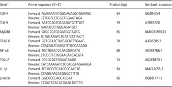
Real-time PCR was performed using SYBR Premix Ex Taq (Tli RNaseH Plus) qPCR kit (TaKaRa Biotechnology Dalian Co., Ltd) with ABI 7900HT (Applied Biosystems, Foster City, CA, USA). The PCR reaction consisted of 5.0 µl SYBR Premix Ex Taq (2×), 0.4 µl forward primer (10 μM), 0.4 µl reverse primer (10 μM), 0.2 µl ROX Reference Dye (50×), 1.0 µl cDNA and 3.0 µl double distilled water in a total volume of 10 µl. Cycling conditions were 95°C for 30 s, followed by 40 cycles of 95°C for 5 s and 60°C for 34 s. The primer pairs used are shown in Table 2. At the end of amplification, melting curve analysis was performed to identify amplification specificity. The
 $${\rm 2}^{{{\minus}\Delta \Delta C_{t} }} $$
method was used to process the real-time PCR results according to Livak and Schmittgen (Reference Livak and Schmittgen2001), with β-actin as the housekeeping gene. All samples were run in triplicate.
Primer sequences of the target and reference genes

1 Gene abbreviations: TLR=toll-like receptor; MyD88=myeloid differentiation factor 88; TRAF-6=TNF receptor-associated factor 6; NF-κB=nuclear factor kappa B; TOLLIP=toll-interacting protein; IL-1β=interleukin-1 beta.
Statistical analysis
All data were analyzed by ANOVA, using the GLM procedure of the SPSS 17.0 (Chicago, IL, USA), as a completely randomized block design with dietary treatments as main effects. Results were expressed as means with their standard errors. Duncan’s multiple range test was used to compare differences among the four treatment groups. Each animal was considered as an experimental unit. Effects were considered significant at P<0.05, whereas 0.05<P<0.10 was considered a tendency.
Results
Growth performance
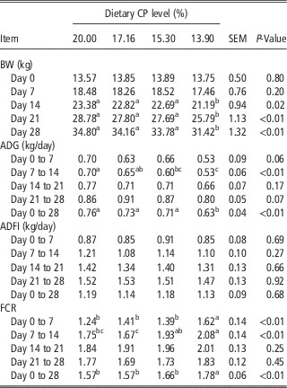
The effects of dietary CP level on growth performance are summarized in Table 3. BW was markedly reduced on day 14, 21 and 28 (P<0.05) as dietary CP level decreased. With the reduction of dietary CP level, ADG was markedly decreased during week 2 and overall (day 0 to 28) (P<0.01), and tended to decrease during week 1 (P=0.06) and week 4 (P=0.07). No significant differences were observed for the ADFI among dietary treatments. As dietary CP level decreased, FCR significantly increased during week 1, 2 and the overall study (P<0.01).
Growth performance of pigs fed low-CP diets supplemented with various AAFootnote 1

ADG=average daily gain; ADFI=average daily feed intake; FCR=feed conversion ratio.
a,b,cMeans in a row without common superscripts differ (P<0.05).
1 Values are means with pooled SEM, n=10 pigs/group.
Relative organ weights and intestinal morphology
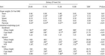
There were no significant effects of dietary treatment on relative organ weights of heart, spleen and kidney (expressed as a percentage of final BW). However, the relative weights of liver and pancreas decreased as the dietary CP level was reduced (P<0.05) (Table 4). There was a significant reduction of the villous height in duodenum (P<0.01) as dietary CP level was decreased. In addition, the crypt depth was markedly reduced (P<0.01) in duodenum, jejunum and ileum. As a result, the ratio of villous height to crypt depth (VCR) was increased in the jejunum (P<0.05), ileum (P<0.01) and tend to increase (P=0.08) in the duodenum.
Relative organ weights and small intestinal morphology of pigs fed low-CP diets supplemented with various AAFootnote 1

VCR=villous height to crypt depth ratio.
a,b,cMeans in a row without common superscripts differ (P<0.05).
1 Values are means with pooled SEM, n=8 pigs/group.
Plasma urea nitrogen, total protein and AA
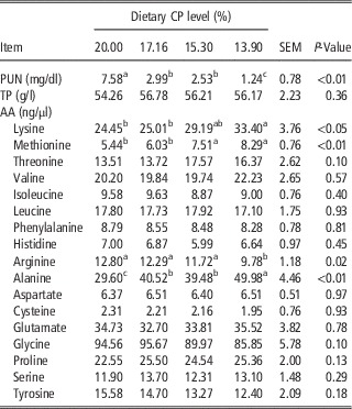
As shown in Table 5, the PUN concentration decreased (P<0.01) as dietary CP level reduced. However, there were no dietary effects on plasma concentration of TP. The reduction in the dietary CP level increased plasma concentrations of methionine, alanine and lysine (P<0.05) and decreased concentration of arginine (P<0.05).
Plasma urea nitrogen, total protein and AA of pigs fed low-CP diets supplemented with various AAFootnote 1

PUN=plasma urea nitrogen; TP=total protein.
a,b,cMeans in a row without common superscripts differ (P<0.05).
1 Values are means with pooled SEM, n=8 pigs/group.
Immunoglobulins and peripheral blood T-cells subsets
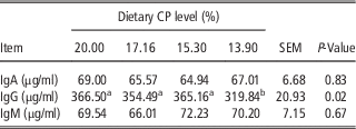
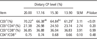
The plasma concentrations of IgA, IgG and IgM are reported in Table 6. During the experimental period, there were no significant differences for plasma concentrations of IgA and IgM among dietary treatments. However, plasma concentration of IgG was decreased with the reducing dietary CP level (P<0.05). As shown in Table 7, low-CP diets markedly decreased the percentage of CD3+T cells (P<0.01). However, there were no significant differences on the percentages of CD3+CD4+, CD3+CD8+Tcells and the CD4+/CD8+ratio among dietary treatments.
Plasma immunoglobulin concentrations of pigs fed low-CP diets supplemented with various AAFootnote 1

IgA=immunoglobulin A; IgG=immunoglobulin G; IgM=immunoglobulin M.
a,bMeans in a row without common superscripts differ (P<0.05).
1 Values are means with pooled SEM, n=8 pigs/group.
Peripheral blood CD3+, CD3+CD4+, CD3+CD8+T-cells percentages and CD4+/CD8+ratio of pigs fed low-CP diets supplemented with various AAFootnote 1

a,b,cMeans in a row without common superscripts differ (P<0.05).
1 Values are means with pooled SEM, n=8 pigs/group.
Gene expression in the intestine and spleen
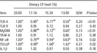
The Low-CP diets decreased the mRNA abundance of TLR-4, MyD88 and NF-κB (P<0.05), but increased the mRNA abundance of TOLLIP (P<0.05) in the ileum (Table 8). However, no significant difference was observed for the innate immunity-related gene expression of spleen (date not shown).
The mRNA abundance of innate immunity-related genes in the ileum of pigs fed low-CP diets supplemented with various AAFootnote 1

TLR=toll-like receptor; MyD88=myeloid differentiation factor 88; TRAF-6=TNF receptor-associated factor 6; NF-κB=nuclear factor kappa B; TOLLIP=toll-interacting protein; IL-1β=interleukin-1 beta.
a,b,cMeans in a row without common superscripts differ (P<0.05).
1 Values are means with pooled SEM, n=8 pigs/group.
Discussion
The current results showed that pig growth performance was similar when dietary CP level was reduced from 20.00% to 15.30%, however, a further decrease from 15.30% to 13.90% caused a lower ADG and higher FCR compared with pigs receiving 20.00% CP diet. Consistently, a recent study by Powell et al. (Reference Powell, Bidner, Payne and Southern2011) demonstrated that growth performance of pigs (20–50 kg) could be maintained when dietary CP level was reduced by ~5% units with the supplementation of IAA. Moreover, Wu (Reference Wu2014) emphasized both IAA and DAA should be taken into consideration in the formulation of balanced diets to maximize protein accretion in animals. It was reported that generation of DAA in pigs would be reduced by a deficiency of total N in low-CP diet (Columbus et al., Reference Columbus, Lapierre, Fuller, Htoo and de Lange2012), and become a limiting factor for normal growth of pigs (Gloaguen et al., Reference Gloaguen, Le Floc’h, Corrent, Primot and van Milgen2014). In this study, the decreasing growth performance of pigs fed 13.90% CP diet may be partially resulting from insufficient contents of total N, which was 30.5% lower than that of pigs fed 20.00% diet. Accordingly, the ratio between N from IAA and N from DAA (IAAN : DAAN) was 62 : 38 in the 13.90% CP diet, it is much higher than the optimal ratio of IAAN : DAAN (50 : 50) reported in growing pigs (Lenis et al., Reference Lenis, van Diepen, Bikker, Jongbloed and van der Meulen1999). Furthermore, low-CP diet supplemented with IAA would cause a deficiency of intact protein and related release of peptides (Deng et al., Reference Deng, Cui, Peng, Fang, Wang, Cui and Liu2007). These intact protein and small peptides could be more effective than a diet containing free AA for protein synthesis of animals (Boza et al., Reference Boza, Moennoz, Vuichoud, Jarret, Gaudard-de-Weck and Ballevre2000).
The decreasing dietary CP level decreased the relative organ weights of liver and pancreas, which is in agreement with the results by Kerr et al. (Reference Kerr, McKeith and Easter1995) and Heo et al. (Reference Heo, Kim, Hansen, Mullan, Hampson and Pluske2010). The reduction in pancreas weight of pigs fed low-CP diets may suggest a reduced amount of pancreatic protease, which would be required to digest the smaller quantity of consumed protein (Heo et al., Reference Heo, Kim, Hansen, Mullan, Hampson and Pluske2010). Besides, the liver weight has been reported to decrease with decreasing dietary protein level, the lower liver may be due to an adaptation of liver to less AA metabolism in pigs receiving low-CP diet (Kerr et al., Reference Kerr, McKeith and Easter1995). The small-intestinal morphology is usually used to reflect intestinal development. The shortening of the villous height may imply the decreased surface area for nutrient absorption, and a deeper crypt may suggest a faster turnover of new villous cells (Xia et al., Reference Xia, Hu and Xu2005). In the current study, the villous height in duodenum and crypt depth in duodenum, jejunum and ileum were both significantly decreased, but VCR was increased in all three intestinal segments when the dietary CP level was reduced from 20.00% to 13.90%. A consistent report showed that a reduction in villous height and crypt depth in the upper small intestine, when dietary CP level was decreased from 24.9% to 6.3% (Gu and Li, Reference Gu and Li2004), while VCR was increased in the jejunum of pigs receiving low-CP diets (Guay et al., Reference Guay, Donovan and Trottier2006). These results may be ascribed to the lower peptide-bound AA in low-CP diets, which decreased mucosal protein content in the small intestine (Guay et al., Reference Guay, Donovan and Trottier2006). However, intestinal morphology was not affected when dietary CP level was reduced from 25.10% to 19.20% (Heo et al., Reference Heo, Kim, Hansen, Mullan, Hampson and Pluske2010), in which 19.20% CP diet may have proper intact protein or peptide-bound AA for pigs, compared with the other studies (Gu and Li, Reference Gu and Li2004; Guay et al., Reference Guay, Donovan and Trottier2006).
The decreasing PUN concentration in pigs fed low-CP diets suggests that AA were excessive in the higher CP diets. PUN concentration is mostly dependent on the amounts and balance of AA (Nyachoti et al., Reference Nyachoti, Omogbenigun, Rademacher and Blank2006; Yue and Qiao, Reference Yue and Qiao2008). Previous studies have also reported that there were reductions in PUN concentration by feeding low-CP diets (Figueroa et al., Reference Figueroa, Lewis, Miller, Fischer, Gómez and Diedrichsen2002; Nyachoti et al., Reference Nyachoti, Omogbenigun, Rademacher and Blank2006). Besides, reducing dietary CP level from 20.00% to 13.90% resulted in higher plasma concentrations of lysine, methionine and alanine, this response is in agreement with previous reports that the higher protein degradation of pigs occurred in pigs fed low-CP diet, thus increased the plasma concentration of some free AA (Guay and Trottier, Reference Guay and Trottier2006). On the other hand, the higher plasma concentrations of lysine and methionine may be related to the higher supplementation of industrial lysine and methionine in low-CP diet (Figueroa et al., Reference Figueroa, Lewis, Miller, Fischer and Diedrichsen2003). The lower plasma concentration of arginine may reflect the lower dietary provision of arginine in pigs fed low-CP diet. In addition, pigs can synthesize arginine with precursors such as glutamine, glutamate and proline (Wu, Reference Wu1997), the lower level of these precursors may be responsible for the lower plasma concentration of arginine. It is implied that dietary protein level can affect immune function of animals and humans (Ruth and Field, Reference Ruth and Field2013). In this study, plasma concentration of immunoglobulins did not markedly differ when dietary CP level was reduced to 15.30%, however, plasma IgG concentration of pigs fed 13.90% CP diet was significantly lower than that of pigs fed 15.30%, 17.16% and 20.00% CP diets, even though all IAA met the requirement of pigs during this stage. The poor humoral immune response may be related to the deficiency of DAA in the 13.90% CP diet, because some DAA (e.g. glutamate and aspartate) have been demonstrated to regulate immune function (Wu, Reference Wu2014). In this study, glutamate and aspartate were only 59% and 54% of that in control diet, respectively. Dietary glutamate has been reported to improve immune status of rats recovering from methotrexate treatment (Lin et al., Reference Lin, Abcouwer and Souba1999). In addition, glutamate is a specific precursor for the intestinal synthesis of glutathione, which is required to protect the intestinal mucosa and optimize immune cell function (Ruth and Field, Reference Ruth and Field2013).
In this study, we also observed the low-CP diet affected cellular immunity-related parameters. The percentage of CD3+T cells was decreased along with the decreasing dietary CP level. CD3+molecules are the surface marker of mature T cells and represent the population of mature T cells (Deng et al., Reference Deng, Huang, Li, Wu, Xie, Tang, Kang, Zhang, Fan, Kong, Ruan, Xiong, Deng and Yin2011). The decreasing CD3+T cells in pigs fed low-CP diet suggested there were lower number of mature T cells in peripheral blood. In the present study, furthermore, we found that the mRNA abundance of TLR-4, MyD88 and NF-κB were markedly decreased in the ileum of pigs fed low-CP diets. Toll-like receptors (TLRs) are typical pattern recognition receptors that play an important role in the activation of the innate immune system following infection (Uematsu and Akira, Reference Uematsu and Akira2006). The TLR4-Myd88-NF-κB signal pathway is involved in the inflammatory response (Kawai and Akira, Reference Kawai and Akira2009). The abnormal expression of TLR-4, MyD88 and NF-κB indicate that feeding extremely low-CP diets may affect the intestinal innate immunity, even though all IAA met the requirement of pigs. It is also reported that some of the traditionally classified DAA (e.g. glutamate and arginine) play important roles in maintaining normal immunity, which is indeed lower in the low-CP diets (Wu, Reference Wu2014). Particularly, 30–50% of AA (e.g. glutamate, aspartate, glutamine and proline) in the diet are utilized by the small intestine during the first pass metabolism (Wu, Reference Wu1998), therefore, the lower DAA resulting from the low-CP diet may be responsible for the changed intestinal innate immunity. In addition, low-CP diet, supplemented with IAA, has been shown to alter intestinal bacterial community (Opapeju et al., Reference Opapeju, Rademacher, Blank and Nyachoti2009), which could be a factor to sensitize the intestinal immune system.
Conclusions
Our results demonstrate that the reduction of dietary CP level from 20.00% to 15.30%, supplemented with industrial AA, had no significant effect on growth performance and limited effect on immunological parameters of 10 to 35 kg pigs. However, poor growth performance and altered immunological parameters were observed when reducing dietary CP level up to 13.90%, even though industrial AA were added to meet IAA requirements of pigs. The lack of intact protein, DAA and/or nitrogen in low-CP diets are all potential factors leading to poor growth performance and immune function.
Acknowledgments
This work was supported by grants from the National Basic Research Program (973 Program) of China (No. 2013CB127306) and the Program for Changjiang Scholars and Innovative Research Team in University, Ministry of Education of China (No. IRT13083).