Case
A 4-year-old previously healthy girl was transferred to our facility with fever of 12 days, respiratory distress, eosinophilia, and neutropaenia, with white blood cell count of 54×109/L, 83% eosinophils, and absolute neutrophil count <500/mm3. She was treated presumptively for pneumonia and covered for occult bacterial infection with broad-spectrum antibiotics. On the 3rd day of hospitalisation, she developed respiratory decompensation with worsening pulmonary oedema and pleural effusions seen on chest radiograph. She was transferred to the paediatric ICU and intubated. The following morning, she was noted to have an accelerated junctional rhythm at 104 beats per minute. An echocardiogram was obtained, which showed dilation of the right atrium and a mass at the apex of the right ventricle. Systolic and diastolic functions by echocardiogram were preserved.
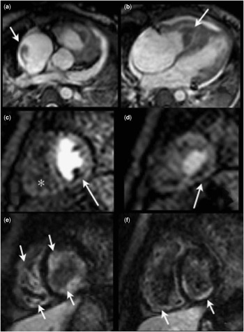
Cardiac MRI was performed on a 1.5 T Achieva MR scanner (Philips Healthcare, Best, the Netherlands) to evaluate for tumour versus thrombus. Cine steady-state free precession sequences in two-chamber, four-chamber, and short-axis orientations were performed before and after intravenous contrast administration. During rapid intravenous injection of 0.1 mmol/kg of Gadolinium contrast agent, gadopentetate dimeglumine (Magnevist, Bayer Healthcare, Whippany, New Jersey, USA), perfusion sequence was performed. The standard T1-scout or Look-Locker sequence was used to determine the inversion time for the viability sequence, Phase-sensitive Inversion Recovery, at the point when normal myocardium is nulled. T1- and T2-weighted imaging were also performed; however, due to very high heart rates of 155 bpm (average), the image quality was sub-optimal and did not contribute to the overall interpretation of the examination. Results were consistent with thrombus in the apex of the right ventricle with an additional thrombus adherent to the lateral superior wall of the right atrium. The patient was started on systemic anti-coagulation. Initial MRI was also notable for decreased perfusion of the inferior medial papillary muscle and adjacent endocardium, as well as late gadolinium enhancement along the endocardial surfaces of the right and left ventricles (Fig 1).
Images from initial cardiac MRI performed on patient who was in respiratory distress with heart rate averaging at 155 beats per minute during the examination. Patient was intubated but freely breathing during examination. ( a ) Images from axial cine SSFP showing thrombus in right atrium (arrow) and ( b ) in right ventricle (arrow); perfusion imaging in short axis at mid ventricle ( c ) showing decrease perfusion at the inferomedial papillary muscle and adjacent endocardium (arrow) and near apex ( d ) showing decrease perfusion in endocardium (arrow); there was no blood pool signal in the right ventricle because of presence of thrombus (*). Viability imaging with PSIR in short axis ( e , f ) showed diffuse areas of LGE along the endocardial surfaces of both right and left ventricles. LGE=late gadolinium enhancement; PSIR=phase sensitive inversion recovery pulse sequence; SSFP=steady-state free precession.

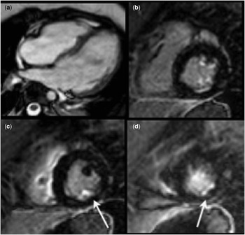
Subsequently, results of infectious and oncologic work-up revealed diagnosis of idiopathic hypereosinophilic syndrome. Her other clinical manifestations included small-vessel vasculitis of the brain, pulmonary parenchymal disease with emboli in the lungs, and eosinophilic infiltration of the bone marrow. She was started on steroids with improvement of symptoms. Follow-up cardiac MRI 46 weeks after the start of appropriate therapy showed resolution of the right atrial and ventricular thrombi, but with late gadolinium enhancement along the endocardial surface of the right ventricular apex. There was also late gadolinium enhancement of the inferomedial papillary muscle and adjacent endocardium corresponding to the areas of decreased perfusion seen on the initial MRI (Fig 2). Systolic and diastolic functions were followed-up on serial echocardiogram examinations and continued to be normal.
Images from follow-up cardiac MRI performed on patient 46 weeks after initial study. Patient’s medical condition was much improved with heart rate averaging 105 beats per minute during examination. Patient was sedated without intubation for the examination. One image from four-chamber cine SSFP ( a ) showed resolution of right ventricle and right atrial thrombi. Viability imaging with PSIR in short axis ( b , c , d ) showed significantly less LGE along the endocardial surface compared to initial examination (Fig 1e, f). Focal areas of LGE (arrows) along the posteromedial papillary muscle and adjacent endocardium matching the perfusion defects in the initial examination (Fig 1c, d). LGE = late gadolinium enhancement; PSIR=phase sensitive inversion recovery pulse sequence; SSFP=steady-state free precession.

Discussion
Endomyocardial fibrosis is a common cardiac complication of hypereosinophilia that has been described in the literature since 1893.Reference Brockington and Olsen 1 Most cases occur in tropical and sub-tropical regions in association with parasitic infections, where it is a common cause of heart failure;Reference Oh, Kim, Youn, Choi and Kang 2 however, cases are also regularly described in non-tropical parts of the world in association with idiopathic hypereosinophilia or hypereosinophilia due to vasculitides – particularly Churg–Strauss – malignancy, connective tissue disorders, or allergy.Reference Kleinfeldt, Nienaber and Kische 3
The pathophysiology of endomyocardial fibrosis is thought to occur in the following three phases: acute necrotic stage, characterised by eosinophilic infiltration of the myocardium; formation of mural thrombi; and finally restrictive cardiomyopathy due to hyaline fibrous tissue formation with progressive valvar incompetence.Reference Kleinfeldt, Nienaber and Kische 3 This progression of disease is founded based on histological study of pathology specimens in various stages of illness.Reference Brockington and Olsen 1 , Reference Tai, Ackerman, Spry, Dunnette, Olsen and Gleich 4
The utility of cardiac MRI in diagnosing cardiac involvement in hypereosinophilic syndrome has been well-described in the literature.Reference Oh, Kim, Youn, Choi and Kang 2 , Reference Kleinfeldt, Nienaber and Kische 3 , Reference Caudron, Arous, Fares, Lefebvre and Dacher 5 – Reference Debl, Djavidani and Buchner 9 Cardiac MRI findings have been described corresponding to these histological stages,Reference Cheung and Chan 6 , Reference Pillar, Halkin and Aviram 7 , Reference Debl, Djavidani and Buchner 9 and the use of cardiac MRI for prognostication and treatment monitoring in endomyocardial fibrosis has been postulated.Reference Cheung and Chan 6
Our findings confirm these reports in the literature – areas of late gadolinium enhancement of the right and left ventricles on initial study indicate tissue inflammation and necrosis, some of which later became fibrotic. Of note, a perfusion examination was also performed given the initial concern for malignancy. This showed resting perfusion defects in the same areas where late gadolinium enhancement developed as seen on the follow-up cardiac MRI examination. This evolution of imaging findings is interesting because studies of acute myocarditis typically have normal first-pass perfusion on cardiac MRI, whereas perfusion defects can be a sign of ischaemia/infarction.Reference Laissy, Hyafil and Feldman 10 This perfusion defect likely represents thrombosis in the ventricular wall segments, which is common in patients with hypereosinophilic syndrome,Reference Kleinfeldt, Nienaber and Kische 3 but not seen in other inflammatory cardiomyopathies. The cause of cardiac thrombosis in hypereosinophilic syndrome is proposed to be due to the pro-coagulant activity of eosinophil granule proteins.Reference Kleinfeldt, Nienaber and Kische 3 The contribution of eosinophilic infiltration versus mural thrombus to eventual formation of endomyocardial fibrosis is unclear.
To our knowledge, this is the first case of endomyocardial fibrosis followed-up by serial cardiac MRI in a child. The evolution of imaging findings also supports the proposed mechanism of disease based on histological study. This case affirms the adult literature that cardiac MRI is a powerful non-invasive tool for diagnosis and monitoring of endomyocardial fibrosis due to hypereosinophilia syndrome.
Acknowledgements
None.
Financial Support
This research received no specific grant from any funding agency, commercial, or not-for-profit sectors.
Conflicts of Interest
None.