Introduction
Fibre-optic nasendoscopy is widely used in clinical practice. Moreover, it can be used to examine the trachea when inserting a percutaneous tracheostomy tube; the nasendoscope is used to inspect the trachea during the procedure to ensure correct placement of the incision and the tube. Originally described by Ciaglia in 1985, percutaneous dilatational tracheostomy is now frequently used worldwide as a technique of airway control. Its advantages include relative speed and simplicity.Reference Koitschev, Paasch and Plinkert1
A protective sheath is sometimes used as a barrier against infection during nasendoscopy.2 When the endoscopic procedure is completed, the sheath is discarded. The sheath is transparent and has an elastic section on the proximal end that fits tightly onto the endoscope to prevent displacement. This provides tension on the sheath tip, causing it to become closely applied to the optic fibres and thus minimising reduction in image quality.Reference Vaz, Ripley, Lim, Kanegaonkar and Harries3 The sheath has the potential to reduce scope ‘downtime’ by eliminating the need for high-level disinfection between procedures. Endoscopes used with and without a sheath are reported to have a similar level of illumination, image clarity and ease of insertion.Reference Colt, Beamis, Harrell and Mathur4
We report an incident of sheath retention which occurred during insertion of a percutaneous tracheostomy in a patient with bilateral vocal fold paralysis and respiratory distress. We also describe a novel method of resolving the problem. As far as we are aware, this is the first time such an incident has been reported.
Case report
The procedure in question commenced with insertion of a Karl Storz 3.5 mm flexible fibre-optic nasendoscope (Karl Storz Endoscopy, Slough, UK) via the nose through the larynx, to view the inner wall of the trachea and to confirm placement of the incision and the percutaneous tracheostomy tube. The endoscope was handled by an experienced clinician who had used it on several previous occasions without reported complication. The endoscope shaft was covered with a protective sterile sheath (Endoshaft Cover®; Xmed, Mirandola, Italy).
Without application of excessive force, the sheath tip became detached and fell into the airway prior to making the tracheal incision. The procedure was abandoned, and a rigid bronchoscope was inserted to examine the airway more thoroughly and to attempt removal of the foreign body.
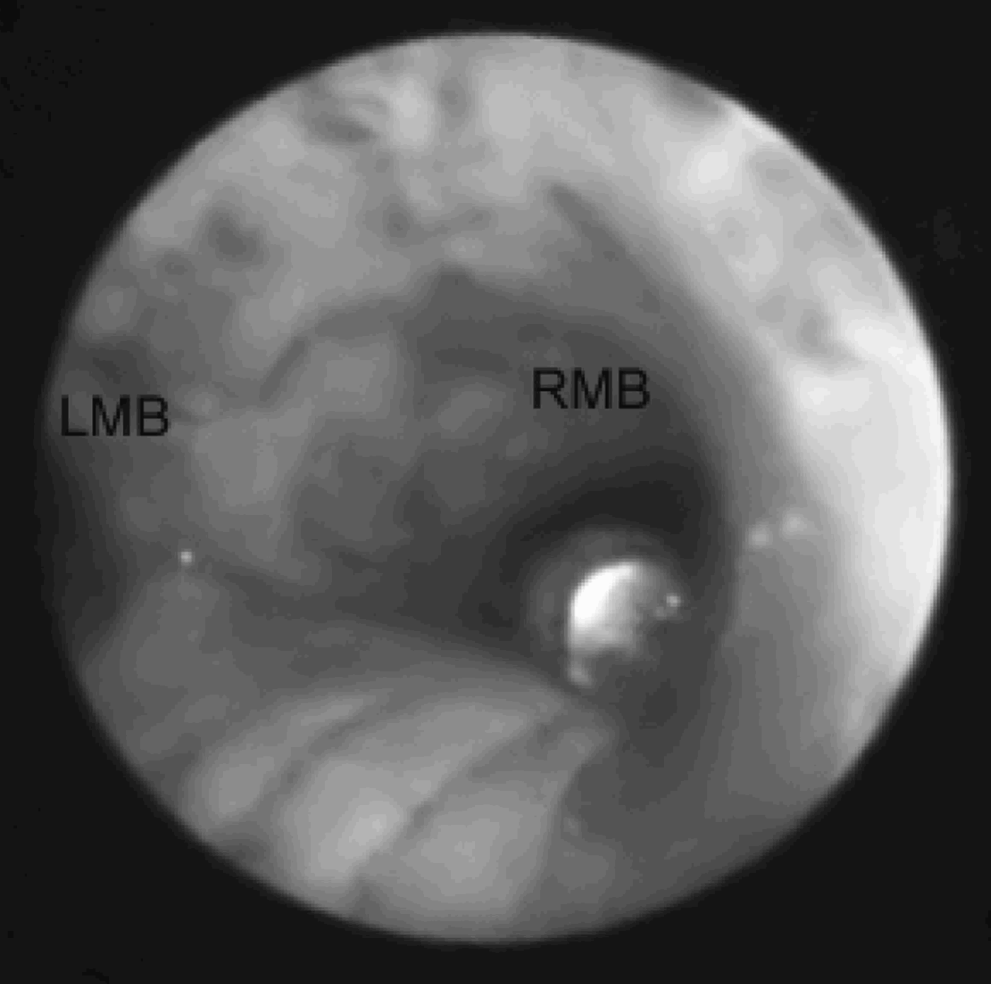
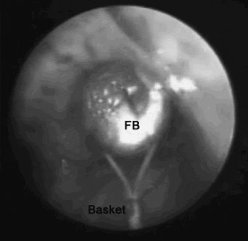
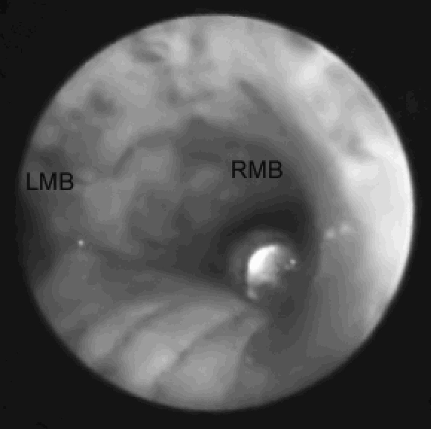
The foreign body was found lying at the level of the carina, near the root of the right main bronchus (Figure 1). Attempts at removal using forceps and suction failed, and these efforts resulted in the foreign body being pushed deeper into the right main bronchus. A SeguraTM HemisphereTM stone retrieval basket (Boston Scientific International, La Garenne Colombes Cedex, France), used to retrieve stones from the urinary tract,Reference Hare5 was available in the operating theatre. This instrument was inserted through the bronchoscope, into the trachea, passed beyond the foreign body and then opened to enclose the foreign body. The basket was tightened around the foreign body, and both were extracted successfully (Figure 2). The procedure had no associated morbidity.
Bronchoscopic view showing foreign body at tracheal bifurcation, near the root of the right main bronchus (RMB). LMB = left main bronchus
Bronchoscopic view showing tightening of the stone retrieval basket around the foreign body (FB). Both were extracted successfully.

Discussion
For a very commonly used procedure, flexible nasendoscopy is considered to be safe, with few serious complications. There are several manufacturers of endoscopes and protective elastic sheaths. Following the above incident, we contacted the manufacturers of the sheath in question, who instituted an investigation. We conducted a literature search, using Pubmed®, EMBASE (1974 to date) and MEDLINE (1950 to date) and including the terms ‘sheath or endosheath’, ‘nasendoscopy or nasendoscope’ and ‘airway or trachea or bronchus near foreign body’. This revealed no similar report in the world literature. Thus, as far as we are aware, the present case represents the first published report of such a complication.
• Nasendoscopy sheaths are widely used to reduce endoscope ‘downtime’, with minimal effect on image quality
• Tip detachment is a potential danger which has not previously been reported
• Retrieving the detached tip is a clinical priority, and should be performed in a controlled environment
• Many instruments are available, including those more popular amongst other specialties, such as urological stone retrieval baskets
We do not feel that this case should change current clinical practice. However, clinicians should be aware that any instrument introduced into the airway has the potential to fail and in the process produce a foreign body, which may cause serious complications. The widespread use of endosheaths further underlines the importance of awareness of this potential complication. A urology basket may be a useful addition to the armamentarium of an ENT surgeon faced with a difficult foreign body in the airway.Reference Dajani6