Ventricular septal defects, as one of the most common congenital heart diseases, can be divided into perimembranous, inlet, muscular, and infundibular defect based on their anatomical location.Reference Minette and Sahn 1 Infundibular ventricular septal defect, locating above the crista supraventricularis and beneath both the aortic and pulmonary valves, is also referred to as supracristal or doubly committed subarterial ventricular septal defect. Higher incidence of doubly committed subarterial ventricular septal defect was reported in Asian populations, accounting for 33% of patients with ventricular septal defects.Reference Warnes, Williams and Bashore 2
Although conventional surgical repair remains the gold standard for treatment of doubly committed subarterial ventricular septal defects,Reference Mavroudis, Baker, Idriss, Mavroudis and Baker 3 , Reference Demirag, Keceligil and Kolbakir 4 it requires cardiopulmonary bypass and total sternotomy, inevitably resulting in scars and likely complications. Due to the unusual location, device closure was not considered suitable for such defects for risks of introducing aortic regurgitation.Reference Kenny, Tometzki and Martin 5 , Reference Hijazi 6 Recently, with the development of asymmetric or eccentric ventricular septal defect occluders, transfemoral device closure of doubly committed subarterial ventricular septal defects has been attempted in several pilot studies.Reference Zhou, Pan, Guan and Ge 7 – Reference Gu, You, Zhao, Zheng and Qin 9 However, the success rate was low mainly due to difficulties in building the delivery track, orienting or positioning the eccentric device intra-procedurally, and suboptimal occluding results.Reference Zhao Yang, Hua and Yuan Ji 8 Until now, there is limited experience about transcatheter closure of doubly committed subarterial ventricular septal defects with the Amplatzer ductal occluder. This study describes our initial experience in using Amplatzer ductal occluder to close doubly committed subarterial ventricular septal defects while addressing its technical points, safety, and efficacy.
Materials and methods
Patient selection
Between March, 2015 and July, 2017, 22 consecutive patients with doubly committed subarterial ventricular septal defects who underwent transcatheter closure using the Amplatzer ductal occluder at our centre were included. Inclusion criteria were age ≥1 year and body weight ≥10 kg; confirmed diagnosis of doubly committed subarterial ventricular septal defect by transthoracic echocardiography; and ventricular septal defect diameter from 2 to 8 mm (<8 mm). Exclusion criteria were grade ≥2 aortic regurgitation; obvious aortic valve prolapse; significant cardiac and non-cardiac comorbidities that could affect the clinical outcome of defect closure; and ventricular septal defects with right-to-left shunting due to severe pulmonary hypertension.
All patients or their guardians had signed a written informed consent. The study was approved by the Chinese Clinical Trial Registry and Ethics Committee of our hospital.
Devices and selection
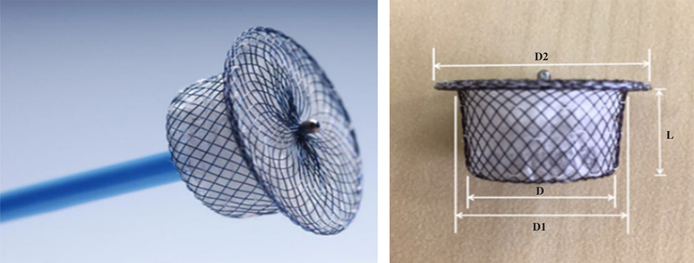
The first-generation Amplatzer ductal occluder device (Starway Medical Technology, Inc) was used in the study (Fig 1). Device size (diameter) at the distal end (D1) is 2 mm larger than at proximal end (D), and the left disc (D2) is 4 mm larger than at distal end (D1). The occluder is sized according to D and D1, which ranges from 4/6 mm (L = 7 mm, D2 = 10 mm), 6/8 mm (L = 7 mm, D2 = 12 mm), 8/10 mm (L = 8 mm, D2 = 14 mm), and 10/12 mm (L = 8 mm, D2 = 16 mm) in the study.
Configuration of the Amplatzer ductal occluder. D, Occluder size (diameter) at the proximal end; D1, occluder size (diameter) at the distal end; D2, left-side disc of the occluder; L, length of the occluder.

Because doubly committed subarterial ventricular septal defect in almost all cases was partially or completely covered by the right coronary cusp, transthoracic echocardiography, instead of ventricular angiography, was used to size the defect. Generally, an Amplatzer ductal occluder device with D end 1–2 mm larger than the maximal defect diameter was selected for first closure attempt. If significant aortic regurgitation existed or the device was liable to dislodge during closure attempt, a smaller or larger device would be used to replace the selected device.
Procedure
Ten patients aged <12 years were operated under general anaesthesia; the other 12 patients were operated under local anaesthesia. Cefazolin was administered half an hour before the procedure. After setting up the femoral arterial and venous accesses, intravenous heparin (100 U/kg) was given. The procedure was monitored under fluoroscopy and transthoracic echocardiography.
Most procedural steps were similar to previous reports,Reference Hijazi 6 , Reference Lee, Song and Choi 10 – Reference Fu, Bass and Amin 12 but some key steps were modified in our study. After right cardiac catheterisation, left ventriculography with 60–80° left anterior oblique plus 25° cranial angulation projection and aortic root angiography were performed to confirm the location of ventricular septal defect and its relation to the aortic valve. Right Judkin’s catheter or shortcut pigtail catheter was advanced into the left ventricle through femoral artery and crossed ventricular septal defect into pulmonary artery or superior vena cava. An exchange guidewire (0.035 inch × 260 cm) was advanced through it and snared with a Gooseneck Snare to exteriorise through femoral vein, establishing a long wire loop from the femoral artery to the femoral vein. A long sheath with a dilator was advanced from the femoral vein across the ventricular septal defect into the ascending aorta. Then the dilator was removed, but the guidewire was maintained in the delivery system. After flushing the long sheath, the Amplatzer ductal occluder was inserted and advanced to the tip of the sheath. The left retention disc of the Amplatzer ductal occluder was half deployed in the ascending aorta. Afterwards, the device and sheath were pulled simultaneously and slowly from the ascending aorta down to the left ventricular outflow tract just below the aortic valve where the left retention disc of the Amplatzer ductal occluder was wholly deployed, with the whole process being precisely controlled by both transthoracic echocardiography and fluoroscopy. When the left disc of the Amplatzer ductal occluder was wholly opened and just reached the subaortic location, the device and the sheath were pulled together to the ventricular septum and then the right disc of the Amplatzer ductal occluder was deployed if the “waist sign” was seen under fluoroscopy. Prior to the final release, left ventriculography and transthoracic echocardiography were performed to confirm the proper position of the Amplatzer ductal occluder device and the absence of new-onset or aggravated aortic regurgitation (Fig 2a–g). The arterial and venous sheaths were removed immediately post-procedurally, and haemostasis was achieved using manual compression. Oral aspirin 100 mg daily was administered for adults and 3 mg/kg daily for children for the next 6 months.
The procedure for transcatheter closure of dcVSD. ( a ) A dcVSD with left-to-right shunting in left ventriculography prior to the procedure. ( b ) Establishing a delivery track by advancing a dilator and a long sheath up to the ascending aorta. ( c ) Deploying the left-side disc of the occluder just below the aortic valve and then simultaneously withdrawing the device and sheath till inter-ventricular septum. ( d ) Deploying the right-side disc when the waist sign of the occluder was seen. ( e and e′ ) Removing the guide wire from the venous side after achieving acceptable closure results by doubly checking with X-ray fluoroscopy (no occluder dislocation seen pulling–pushing the delivery system [ e ]) and TTE (no significant residual shunting and tricuspid regurgitation [ e′ , upper] or aortic regurgitation [ e′ , lower] on the apical five-chamber view); ( f ) further confirming no significant residual shunting before releasing the occluder by left ventriculography; ( g and g′ ) good position and configuration of the released occluder under X-ray fluoroscopy ( g ) and good occluding result with neither systolic residual shunting and tricuspid regurgitation on the apical five-chamber view ( g′ , upper) nor aortic valve/sinus deformation on parasternal long-axis view ( g′ , lower) by TTE. dcVSD = doubly committed subarterial ventricular septal defect; TTE = transthoracic echocardiography.

Echocardiographic study
Besides estimating defect size and grading aortic regurgitation routinely, transthoracic echocardiography was performed to measure the width of residual shunts and aortic regurgitation intra-procedurally, post-procedurally, and at follow-up visit by measuring the maximal width of residual shunts or aortic regurgitation jets with vena contracta method.Reference Zoghbi, Adams and Bonow 13
Follow-up
Follow-up was scheduled at 1, 3, and 12 months in the first year, thereafter annually. Electrocardiography and transthoracic echocardiography were mandatory in each follow-up visit. No patents lost to the follow-up.
Definition of complications and occlusion success
Device-associated major complications were composed of death, aortic valve or sinus rupture, device dislocation/embolisation, moderate-severe new-onset aortic regurgitation (grade ≥2), moderate-severe device-caused ventricular outflow obstruction (local gradient ≥30 mmHg), new-onset atrioventricular block (≥2°), uncontrollable haemolysis, infectious endocarditis, or any reasons leading to surgical operation; while minor complications included mild new-onset aortic regurgitation (grade <2), mild device-caused outflow obstruction (local gradient <30 mmHg), new-onset atrioventricular block (<2°), or controllable haemolysis.
Any detectable shunting after device release was considered as residual shunt, which was further categorised echocardiographically as mild residual shunt when shunting width was <2 mm, or as moderate-severe when shunting width was ≥2 mm.
Clinical occlusion success was defined as neither major complications nor moderate-severe residual shunts peri-procedurally and during follow-up.
Statistical analysis
All analyses were performed with SPSS 16.0 for Windows (SPSS Inc, Chicago, Illinois, USA). Continuous variables were expressed as mean ± standard deviation or median with range, and categorical variables as percentage. For the analysis of parameter differences among follow-up stages, one-way ANOVA followed by LSD test was used for continuous variables, and chi-square or Fisher’s exact probability test for categorical variables. A p-value <0.05 was considered statistically significant.
Results
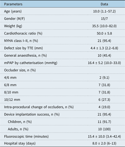
The baseline clinical and procedural characteristics of patients are summarised in Table 1. There were 15 males and 7 females, and 54.5% being children aged <14 years. The defect size was relatively small (4.4 ± 1.3 mm) with relatively small occluders implanted. Intra-procedural occluder change was 19.0%. One case (a 2.8-year-old boy with a doubly committed subarterial ventricular septal defect of 4 mm) failed during device implantation due to device-induced significant aortic regurgitation, resulting in a total implantation success of 95.4% with similar success between children and adults (91.7% versus 100%).
Baseline clinical and procedural characteristics

F = female; M = male; mPAP = mean pulmonary artery pressure; NYHA = New York Heart Association; TTE = transthoracic echocardiography.
Note: Age and weight were expressed as median with range; other continuous variables as mean ± standard deviation; categorical data as percentages.
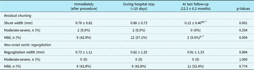
Changes in residual shunts and new-onset aortic regurgitation are listed in Table 2. Quantitative analysis showed most residual shunts were mild, became smaller, and finally disappeared; all new-onset aortic regurgitations were mild and unchanged during the follow-up period. Qualitative analysis showed that there were moderate-severe residual shunts in two patients (9.5%) and mild residual shunts in nine patients (42.8%) immediately after operation. During hospital stay, moderate-severe residual shunts were noted in 2 patients (9.5%) and mild residual shunts in 12 patients (57.1%). At the last follow-up, all mild residual shunts disappeared, and the two moderate residual shunts became mild. There were mild new-onset aortic regurgitations in nine patients (42.8%), and no moderate-severe new-onset aortic regurgitation was observed during hospital stay. During the follow-up, aortic regurgitation completely resolved in two patients (9.5%), didn’t worsen in seven patients (33.3%) of the above nine patients, and another four patients (19.0%) newly developed mild aortic regurgitation (grade <1).
Changes in residual shunt and new-onset aortic regurgitation during follow-up

Compared to the stage immediately after procedure, ##p < 0.01,#p < 0.05. Compared to the stage during hospital stay, **p < 0.01.
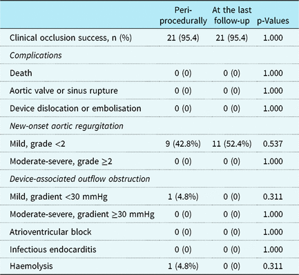
Clinical outcomes peri-procedurally and at the last follow-up are listed in Table 3. The defined clinical occlusion success remained 95.4% at follow-up of mean 12.3 months. Although there were no major device-associated complications, minor complications (mainly due to mild new-onset aortic regurgitation) were not infrequent with occasional mild residual shunts. However, such minor complications or mild residual shunts kept stable without hemodynamic significance during the follow-up period.
Clinical occlusion success and device-associated complications during follow-up

Discussion
Owing to its special anatomy, doubly committed subarterial ventricular septal defect has a high risk of aortic valve prolapse and regurgitation and a low rate of spontaneous closure.Reference Minette and Sahn 1 Early treatment of such type of ventricular septal defect is considered justifiable.Reference Tweddell, Pelech and Frommelt 14 Though a number of devices and methods have been attempted, there is limited data about using Amplatzer ductal occluder for the closure of doubly committed subarterial ventricular septal defects. In this study, we found that transcatheter closure of doubly committed subarterial ventricular septal defects using the Amplatzer ductal occluder was technically feasible. The occlusion success rate was high immediately and at follow-up of mean 12.3 months without major complications and significant residual shunts.
Advantages and disadvantages in using the Amplatzer ductal occluder for closure of doubly committed subarterial ventricular septal defects
Although the use of occluders to close doubly committed subarterial ventricular septal defects is considered a more feasible technique, it still has lots of technical difficulties with lower success rate.Reference Zhao Yang, Hua and Yuan Ji 8 In this study, transcatheter closure was attempted using the first-generation Amplatzer duct occluder. Although not designed to be used for the closure of doubly committed subarterial ventricular septal defects, the first-generation Amplatzer ductal occluder has several advantages compared with various types of asymmetric ventricular septal defect occluders.Reference Zhou, Pan, Guan and Ge 7 , Reference Zhao Yang, Hua and Yuan Ji 8 First, the D1 end and D2 end is symmetrically larger than the D end, so the left disc does not easily slide into the right ventricle, which increases device stability during the releasing process. Second, the Amplatzer ductal occluder device has a retention skirt only on one side as well as a long waist (7–8 mm) with a tapered configuration, which helps to hold the device firmly in place because the device stability is determined primarily by pressure gradients between the left and right chambers.Reference Mahimarangaiah, Subramanian and Kikkeri Hemannasetty 15 Third, the Amplatzer ductal occluder design enables easy manipulation, since there is no need to adjust the orientation of the device during operation. We found that the fluoroscopic time in our study was half shorter than that in a previous report when using asymmetric ventricular septal defect occluders.Reference Zhao Yang, Hua and Yuan Ji 8 Fourth, the connecting waist of the Amplatzer ductal occluder is much longer than that of the asymmetric ventricular septal defect occluder (7–8 mm versus 1.5–3 mm).Reference Zhou, Pan, Guan and Ge 7 When the device is just in the right place, a definite “waist sign” is easily seen to help check device positioning. Fifth, the absence of a right-sided disc might decrease chances of tricuspid regurgitation and pulmonary insufficiency, as well as imparts less compression by the two discs on the conduction system, thereby decreasing device-associated conduction blocks or arrhythmias. At last, the Amplatzer ductal occluder device is also cost-effective compared with asymmetric ventricular septal defect occluders.
In spite of so many advantages, there are several potential shortcomings in using the Amplatzer ductal occluder for the closure of doubly committed subarterial ventricular septal defects. For instance, a left-side symmetric skirt of the Amplatzer ductal occluder may cause distortion, erosion, or rupture of the aortic sinus or valve, resulting in aortic regurgitation; a longer waist of the Amplatzer ductal occluder may cause obstruction of the ventricular outflow track. In our study, new-onset aortic regurgitation or right ventricular outflow track obstruction did occur, but these were usually trivial and recoverable without hemodynamic relevance.
Tricks and points when using the Amplatzer ductal occluder for the closure of doubly committed subarterial ventricular septal defects
Several technical difficulties were described previously by Chen et al when using asymmetric occluders for transcatheter closure of doubly committed subarterial ventricular septal defects.Reference Zhao Yang, Hua and Yuan Ji 8 First, it may not be easy for a catheter to engage the defect entrance for a wire to pass through the ventricular septal defect. We suggest that using a pigtail catheter with proper shortcut tip and a soft PTCA wire (subsequently switching to standard guide wire after passing the pigtail catheter) will be greatly helpful in solving the problem. Also, the guide wire is always maintained in the delivery system once the delivery pathway is established as described by Gu et al,Reference Gu, You, Zhao, Zheng, Li and Qin 16 which may need a relatively larger delivery catheter. Second, it may be difficult to push the wire loop and delivery catheter into the left ventricle. We suggest using the “supravalvular aortic release method” under precise control of fluoroscopy and transthoracic echocardiography as described above. Third, it may be difficult to accurately measure the defect size by ventriculography since the doubly committed subarterial ventricular septal defect might be almost partially or entirely covered by the coronary cusp. We suggest using transthoracic echocardiography rather than ventriculography to avoid underestimation of defect size.
Limitations
This was an observational pilot study involving patients weighing >10 kg with defect size <8 mm; patients with obvious aortic valve prolapse or grade ≥2 aortic regurgitation were excluded. The small case number as well as the relatively short follow-up time were also the limitations of this study. Thus, the findings cannot be applied to little children and subarterial ventricular septal defects of larger sizes. Further studies with long-term follow-up are required to further confirm the safety and efficacy of the Amplatzer ductal occluder for transcatheter closure of doubly committed subarterial ventricular septal defects.
Conclusion
Transcatheter closure of doubly committed subarterial ventricular septal defects in the selected patients using the Amplatzer ductal occluder is feasible. However, the superiority of the Amplatzer ductal occluder for transcatheter closure of doubly committed subarterial ventricular septal defects can be proved with long-term follow-up studies on a larger number of patients.
Acknowledgement
The authors thank all the staff of the Catheter Lab, and Department of Cardiology at Union Hospital for their kind support and enthusiasm for this study.
Financial Support
This study was funded by the National Natural Science Foundation of China (grant no. 81670332).
Conflict of Interest
The authors declare that they have no conflict of interest.
Ethical Approval
All procedures performed in studies involving human participants were in accordance with the ethical standards of the Chinese Clinical Trial Registry and the Ethics Committee of the Union Hospital Affiliated to Fujian Medical University and complied with the 1964 Helsinki Declaration and its later amendments.
Informed Consent
Signed informed consent was obtained from all patients or their guardians.