Introduction
The genus Aspidothelium Vain. was established by Vainio (Reference Vainio1890) to accommodate a lichenized species with whitish to pinkish perithecia and muriform ascospores. Santesson (Reference Santesson1952) emended the genus to also include species with transversely septate ascospores and with perithecia covered with diverse appendages. The genus is otherwise characterized by a crustose thallus, a chlorococcoid photobiont, sessile, exposed perithecia, a paraplectenchymatous involucrellum, dense, unbranched paraphyses, short abundant periphyses, apically thick-walled, cylindrical to clavate asci and hyaline, fusiform, thin-walled, non-amyloid ascospores. The genus is restricted mainly to tropical regions, growing on leaves and bark (Nelsen et al. Reference Nelsen, Lücking, Cáceres, Aptroot and Lumbsch2017). The systematic placement of Aspidothelium remained uncertain for a long time, with multiple reclassifications, since its establishment by Vainio (Reference Vainio1890). It was once merged with Aspidopyrenium Vain., Porinopsis Malme, Patellonectria Speg. and Thelenella Nyl. The genus has also been considered as a member of families such as Verrucariaceae Eschw., Strigulaceae A. B. Frank, Aspidotheliaceae Räsänen ex J. C. David & D. Hawksw. or Thelenellaceae O. E. Erikss. ex H. Mayrhofer (Nelsen et al. Reference Nelsen, Lücking, Cáceres, Aptroot and Lumbsch2017). The genus differs from Thelenella in having sessile, exposed perithecia and distinctive fusiform ascospores, whereas species of Porina Ach. and Trichothelium Müll. Arg. differ in their perithecial anatomy and ascospore morphology. Recent molecular phylogenetic analyses by Nelsen et al. (Reference Nelsen, Lücking, Cáceres, Aptroot and Lumbsch2017) using nuclear and mitochondrial ribosomal markers (nuSSU, nuLSU, mtSSU) have firmly placed Aspidothelium within the subclass Ostropomycetidae Reeb et al. in the Lecanoromycetes O. E. Erikss. & Winka, forming a monophyletic clade nested within the family Thelenellaceae. Kraichak et al. (Reference Kraichak, Huang, Nelsen, Leavitt and Lumbsch2018) placed the family Thelenellaceae in the newly established order Thelenellales Lumbsch & Leavitt.
There are currently 18 species recognized in Aspidothelium (Nelsen et al. Reference Nelsen, Lücking, Cáceres, Aptroot and Lumbsch2017). Of these, 13 are foliicolous: A. arachnoideum Lücking, A. cinerascens Vain., A. fugiens (Müll. Arg.) R. Sant., A. geminiparum (Malme) R. Sant., A. gemmiferum Sérus. & Lücking, A. hirsutum Yeshitela et al., A. macrosporum (Müll. Arg.) Lücking, A. mirabile Lücking, A. ornatum Lücking, A. papillicarpum Lücking, A. scutellicarpum Lücking, A. trichothelioides Sérus. & Vězda, and A. verruculosum R. Sant. The remaining five are corticolous: A. cuyabense (Malme) P. M. McCarthy, A. glabrum Lücking et al., A. lueckingii Flakus, A. silverstonei Soto Medina et al., and A. submuriforme Aptroot et al. Substratum preference is not completely restricted since some foliicolous taxa, such as Aspidothelium cinerascens, have also been reported from smooth bark (McCarthy et al. Reference McCarthy, Kantvilas and Vězda2001; Aptroot et al. Reference Aptroot, Lücking, Sipman, Umaña and Chaves2008). To date, only two species of Aspidothelium are known to occur in India, A. scutellicarpum and A. fugiens (≡ Thelenella fugiens; Sinha et al. Reference Sinha, Nayaka and Mishra2024).
Arunachal Pradesh, situated within the Himalayan biodiversity hotspot, represents one of the most ecologically diverse regions of India. The state encompasses a wide range of vegetation types, ranging from tropical lowland to alpine zones, supporting a high level of biotic diversity. Despite occupying only c. 2.54% of India’s total geographical area, the state is recognized for its rich plant diversity contributing up to 23.52% of the country’s flora (Halder et al. Reference Halder, Tag, Rawat, Shenoy, Ghosh, Dash and Mao2024). Floristic assessments of Arunachal Pradesh have documented a total of 5192 species of angiosperms, 25 gymnosperms, 656 ferns and fern-allies, 483 bryophytes and 11 species of macrofungi (Roy et al. Reference Roy, Jha and Dutta2022; Halder et al. Reference Halder, Tag, Rawat, Shenoy, Ghosh, Dash and Mao2024; Rawat et al. Reference Rawat, Paul and Sahu2025). Arunachal Pradesh is the third most lichen-rich state in India with 884 species (Sinha et al. Reference Sinha, Nayaka and Mishra2024). In addition to its plant wealth, the region also harbours remarkable animal diversity, contributing to 20% of the country’s total fauna with 210 species of mammals, 10 primates, 850 birds, 115 reptiles, 50 amphibians, 215 fishes and 900 species of butterflies (Hagio & Saikia Reference Hagio and Saikia2026). Hence, Arunachal Pradesh is an undoubted biodiversity hotspot in the Eastern Himalayan region.
During a recent expedition to the East Kameng district of the state, leaf samples bearing foliicolous lichens were collected. A detailed observation revealed one of the taxa to be a new species of Aspidothelium, its perithecia furnished with a corona of lobes. This represents a notable addition to the lichen diversity of India.
Materials and Methods
Microscopy and morphological observations
The morphological and anatomical features of thalli were examined using a Leica S8APO stereo zoom microscope and a Leica DM500 compound microscope, equipped with a digital camera and calibrated image analysis software. Thin hand-cut sections were mounted in water to measure the perithecia, hamathecium, exciple, ascospores and asci. Measurements of ascospores are presented in the format (a–)b–c(–d) where b–c represents the range containing 90% of the measured values centred on the mean and a and d denote the extreme values (n = 35). Spot tests were performed on the lichen thalli using 10% aqueous potassium hydroxide (K), Steiner’s stable paraphenylenediamine (P), and 50% aqueous calcium hypochlorite (C). Secondary metabolites were analyzed using thin-layer chromatography (TLC) in solvent system ‘A’, following Orange et al. (Reference Orange, James and White2001). Specimen coordinates were georeferenced using Google Earth Pro (v. 7.3) and a spatial distribution map was subsequently generated in QGIS (v. 3.10) (Fig. 1).
Distribution of Aspidothelium coronatum. A, map of India showing Arunachal Pradesh. B, map of Arunachal Pradesh highlighting the East Kameng district. C, holotype locality within the East Kameng district. In colour online.

Figure 1 Long description
Panel A is a map of India in the top-left. The state of Arunachal Pradesh is highlighted in red in the Northeast. A North arrow is in the top-right and a scale bar of 0 to 1,000 k m is at the bottom-left.
Panel B is a map of Arunachal Pradesh in the bottom-left. The East Kameng District is highlighted in orange in the Southwest. A North arrow is in the top-right and a scale bar of 0 to 100 k m is at the bottom-left.
Panel C is a large topographic map of East Kameng District on the right. The map uses a color gradient to show elevation. Red represents 5,000 m in the North, transitioning through orange, yellow, and green to blue, which represents 500 m in the South and along river valleys. A yellow triangle at the center-right marks the Holotype locality. A North arrow is in the top-right and a scale bar of 0 to 15 k m is at the bottom-left. The legend for elevation includes intervals of 500 m, 1,250 m, 2,000 m, 2,750 m, 3,500 m, 4,250 m, and 5,000 m.
Results and Discussion
India exhibits significant diversity in foliicolous lichens, with 186 taxa (Sinha et al. Reference Sinha, Nayaka and Mishra2024). They are prevalent in tropical and subtropical forests, particularly in North-East India including Himalaya, the Western Ghats and the Andaman and Nicobar Islands, below an altitude of 1500 m (Singh & Pinokiyo Reference Singh and Pinokiyo2008). The regions of North-East India and the Western Ghats are notable for their rich biodiversity, hosting over 10 000 and 7400 species of flowering plants, respectively (Nayar et al. Reference Nayar, Sibi and Beegam2014; Shankar Reference Shankar2021). The humid climate along with the high diversity of plants in these regions support luxuriant growth of foliicolous lichens. Martínez-Colín et al. (Reference Martínez-Colín, Lücking and Herrera-Campos2021) showed that diversity of foliicolous lichens is strongly influenced by the diversity of phorophytes and the microsite conditions where leaves grow. Different plant species, because of differences in leaf texture, longevity, hairs, glands and microclimate, support different lichen communities. The foliicolous lichens are excellent indicator organisms for tropical rainforest ecology because they respond rapidly to minor environmental changes. Their short life cycle, small size, high diversity and sensitivity to humidity, light, rainfall and leaf longevity make them useful for monitoring forest conditions (Lücking Reference Lücking2008a). Aspidothelium is an interesting but overlooked genus that occurs both on leaves and bark in tropical forests. It can be an indicator of humid old-growth forest or relatively undisturbed forest because some species prefer stable, moist forest microclimates. For example, A. lueckingii Flakus was described from mountain cloud forest in Bolivia, indicating its occurrence in persistently humid forest systems (Flakus Reference Flakus2009).
In recent years, several new species of lichens have been reported from North-East India, especially from the East Kameng district of Arunachal Pradesh. For example, the basidiolichens Cyphellostereum indicum Nayaka & A. Debnath (Nayaka & Debnath Reference Nayaka and Debnath2023) and Sulzbacheromyces arunachalensis A. Debnath & Nayaka (Debnath et al. Reference Debnath, Nayaka, Joseph and Ngangom2025) represent new discoveries from the region. The discovery of yet another new species, A. coronatum, indicates the rich biodiversity of the state and the occurrence of several undiscovered species. A worldwide identification key to the species of Aspidothelium is provided below.
Taxonomy
Aspidothelium coronatum A. Debnath & Nayaka sp. nov.
MycoBank No.: MB 862794
This species is distinguished from other taxa in the genus by the presence of 5–7 subapical, lacerated, concolorous, rather short, subulate, lobe-like appendages around the perithecia, in combination with large, submuriform ascospores 80–105 × 14–22 μm, with 15–17 transverse septa and 1–2 longitudinal septa.
Type: India, Arunachal Pradesh, East Kameng District, Pakke Village, on a leaf, c. 570 m alt., 27°33′38.14″N, 92°58′47.12″E, 8 November 2022, S. Nayaka & A. Debnath 22-048487 (LWG—holotype).
(Fig. 2)
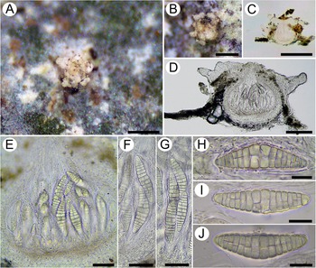
Aspidothelium coronatum (holotype) A, perithecium on a leaf substratum. B, detail of perithecium showing corona of lobes around the ostiole. C–E, anatomical details of perithecia. F, ascus with two ascospores. G, ascus with four ascospores. H–J, hyaline submuriform ascospores. Scales: A–C = 500 μm; D = 100 μm; E = 50 μm; F & G = 40 μm; H–J = 20 μm. In colour online.
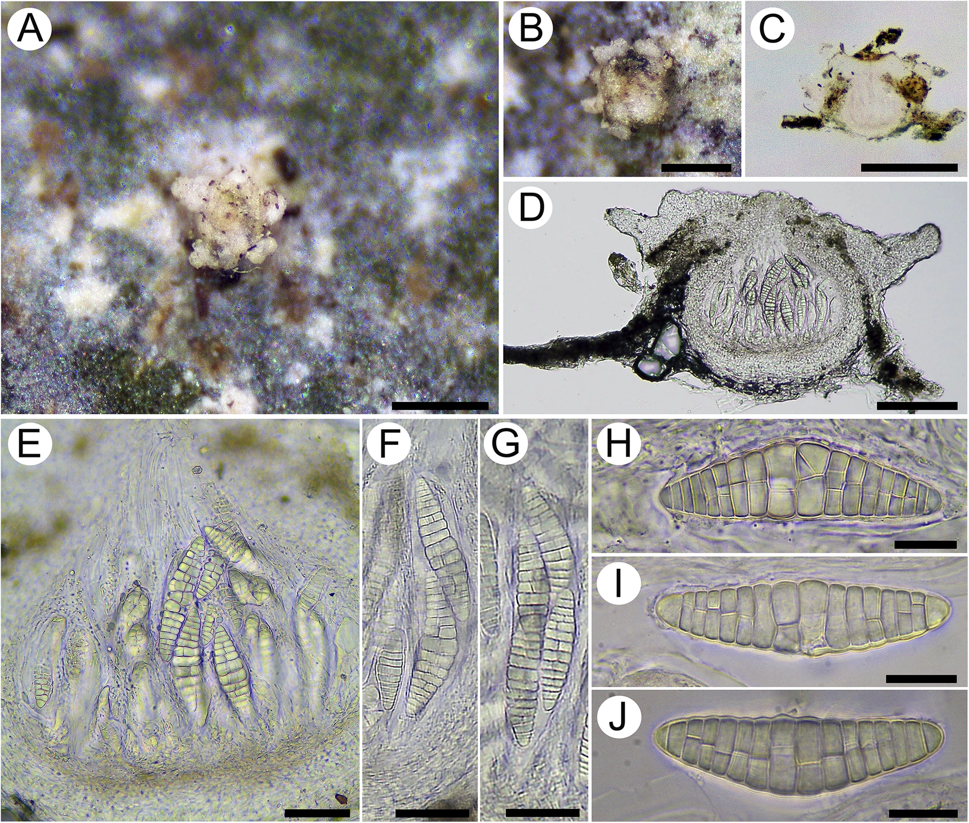
Figure 2 Long description
A multi-panel micrograph series organized as follows.
* Panel A. A top-down macro photo of a pale, star-shaped perithecium anchored to a mottled green and brown leaf surface. Scale bar represents 500 micrometers.
* Panel B. A close-up of the perithecium showing a central ostiole surrounded by a distinct corona of rounded lobes. Scale bar represents 500 micrometers.
* Panel C. A low-magnification cross-section of the perithecium showing a circular internal cavity. Scale bar represents 500 micrometers.
* Panel D. A higher magnification cross-section revealing the internal structure with multiple elongated asci packed inside the central chamber. Scale bar represents 100 micrometers.
* Panel E. A detailed anatomical view of the perithecium base with several asci containing visible spores. Scale bar represents 50 micrometers.
* Panel F. A single elongated ascus containing two large, segmented ascospores. Scale bar represents 40 micrometers.
* Panel G. An ascus containing four elongated, segmented ascospores. Scale bar represents 40 micrometers.
* Panels H, I, and J. High-magnification views of individual hyaline submuriform ascospores. Each spore is spindle-shaped with multiple transverse and longitudinal septa creating a grid-like internal pattern. Scale bars represent 20 micrometers.
Thallus epiphyllous, crustose, smooth, forming scattered, rounded to irregular patches, 2–5 mm across, 14–27 μm thick, white to pale greenish grey, lacking crystals, occasionally peeling off from the substratum. Soredia, isidia and prothallus absent. Photobiont chlorococcoid with globose to subglobose cells, 8–10 μm diam.
Ascomata perithecioid, sessile, wart-shaped, prominent, solitary, 0.3–0.6 mm diam. and 0.2–0.4 mm high, not constricted at the base, white to pinkish, with 5–7 subapical, lacerated, concolorous, lobe-like appendages around the perithecium, 100–150 μm long, somewhat similar to those found in A. fugiens (Lücking Reference Lücking1999). Ostiole apical, punctiform, brown. Exciple 10–40 μm thick, prosoplectenchymatous, colourless, entire. Involucrellum paraplectenchymatous, 50–110 μm thick, laterally spreading, colourless to pale yellowish with outermost part pale brownish, I+ orange-red, KI+ yellow. Centrum subglobose to depressed-ovate, 250–400 μm wide. Hamathecium colourless, not inspersed, I− and K/I− or slightly yellowish. Periphyses, short, c. 1.5 μm thick, unbranched, lining the ostiolar canal. Paraphyses unbranched, loosely embedded in hymenial gel, up to 1.5 μm thick. Asci bitunicate, clavate-cylindrical, 2–4-spored, with well-defined ocular chamber, 120–130 × 15–28 μm. Ascospores fusiform, hyaline, submuriform (80–)90–100(–105) × (14–)16–18(–22) μm with 15–17 transverse septa and around half of the segments with 1–2 longitudinal septa, slightly constricted at septa, median cells somewhat broadened (14–19 × 6.4–4.5 μm).
Conidiomata not seen.
Chemistry
Thallus K−, C−, KC−, P−, UV−; no substances detected by TLC.
Etymology
The species epithet refers to the corona of lobules around the perithecia.
Habitat and distribution
Aspidothelium coronatum is known from the East Kameng district in Arunachal Pradesh, found at an elevation of 570 m, growing on broad-leaved foliage in shaded and humid conditions.
Remarks
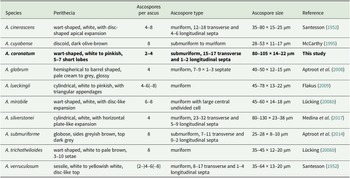
The new species is distinct due to the combination of lobe-like appendages on the perithecia and rather large, submuriform ascospores. Among the 13 foliicolous species of Aspidothelium, 10 produce submuriform or muriform ascospores (Table 1), with A. cinerascens, A. mirabile, A. trichothelioides and A. verruculosum producing muriform ascospores only. Aspidothelium cinerascens differs from the new species by having regularly muriform ascospores 35–80 × 15–25 μm with 12–18 transverse septa and 4–6 longitudinal septa, and perithecia featuring an irregular, disc-like expansion. The ascospores of Aspidothelium mirabile have a large, undivided middle cell. Aspidothelium trichothelioides has perithecia furnished with irregular, long setae while the ascospores are much shorter (35–45 μm long) and regularly muriform. Aspidothelium verruculosum differs by its coarsely verrucose thallus and the shorter, regularly muriform ascospores. The corticolous species Aspidothelium submuriforme is similar to the new species in having submuriform ascospores but differs by lacking perithecial ornamentation and by having smaller ascospores, 25–28 μm in length. Aspidothelium cuyabense differs by having a pale, greenish white, granular-isidiate thallus and smaller submuriform to muriform ascospores, 28–53 μm long. Aspidothelium lueckingii is distinguished by perithecia bearing triangular seta-like appendages at the apex and narrowly ellipsoid, regularly muriform, smaller ascospores (45–78 μm long).
Comparison of diagnostic characters between Aspidothelium coronatum sp. nov. (in bold) and other Aspidothelium taxa with submuriform to muriform ascospores.
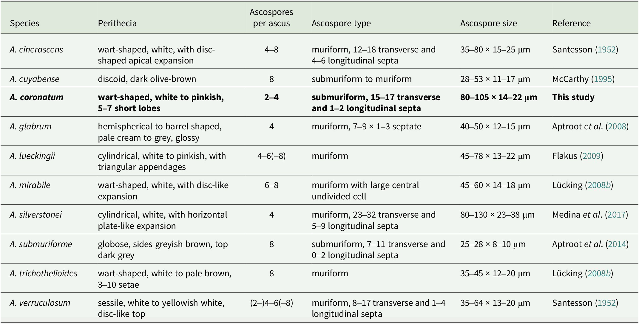
Table 1. Long description
The table contains six columns and ten rows of data.
* Aspidothelium cinerascens: Perithecia are wart-shaped, white, with disc-shaped apical expansion. Ascospores per ascus: 4 to 8. Ascospore type: muriform, 12 to 18 transverse and 4 to 6 longitudinal septa. Ascospore size: 35 to 80 by 15 to 25 mu m. Reference: Santesson 1952.
* Aspidothelium cuyabense: Perithecia are discoid, dark olive-brown. Ascospores per ascus: 8. Ascospore type: submuriform to muriform. Ascospore size: 28 to 53 by 11 to 17 mu m. Reference: McCarthy 1995.
* Aspidothelium coronatum: Perithecia are wart-shaped, white to pinkish, 5 to 7 short lobes. Ascospores per ascus: 2 to 4. Ascospore type: submuriform, 15 to 17 transverse and 1 to 2 longitudinal septa. Ascospore size: 80 to 105 by 14 to 22 mu m. Reference: This study.
* Aspidothelium glabrum: Perithecia are hemispherical to barrel shaped, pale cream to grey, glossy. Ascospores per ascus: 4. Ascospore type: muriform, 7 to 9 by 1 to 3 septate. Ascospore size: 40 to 50 by 12 to 15 mu m. Reference: Aptroot et al. 2008.
* Aspidothelium lueckingii: Perithecia are cylindrical, white to pinkish, with triangular appendages. Ascospores per ascus: 4 to 6 or 8. Ascospore type: muriform. Ascospore size: 45 to 78 by 13 to 22 mu m. Reference: Flakus 2009.
* Aspidothelium mirabile: Perithecia are wart-shaped, white, with disc-like expansion. Ascospores per ascus: 6 to 8. Ascospore type: muriform with large central undivided cell. Ascospore size: 45 to 60 by 14 to 18 mu m. Reference: Lucking 2008b.
* Aspidothelium silverstonei: Perithecia are cylindrical, white, with horizontal plate-like expansion. Ascospores per ascus: 4. Ascospore type: muriform, 23 to 32 transverse and 5 to 9 longitudinal septa. Ascospore size: 80 to 130 by 23 to 38 mu m. Reference: Medina et al. 2017.
* Aspidothelium submuriforme: Perithecia are globose, sides greyish brown, top dark grey. Ascospores per ascus: 8. Ascospore type: submuriform, 7 to 11 transverse and 0 to 2 longitudinal septa. Ascospore size: 25 to 28 by 8 to 10 mu m. Reference: Aptroot et al. 2014.
* Aspidothelium trichothelioides: Perithecia are wart-shaped, white to pale brown, 3 to 10 setae. Ascospores per ascus: 8. Ascospore type: muriform. Ascospore size: 35 to 45 by 12 to 20 mu m. Reference: Lucking 2008b.
* Aspidothelium verruculosum: Perithecia are sessile, white to yellowish white, disc-like top. Ascospores per ascus: 2, 4 to 6, or 8. Ascospore type: muriform, 8 to 17 transverse and 1 to 4 longitudinal septa. Ascospore size: 35 to 64 by 13 to 20 mu m. Reference: Santesson 1952.
Additional specimen examined
India: Arunachal Pradesh: East Kameng District, Pakke Village, on leaf, c. 564 m alt., 27°33′34.62″N, 92°58′46.01″E, 08 xi 2022, S. Nayaka & A. Debnath 22-048486 (LWG).
Worldwide key to the species of Aspidothelium
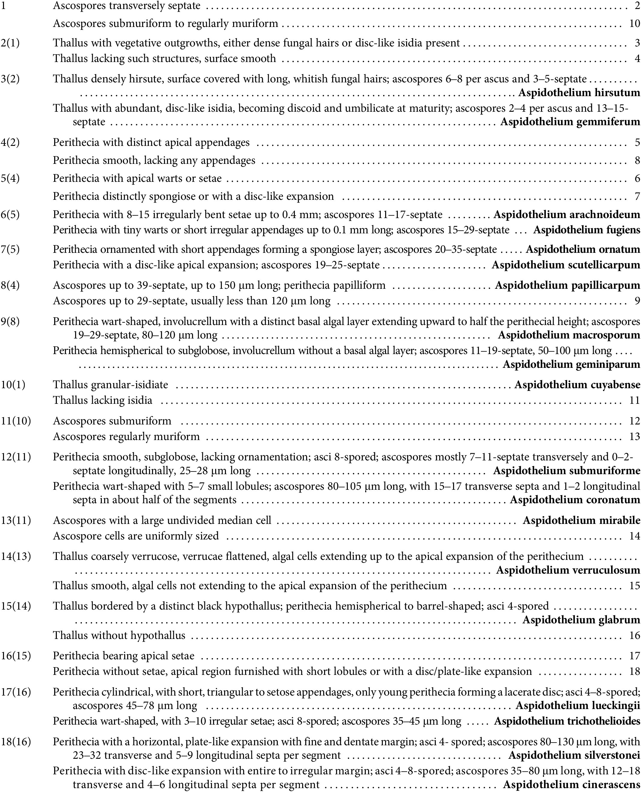
Long description
A taxonomic identification table for the genus Aspidothelium.
* Step 1: Ascospores transversely septate leads to step 2. Ascospores submuriform to regularly muriform leads to step 10.
* Step 2: Thallus with vegetative outgrowths leads to step 3. Thallus lacking such structures leads to step 4.
* Step 3: Densely hirsute thallus with 3 to 5 septate ascospores identifies Aspidothelium hirsutum. Thallus with disc-like isidia and 13 to 15 septate ascospores identifies Aspidothelium gemmiferum.
* Step 4: Perithecia with apical appendages leads to step 5. Perithecia smooth leads to step 8.
* Step 5: Perithecia with apical warts or setae leads to step 6. Perithecia spongiose or with disc-like expansion leads to step 7.
* Step 6: 8 to 15 bent setae and 11 to 17 septate ascospores identifies Aspidothelium arachnoideum. Tiny warts and 15 to 29 septate ascospores identifies Aspidothelium fugiens.
* Step 7: Spongiose layer and 20 to 35 septate ascospores identifies Aspidothelium ornatum. Disc-like apical expansion and 19 to 25 septate ascospores identifies Aspidothelium scutellicarpum.
* Step 8: Ascospores up to 39 septate and 150 micrometers long identifies Aspidothelium papillicarpum. Ascospores up to 29 septate and under 120 micrometers long leads to step 9.
* Step 9: Wart-shaped perithecia with basal algal layer identifies Aspidothelium macrosporum. Hemispherical perithecia without basal algal layer identifies Aspidothelium geminiparum.
* Step 10: Granular-isidiate thallus identifies Aspidothelium cuyabense. Lacking isidia leads to step 11.
* Step 11: Submuriform ascospores leads to step 12. Regularly muriform ascospores leads to step 13.
* Step 12: Smooth subglobose perithecia and 25 to 28 micrometer ascospores identifies Aspidothelium submuriforme. Wart-shaped perithecia with lobules and 80 to 105 micrometer ascospores identifies Aspidothelium coronatum.
* Step 13: Large undivided median cell identifies Aspidothelium mirabile. Uniformly sized cells lead to step 14.
* Step 14: Coarsely verrucose thallus identifies Aspidothelium verruculosum. Smooth thallus leads to step 15.
* Step 15: Black hypothallus and 4 spored asci identifies Aspidothelium glabrum. Without hypothallus leads to step 16.
* Step 16: Perithecia with apical setae leads to step 17. Perithecia without setae leads to step 18.
* Step 17: Cylindrical perithecia and 45 to 78 micrometer ascospores identifies Aspidothelium lueckingii. Wart-shaped perithecia and 35 to 45 micrometer ascospores identifies Aspidothelium trichothelioides.
* Step 18: Plate-like expansion with dentate margin and 80 to 130 micrometer ascospores identifies Aspidothelium silverstonei. Disc-like expansion with irregular margin and 35 to 80 micrometer ascospores identifies Aspidothelium cinerascens.
Acknowledgements
The authors are grateful to the Director, CSIR–National Botanical Research Institute, Lucknow, for providing essential infrastructure and laboratory facilities. We also thank the Department of Environment and Forests, Itanagar, for granting permission to conduct fieldwork and access forest areas in Arunachal Pradesh. This manuscript has been approved by the Institute’s publication Ethics Committee under approval number CSIR-NBRI_MS/2026/01/14. The study is funded by the Anusandhan National Research Foundation (ANRF; formerly Science and Engineering Board, SERB, New Delhi) under file number CRG/2021/002263.
Author ORCID
Sanjeeva Nayaka, 0000-0001-6541-2362.
Competing Interests
The authors declare none.