Introduction
Pallisentis (Pallisentis) nandai Sarkar (1953) was placed in the subgenus Pallisentis Van Cleave, 1928, sensu stricto established by Amin et al. (Reference Amin, Heckmann, Ha, Luc and Doanh2000). The subgenus is characterized by proboscis hooks gradually declining in size posteriorly and by a long cement gland with many giant nuclei. Two other subgenera had different proboscis hook and cement gland patterns (Amin et al., Reference Amin, Heckmann, Ha, Luc and Doanh2000). The distribution of P. nandai appears to extend throughout the length of the Ganga River and its tributaries from its northern sources in China to its lower delta in eastern India and Bangladesh. Its description from Nandus nandus (Hamilton) in the Ganga delta from a Calcutta fish market (Sarkar, Reference Sarkar1953) was repeated with some variations by other observers, including Soota & Bhattacharya (Reference Soota and Bhattacharya1982), Bhattacharya (Reference Bhattacharya2007) and Naidu (Reference Naidu2012). Since its description from N. nandus (Nandidae), P. nandai has also been reported from the tank goby, Glossogobius giuris (Hamilton) (Gobiidae), from other tributaries of the Ganga in Uttar Pradesh (UP), India, and from many tributaries of the Ganga in the Bangladesh delta (see Naidu, Reference Naidu2012 for references). Our finding of P. nandai in Bijnor near the northern reaches of the Ganga adds new geographical dimensions and morphological and descriptive information that expand our understanding of this interesting acanthocephalan. Scanning electron microscopy (SEM), energy-dispersive X-ray analysis, micropore and related studies, and DNA analysis expand the body of knowledge about P. nandai in particular and of the genus Pallisentis Van Cleave, 1928 in general.
In India, the validity of many species of acanthocephalans is questionable because descriptions over the past few decades have been based on morphological observations lacking detailed descriptive information and illustrations, type specimens were not deposited in recognized museums and previously published data were ignored by the authors (Tadros, Reference Tadros1966; Mital & Lal, Reference Mital R and Lal1976; Pichelin & Cribb, Reference Pichelin and Cribb2001; Gupta et al., Reference Gupta, Gupta and Singhal2015a; Gautam et al., Reference Gautam, Upadhyay, Maurya, Verma and Saxena2017). Molecular data are very scarce for Indian acanthocephalans. We supplement our study with the molecular profile of P. nandai. Thirty sequences are available for the genus Pallisentis in the GenBank database. In all, 24 sequences are available for the 18S ribosomal RNA (rRNA), one for the internal transcribed spacer (ITS) 1 region and three for the ITS1-5.8S-ITS2 cluster, while two sequences are available for the 28S rRNA gene, which demonstrates the scarcity of molecular data for the species of Pallisentis in comparison to the species diversity available (Amin, Reference Amin2013). In the present study, we inferred the phylogenetic relationship of P. nandai based on the 18S and ITS1-5.8S-ITS2 rRNA gene sequences within the genus Pallisentis (Quadrigyridae) in the class Eoacanthocephala.
Materials and methods
Collections
We collected 69 worms from the livers of 20 of 58 examined fish between March and April 2019 in the Ganga River at Bairaj, Bijnor (29°01′N, 77°45′E) in the state of UP, India (table 1). The fish were obtained from local fishermen in a small fish market in Bairaj. Of the 49 extended specimens (not all specimens were extended) that were used, 23 were processed for microscopical studies, 16 were used for SEM, gallium-cut hooks and energy dispersive X-ray analysis (EDXA), five were used for molecular studies and five remain in the Omar Mohamed Amin (OMA) collection. Specimens were deposited in the University of Nebraska's State Museum's Harold W. Manter Laboratory (HWML) collection, Lincoln, Nebraska, USA. Freshly collected specimens were extended in water until proboscides everted, then fixed in 70% ethanol for transport to our Arizona, USA laboratory for processing and further studies. Twenty contorted specimens collected from dead fish on April 5 were discarded.
Collections of Pallisentis nandai made from Nandus nandus in the Ganga River at Bairaj, Bijnor, in 2019.

Methods for microscopical studies
Worms were punctured with a fine needle and subsequently stained in Mayer's acid carmine, destained in 4% hydrochloric acid in 70% ethanol, dehydrated in ascending concentrations of ethanol (24 h each), and cleared in 100% xylene then in 50% Canada balsam and 50% xylene (24 h each). Whole worms were then mounted in Canada balsam. Measurements are in micrometres, unless otherwise noted; the range is followed by the mean values between parentheses. Width measurements represent maximum width. Trunk length does not include proboscis, neck or bursa.
Line drawings were created using a Ken-A-Vision micro-projector (Ward's Biological Supply Co., Rochester, NY, USA), which uses cool quartz iodine 150 W illumination with 10×, 20× and 43× objective lenses. Images of stained whole-mounted specimens were projected vertically on 300 series Bristol draft paper (Starthmore, Westfield, MA, USA), then traced and inked with India ink. Projected images were identical to the actual specimens being projected. Microscope images were created using 10× and 40× objective lenses of a BH2 light Olympus microscope (Olympus Optical Co., Osachi-shibamiya, Okaya, Nagano, Japan) attached to an AmScope 1000 video camera (United Scope LLC, dba AmScope, Irvine, CA, USA), linked to an ASUS laptop equipped with HDMI high-definition multimedia interface system (Taiwan-USA, Fremont, CA, USA). Images from the microscope are transferred from the laptop to a USB and stored for subsequent processing on a computer. Forty two images were made to create the figures.
SEM
Specimens fixed and stored in 70% ethanol were processed for SEM following standard methods (Lee, Reference Lee1992). These included critical-point drying in sample baskets and mounting on SEM sample mounts (stubs) using conductive double-sided carbon tape. Samples were coated with gold and palladium for 3 min using a Polaron #3500 sputter coater (Quorum (Q150 TES); www.quorumtech.com) establishing an approximate thickness of 20 nm. Samples were placed and observed in an FEI Helios Dual Beam Nanolab 600 (FEI, Hillsboro, OR, USA) scanning electron microscope, with digital images obtained in the Nanolab software system (FEI, Hillsboro, OR, USA) and then transferred to a USB for future reference. Samples were received under low vacuum conditions using 10 KV, spot size 2, 0.7 Torr using a Gas Sensitive Electrochemical (GSE) detector.
EDXA
Standard methods were used for preparation similar to the SEM procedure. Specimens were examined and positioned with the above SEM instrument, which was equipped with a Phoenix energy-dispersive X-ray analyser (FEI, Hillsboro, OR, USA). X-ray spot analysis and live scan analysis were performed at 16 Kv with a spot size of 5, and results were recorded on charts and stored with digital imaging software attached to a computer. The TEAM (Texture and Elemental Analytical Microscopy) software system (FEI, Hillsboro, OR, USA) was used. Data were stored in a USB for future analysis. The data included weight percent and atom percent of the detected elements following correction factors.
Ion sectioning of hooks
A dual-beam SEM with a gallium ion source (GIS) is used for the LIMS (Liquid Ion Metal Source) part of the process. The hooks of the acanthocephalans were centred on the SEM stage and cross-sectioned using a probe current between 0.2 nA and 2.1 nA according to the rate at which the area was cut. The time of cutting was based on the nature and sensitivity of the tissue. Following the initial cut, the sample also underwent a milling process to obtain a smooth surface. The cut was then analysed with X-ray at the tip, middle and base of hooks for chemical ions with an electron beam (Tungsten) to obtain an X-ray spectrum. Results were stored with the attached imaging software. The intensity of the GIS was variable according to the nature of the material being cut.
Molecular methods
Genomic DNA was isolated from two specimens fixed in ethanol using a DNeasy® Blood & Tissue Kit (QIAGEN, Hilden, Germany) following the manufacturer's instructions. The 18S ribosomal DNA (rDNA) region V2-V8 was amplified using primers Worm A (5′-GCGAATGGCTCATTAAATCAG-3′); 1270R (5′-CCGTCAATTCCTTTAAGT-3′) (Littlewood & Olson, Reference Littlewood, Olson, Littlewood and Bray2001) and 930F (5′-GCATGGAATAATGGAATAGG-3′); Worm B (5′-CTTGTTACGACTTTTACTTCC-3′) (Littlewood & Olson, Reference Littlewood, Olson, Littlewood and Bray2001) and ITS1-5.8S-ITS2 by primers BD1 (5′-GTCGTAACAAGGTTTCCGTA-3′); BD2 (5′-TATGCTTAAATTCAGCGGGT-3′) (Luton et al., Reference Luton, Walker and Blair1992) and D1 (5′-AGGAATTCCTGGTAAGTGCAAG-3′); (5′-CGTTACTGAGGGAATCCTGGT-3′) (Galazzo et al., Reference Galazzo, Dayanandan, Marcogliese and McLaughlin2002). The amplification reaction was performed as follows: denaturation at 95°C for 3 min for 40 cycles of 94°C for 40 s, 55°C for 45 s, 72°C for 1 min and termination at 72°C for 10 min. The polymerase chain reaction (PCR) products were checked on 1% agarose gel and purified with the PureLink™ Quick Gel Extraction and PCR Purification Combo Kit (Thermo Fisher Scientific, Waltham, MA, USA). Sequencing was done with a Big Dye Terminator version 3.1 cycle sequencing kit in an ABI 3130 Genetic Analyzer (Thermo Fisher Scientific, Waltham, MA, USA) using the above-mentioned primers.
Contiguous sequences of 18S and ITS1-5.8S-ITS2 rDNA sequences of P. nandai were assembled with MEGA7 (Kumar et al., Reference Kumar, Stecher and Tamura2016) and submitted to the GenBank database. To assess the phylogenetic relationship of P. nandai within Pallisentis in the class Eoacanthocephala, the newly generated sequences with other species data available on the GenBank database were aligned together using the program ClustalW (Thompson et al., Reference Thompson, Higgins and Gibson1994), which is implemented in MEGA7. For molecular phylogeny, 18S and ITS1-5.8S-ITS2 sequences of species of Archiacanthocephala and Eoacanthocephala were included. Maximum likelihood (ML) analysis was performed by MEGA7 with 1000 bootstrap replicates. A Bayesian inference (BI) phylogenetic tree was generated by Topali 2.5 (Milne et al., Reference Milne, Lindner, Bayer, Husmeier, Mcguire, Marshall and Wright2009), in which four independent Markov Chain Mont Carlo (MCMCMC) runs with every 100th tree saved and ‘burn-in’ was set to 25%. The evolutionary model GTR + G + I for both ML and BI analyses was estimated with the program jModelTest v2.1.10 (Darriba et al., Reference Darriba, Taboada, Doallo and Posada2012) using corrected Akaike Information Criterion. Genetic distances (uncorrected p-distance) were estimated with MEGA7.
Results
The known distribution of P. nandai in the Gangetic Asian leaffish N. nandus appears to be limited to the Ganga River and its tributaries in India and Bangladesh (fig. 1; Naidu, Reference Naidu2012). The fish host is native to South Asia and Indochina (Pakistan to Thailand). It is common in slow-moving or stagnant bodies of water, including ponds, lakes, ditches and flooded fields, and feeds on aquatic insects and fish (Bhuiyan, Reference Bhuiyan1964; Hossain et al., Reference Hossain, Parween and Rahman1992; Rainboth, Reference Rainboth1996). There have been no reports of P. nandai within the natural range of distribution of N. nandus outside of India and Bangladesh in Bhutan, Cambodia, Laos, Malaysia, Myanmar, Nepal, Pakistan, Thailand and Vietnam (Anonymous, 2019).
Map of collecting localities of Pallisentis nandai in the Ganga River at Bijnor, Aligarh, Chandpur and Calcutta (India), and Sylhet, Dhaka, Barisal and Chittagong (Bangladesh).

We provide a complete description of P. nandai and include new morphological information, SEM and microscope images, molecular analysis and EDXA of hooks of our specimens for the first time. We also report for the first time the presence of para-receptacle structure (PRS) in any member of the genus Pallisentis; all previous records were from acanthocephalans in the genera Neoechinorhynchus Stiles & Hassall, 1905 and Acanthogyrus (Acanthosentis) Verma & Datta, 1929. Additional details of proboscis hook roots, trunk spines, micropores and micropore distribution are described. The original description was incomplete and inaccurate, and some measurements were lumped together for both sexes.
The following morphological description is based on the microscopical examination of 23 specimens (11 males, 12 females) and others used in the SEM studies. These specimens were collected from the livers of 20 of 58 examined leaffish, N. nandus, between March and April 2019 in the Ganga River at Bairaj, Bijnour, UP, India (table 1). The liver has been reported as a site of infection for some other adult acanthocephalans of the genus Pallisentis.
Morphological description of our specimens from Bairaj
Pallisentis nandai Sarkar, 1953
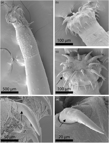
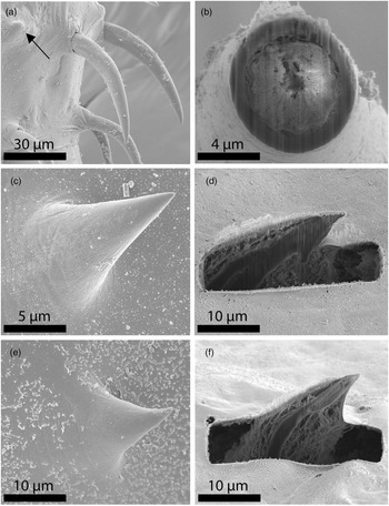
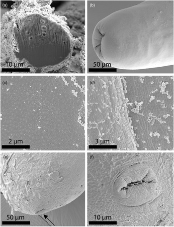
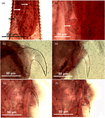
General (figs 2–8). With characters of the family Quadrigyridae, genus Pallisentis and subgenus Pallisentis as diagnosed by Amin et al. (Reference Amin, Heckmann, Ha, Luc and Doanh2000). Shared structures and spine counts larger and more numerous in females than in males (tables 2 and 3). Trunk spinose, curved ventral mostly posteriorly, small, slender, cylindrical with anterior swelling more prominent in females than in males (figs 2a, c and 6a). Body wall even dorsoventrally, with electron dense micropores with different diameters and distributions in different trunk regions (fig. 5c, d), and up to three large hypodermal giant nuclei in occasional females (fig. 7d). Transverse lacunar canals connect two major lateral longitudinal canals at regular intervals, creating appearance of segmentation (fig. 7c). Trunk with slim triangular spines in two zones separated by a spine-free zone (figs 2a and 3a). Anterior collar spines (fig. 4c, d) heavily strengthened by two or three internal support rods of equal length to dermal spines (fig. 7a). Posterior trunk spines (fig. 4e, f) with single much longer and deeply embedded support rod each (fig. 7b). Collar spines in crowded complete circles closely set, beginning slightly posterior to anterior end of trunk and extending posteriorly to level of posterior end of receptacle in both sexes (figs 2b and 6a). Trunk spines in complete circles aligned with transverse lacunar canals at regular intervals widening posteriorly to level of male reproductive system (from anterior testis to mid-cement gland) (fig. 2a) and to level of anterior end to female reproductive system. All spines larger in females than males, with collar spines longer posteriorly than anteriorly and trunk spines smaller and more widely spaced posteriorly than anteriorly (table 3). Proboscis truncated, wider anteriorly and triangulating posteriorly into neck; with ten rows of four hooks each and unusual protruding bumps between larger hooks (fig. 3b, c). Hooks most robust anteriorly, gradually smaller and more slender posteriorly (figs 3d, e and 4a), with solid core and moderate cortical layer (fig. 4b), and with simple posteriorly directed and slightly curved roots, shorter than blades (table 2). Roots of apical and subapical hooks most robust; those of middle and posterior hooks slender (fig. 6c–f). Proboscis receptacle single-walled, about four times as long as proboscis, with dissimilar dorsoventral walls lined with undulating cell layer, and with prominent round cephalic ganglion near its base. Dorsal receptacle wall pinched close to posterior end at point of attachment of shorter retractor muscle and of insertion of anterior limb of PRS (fig. 6b). Other retractor muscle longer, extending along whole internal ventral wall of receptacle to its posterior end (figs 2b and 6a, b). Two sets of protractor muscles emerging from two posterior sites of receptacle, where long and short retractor muscles attach internally to receptacle wall, fan out to attach to body wall posteriorly (fig. 2a). Lemnisci unequal, long, extending well posterior to receptacle (fig. 2a). Gonopore terminal in males and ventro-terminal in females.
Line drawings of specimens of Pallisentis nandai from Nandus nandus collected in the Ganga River at Bairaj, Bijnor, state of Uttar Pradesh, India. (a) A male specimen (in two sections). Note the distribution of trunk spines along the transverse lacunar canals characteristic of this species. (b) The anterior end of a female specimen. Note the inner receptacle lining. (c) A female. (d) Female reproductive system. Note the underdeveloped vulva, bent vagina, thick uterine wall, very thin uterine bell and the four long para-vaginal ligaments. Abbreviations: AT, anterior testis; B, bursa; CG, cement gland; CR, cement reservoir; CRD, cement reservoir duct; L, Lemniscus; LRM, long retractor muscle; PM, protractor muscle; PT, posterior testis; SD, sperm duct; SRM, short retractor muscle; SV, sperm vesicle; LRM, long retractor muscle; PM, protractor muscle; PR, proboscis receptacle; PRS, para-receptacle structure; SRM, short retractor muscle.

SEM of specimens of Pallisentis nandai from Nandus nandus collected in the Ganga River at Bairaj, Bijnor, state of Uttar Pradesh, India. (a) Anterior part of a male specimen showing the proboscis, neck, close circles of collar spines starting posterior to anterior trunk and the more widely spaced trunk spines. (b) A profile of the proboscis and neck. Note the massive size of the apical hooks. (c) A near apical view of a proboscis showing the ten hook rows and the inter-hook pumps (arrow). (d) A lateral view of an apical hook. Note the elevated streaks at its base (arrow). (e) A near frontal view of another apical hook demonstrating a different perspective of the basal elevated streaks (arrow).

SEM of specimens of Pallisentis nandai from Nandus nandus collected in the Ganga River at Bairaj, Bijnor, state of Uttar Pradesh, India. (a) A lateral view of sub-apical hooks with apparent inter hook pumps. (b) A gallium cut of an anterior hook showing its thick cortical layer and its almost solid core depicting its unique metal composition. (c) A collar spine. (d) A gallium-cut section of a collar spine. (e) A trunk spine. (f) A gallium-cut trunk spine. Note the difference in the degree of strength of the internal support rods.

SEM of specimens of Pallisentis nandai from Nandus nandus collected in the Ganga River at Bairaj, Bijnor, state of Uttar Pradesh, India. (a) A high magnification of the posterior circle of collar spines and the anterior circles of trunk spines. (b) The posterior end of a male specimen showing the terminal gonopore with unseen invaginated bursa. (c) Micropores at mid-trunk. (d) Micropores at anterior trunk. (e) The posterior end of a female specimen showing the sub-ventral position of the gonopore (arrow). (f) The orifice of the gonopore of a female specimen.

Microscope images of specimens of Pallisentis nandai from Nandus nandus collected in the Ganga River at Bairaj, Bijnor, state of Uttar Pradesh, India. (a) The anterior end of a female specimen. Note the para-receptacle structure (PRS) (black arrow) inserted at juncture of the short retractor muscle (left white arrow) with the receptacle wall compared to the long retractor muscle (right white arrow). (b) A higher magnification of the nucleated PRS (white arrow) showing its insertion (opposite the junction of the short retractor muscle) and its anterior and posterior limbs. (c) Large anterior proboscis hook with robust root. (d) Second proboscis hook with broad root. (e) Third proboscis hook with more slender and shorter root. (f) Posterior proboscis hook with slender root. All hooks and roots are of female specimens.

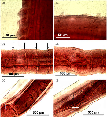
Microscope images of specimens of Pallisentis nandai from Nandus nandus collected in the Ganga River at Bairaj, Bijnor, state of Uttar Pradesh, India. (a) Collar spines of a female specimen. Note the short multi-lobed rod supports. (b) Trunk spines of a female specimen. Note the long single rod support. (c) A mid-trunk segment of a female specimen showing the two heavily stained lateral lacunar canals and a few transverse lacunar canals aligned with trunk spines (arrows). (d) Two giant hypodermal giant nuclei in one female specimen. (e) The posterior part of cement gland with crescent-shaped giant nuclei and the anterior end of the cement gland reservoir (arrow). (f) The posterior part of the male reproductive system showing the posterior half of the cement gland reservoir (right) and its two ducts (white arrows), and the seminal vesicle (black arrow).

Microscope images of specimens of Pallisentis nandai from Nandus nandus collected in the Ganga River at Bairaj, Bijnor, state of Uttar Pradesh, India. (a) The posterior part of a female reproductive system showing the curved vagina, moderately developed sphincter and the thick-walled uterus. Note the four paravaginal ligaments (white arrows). (b) The distal part of the female reproductive system shown in (a), showing the funnel-shaped, thin-walled, unattached uterine bell (black arrow) and the anterior extension of the four paravaginal ligaments.

Morphometric comparisons between our specimens of Pallisentis nandai from Nandus nandus in the Ganga River at Bairaj, Bijnor, Uttar Pradesh, India, and those originally described by Sarkar (Reference Sarkar1953) from the same host in the Ganga River at, Calcutta, West Bengal.

L: length, W: width, H/row: hooks/row.
a Sarkar's (Reference Sarkar1953) measurements of proboscis, receptacle and lemnisci in male and female specimens were the same.
b Range (mean) in μm unless otherwise stated.
c Measurements and numbers in bold represent markedly higher figures in our specimens compared to those in the original description.
Distribution and length of trunk spines of 11 male and 12 female specimens of Pallisentis nandai collected from Nandus nandus in the Ganga River at Bairaj, Bijnor, Uttar Pradesh, India.

a Range (mean).
Males. (Based on 11 mature adults with sperm.) See tables 2 and 3 for measurements and counts of anatomical structures and spines. Reproductive system in posterior half of trunk. Testes contiguous elliptical elongate, with anterior testis slightly longer than posterior testis. Sperm ducts drain each testis dorsally, turning ventral as they unite just anterior to cement gland then joining into large thin-walled common seminal vesicle (fig. 2a). Cement gland as long as both testes with many crescent shaped giant nuclei and ducted connection to pear-shaped cement reservoir. Cement reservoir with two long and narrow ducts surrounding seminal vesicle to join base of penis posteriorly (figs 2a and 7e, f). Bursa not observed.
Females. (Based on 12 adults with ovarian balls.) See tables 2 and 3 for measurements and counts of anatomical structures and spines. Gonopore ventro-terminal with thick-lipped vulva. Vagina bent, with well-developed sphincter and two pairs of paravaginal ligaments extending anteriorly past uterine bell (fig. 2d and arrows in figs 5e, f and 8a). Uterus somewhat short, thick-walled, with two bands of long muscles. Uterine bell thin-walled, funnel-shaped, unattached to body wall, with few gland cells at base (fig. 8b). Eggs not seen.
Taxonomic summary
-
Type and present host. The leaffish, N. nandus (Hamilton).
-
Type locality. The Ganga River at a Calcutta fish market (22.5726°N, 88.3639°E).
-
Present locality. The Ganga River at Bairaj, Bijnor (29°01′N, 77°45′E), UP, India.
-
Site of infection. Liver.
-
Materials deposited. HWML collection number 216362 (male and female voucher specimens on three slides).
-
Representative DNA sequence. The 18S rDNA sequence of P. nandai was deposited in GenBank under the accession numbers MW164853 (1775 bp) and MW164854 (1770 bp), while the ITS1-5.8S-ITS2 region was deposited in GenBank under the accession numbers MW1825515 (785 bp) and MW1825514 (783 bp).
Remarks
The description of P. nandai has not been revised since its original description by Sarkar (Reference Sarkar1953), which was repeated with some variations by other observers, including Soota & Bhattacharya (Reference Soota and Bhattacharya1982), Bhattacharya (Reference Bhattacharya2007) and Naidu (Reference Naidu2012). Since its description from N. nandus, P. nandai has also been reported from the tank goby, G. giuris (Hamilton) (Gobiidae), from other tributaries of the Ganga in UP, India, and from many tributaries of the Ganga in the Bangladesh delta (see Naidu, Reference Naidu2012 for references). For all practical purposes, the 66-year-old description by Sarkar (Reference Sarkar1953) remains the only source of taxonomic information about P. nandai. Our collection of the same acanthocephalan from the same host species in the same river system, albeit about 1500 km away from Calcutta in Bairaj, Bijnor, UP, revealed the extent of the incompleteness of the original description to which we added new information about unreported morphological structures and results of recent technologies such as SEM, gallium cuts, not to mention molecular analysis. We present an updated version of a comprehensive description of that species.
The morphological structures not included in the original description include line drawings of whole males and females, complete proboscis, hooks and roots, female reproductive system, collar and trunk spines, and detail of the proboscis receptacle and associated retractor muscles that are related to the discovery of the PRS not known at that time. We supplement this absent qualitative description with our detailed systematic treatment, line drawings, SEM and microscope images.
The EDXA and the molecular study add new dimensions not available to Sarkar in Reference Sarkar1953. Comparative measurements (table 2) show that the size of hooks, receptacle, lemnisci in males and females, as well as size of testes and trunk spines in females was markedly larger in our specimens from Bairaj than those of Sarkar (Reference Sarkar1953) from Calcutta. This is an interesting finding considering the fact that compared populations were collected from the same host species and from the same river system. Collection localities were, however, about 1500 km apart – one at the northern headwaters and the other near the southern delta of the Ganga.
The PRS
The PRS is reported here in a species of Pallisentis for the first time in an unusual arrangement where, as its insertion point into the receptacle, it is associated with the posterior attachment site of the shorter retractor muscles to the receptacle. Like species of Neoechinorhynchus and Acanthosentis, P. nandai also has a weak single-walled proboscis receptacle. We have examined specimens of four other species of Pallisentis in OMA's personal collection. Only specimens of one of them – Pallisentis indica Mital & Lal, 1976 – had a PRS similar to that described in P. nandai. This brings the total number of species of Pallisentis with PRS to two. Specimens of the other three species that we examined that did not have PRS are Pallisentis (Pallisentis) celatus (Van Cleave, 1928) Baylis, 1933, Pallisentis (Brevitritospinus) vietnamensis Amin, Heckmann, Ha, Luc, Donah (2000) and Pallisentis (Pallisentis) rexus Wongkham & Whitfield (1999).
EDXA
The unique metal composition of hooks (EDXA) demonstrated a considerably high but variable level of sulphur and negligible level of calcium in collar and trunk spines (table 4 and figs 9 and 10) and hook tips (table 6 and fig. 12), but a higher level of sulphur and calcium at the hook basal arch area (table 5 and fig. 11) than at the hook tip and edge. A comparison with the EDXA pattern of another species of Pallisentis, P. indica Mital & Lal, 1976, was considerably different (table 7).
Energy-dispersive X-ray spectrum of gallium-cut collar spines showing high level of sulphur. The X-ray data present the elemental analysis of the spine (see boldfaced figures of collar spines in table 4). Insert: SEM of a gallium-cut collar spine.

Energy-dispersive X-ray spectrum of gallium-cut trunk spines showing high level of sulphur. The X-ray data present the elemental analysis of the spine (see boldfaced figures of trunk spines in table 4). Insert: SEM of a gallium-cut trunk spine.

Energy-dispersive X-ray spectrum of gallium-cut hook arch showing high level of calcium and sulphur. The X-ray data present the elemental analysis of the hook (see boldfaced figures of hook arch in table 5). Insert: SEM of a hook arch and a gallium-cut hook.

Energy-dispersive X-ray spectrum of gallium-cut hook tip showing high level of sulphur and low calcium. The X-ray data present the elemental analysis of the hook (see boldfaced figures of hook tip in table 6). Insert: SEM of a cross-section of a gallium-cut hook.

Chemical composition of the gallium-cut intact spines of Pallisentis nandai.

a The common elements for protoplasm (C, N, O) and those for sample processing (Au, Pd, Ga) are omitted.
b Given in weight%.
c Figures in bold are used in corresponding EDXA spectra.
Chemical composition of the longitudinal gallium-cut hook of Pallisentis nandai.

a The common elements for protoplasm (C, N, O) and those for sample processing (Au, Pd, Ga) are omitted.
b Given in WT%. Figures in bold are used in the corresponding EDXA spectrum.
Chemical composition of the gallium-cut and intact hooks for Pallisentis nandai.

a The common elements for protoplasm (C, N, O) and those for sample processing (Au, Pd, Ga) are omitted.
b Given in WT%.
Chemical composition of the gallium-cut hook tip for two species of Pallisentis.

a The common elements for protoplasm (C, N, O) and those for sample processing (Au, Pd, Ga) are omitted.
b Given in WT%. Figures in bold are used in corresponding EDXA spectra.
Micropores
The electron-dense micropores present throughout the epidermal surface of the trunk of P. nandai are described. They have been found in various regions of the trunk in different diameters and distributions (fig. 5c, d).
Molecular results
Two identical sequences for the 18S and ITS1-5.8S-ITS2 region were generated from isolates of P. nandai collected from N. nandus in India. Phylogenetic relations estimated by ML and BI methods resulted in consensus trees with identical topologies (figs 13 and 14). The phylogenetic relationships of the Pallisentis species were very similar to the phylogenetic relationship obtained in previous studies (e.g. Chaudhary et al., Reference Chaudhary, Amin and Singh2019) within the Eoacanthocephala, which included members of the Quadrigyridae and the Neoechinorhynchidae, The 18S and ITS1-5.8S-ITS2 region of rDNA dataset alignment includes sequences of acanthocephalans representing species of the Quadrigyridae, Neoechinorhynchidae, Moniliformidae and Qligacanthorhynchidae (table 8). 18S sequences obtained from the isolates of P. nandai were placed in a well-supported clade (89/0.91) with other species of Pallisentis reported from India (fig. 13). The clade formed by the members of the Pallisentis was found to be sister of a clade including members of Acanthosentis and Neoechinorhynchus, with good support (fig. 13). Within Pallisentis, the phylogenetic analyses showed that P. nandai was closest to P. indica, a species from Channa punctata (fig. 13). Intra-generic pairwise differences based on partial 18S rDNA sequence between P. nandai with P. indica, Pallisentis sp. BR-2017 and Pallisentis sp. 2 NKG-2016 were 0.56%, 0.86% and 1.02%, respectively. The present phylogenetic analysis based on the ITS1-5.8S-ITS2 data set predicts that P. nandai forms a clade with Pallisentis nagpurensis and P. indica with good bootstrap support (99/1), both reported from India (fig. 14). The clade formed by the members of the Acanthosentis and Neoechinorhynchus was found to be sister of a clade formed by species of Pallisentis.
Phylogenetic reconstruction based on the maximum likelihood (ML) and Bayesian inference (BI) analysis using 18S rDNA sequences of Pallisentis nandai and sequences of Acanthocephala deposited in GenBank. The numbers indicate values of bootstrap >70%. Numbers above branches indicate nodal support as ML and posterior probabilities from BI. Species of Archiacanthocephala are used as an outgroup. The scale bar indicates the expected number of substitutions per site.

Maximum likelihood (ML) and Bayesian inference (BI) tree for Pallisentis nandai based on ITS1-5.8S-ITS2 rDNA sequences. Numbers above branches indicate nodal support as ML analysis followed by bootstrap values of posterior probabilities from BI. Support values lower than 70% are not shown. The scale bar indicates the expected number of substitutions per site.

Acanthocephalan species included in the phylogenetic analysis with information on the host, locality and GenBank accession number.

a Unpublished sequences are available on the GenBank database. HNA, host name not available.
Species in bold sequenced during the present study.
Furthermore, pairwise intra-generic differences based on ITS1-5.8S-ITS2 sequence between P. nandai with P. nagpurensis and isolates of P. indica were 0.20% and 0.26%, respectively. In addition, Neoechinorhynchus brentnickoli and three unidentified species of Neoechinorhynchus clustered as a sister group with strong support (fig. 14).
Discussion
Completeness of life cycle
All our female specimens were gravid with only ovarian balls. Eggs were not found. Reported specimens did not reach sufficient sexual maturity in the liver, where they have been found, to produce eggs. This unusual site of infection opens questions about completing the life cycle with specimens trapped in the liver without egg production and access to the external environment. Sarkar (Reference Sarkar1953) did not find eggs either; all his specimens were collected from ‘the alimentary canal and liver’ of 16 infected fishes from a Calcutta fish market in June 1952. The question of completing the life cycle remains an open question, especially in light of the reporting of P. nandai from the many collecting sites along the Ganga River (fig. 1). Only occasional references were made to intestinal sites of infection – for example, Parveen & Sultana (Reference Parveen and Sultana2014) collected four specimens of P. nandai from the liver, one from the stomach and one from the intestine of N. nandus in Bangladesh. Ahmed (Reference Ahmed1981) also collected specimens of P. nandai from the liver, intestine and mesenteries of N. nandus. Alam & Alam (Reference Alam and Alam2014) found specimens only in the liver of Oreochromis nilotiocus (Lin.), as in our case, but Naidu (Reference Naidu2012) collected specimens only from the intestine of N. nandus and G. giuris (Hamilton). It appears that only intestinal sites support reproductive infrapopulations contributing to the continuation of the life cycle of that species. Worms in the liver do not produce eggs.
Morphological variability
The marked differences in the size of proboscis hooks, receptacle, lemnisci, testes and female spines between our specimens and those described by Sarkar (Reference Sarkar1953) still fall within the range of intraspecific variations not related to host species. Our specimens and those of Sarkar (Reference Sarkar1953) were collected from the headwaters and the lower delta of the Ganga River, respectively, about 1500 km apart. Intraspecific variability related to geographical factors has been repeatedly demonstrated in other species of Acanthocephala. The observed differences in our Rhadinorhynchus trachuri Harada, 1935 (Rhadinorhynchidae) specimens also appear to represent intraspecific variations among Asian and American geographical populations, which may be affected by changes in feeding behaviour (Amin, Reference Amin2019), as has been previously demonstrated in the comparable case of Mediorhynchus papillosus Van Cleave, 1916 (Gigantorhynchidae) by Amin & Dailey (Reference Amin and Dailey1998). Amin & Dailey (Reference Amin and Dailey1998) studied key taxonomic characteristics in various geographical populations of M. papillosus, which has a wide range of distribution in at least 73 species of birds outside of North and South America, in Asia from Taiwan to the east into China, many of the former Soviet Republics and to Eastern Europe to the west. Amin & Dailey (Reference Amin and Dailey1998) compared measurements of specimens from two species of birds in Maryland, one from Colorado (their study material), six from Taiwan, two from Yakutia, Trans-Baikal, three from Lower Yansi River basin, 45 from the Volga basin and Oren Byreg, seven from Ukraine, seven from Bulgaria, three from China and one from Brazil, and demonstrated a distinct geographically based variability, especially in the size of proboscis and its armature, neck, receptacle and testes, which appeared related to geographical restrictions, intermediate and definitive host specificity and distribution, and host feeding behaviour. ‘The U.S. population from Colorado and the Taiwanese population (were shown to be) at the opposite ends of the spectrum’ by Amin & Dailey (Reference Amin and Dailey1998) who dismissed the possibility of elevating them to a specific status. The population variants of P. nandai, like those of R. trachuri, are comparable to the east–west-intraspecific clinal variants of M. papillosus and could have been considered as distinct species, but this notion is dismissed here also for the same reasons.
The PRS
The PRS inserts anteriorly in the body wall near the neck and posteriorly at the posterior end of the receptacle. The presence of the PRS in eoacanthocephalan species with a weak single proboscis receptacle wall was first demonstrated in Neoechinorhynchus (N.) qatarensis Amin, Saoud, Alkuwari, 2002 by Amin et al. (Reference Amin, Saoud and Alkuwari2002). It had since been reported in other eoacanthocephalan species of Neoechinorhynchus Stiles & Hassall, 1905 and Acanthogyrus (Acanthosentis) Verma & Datta, 1929 by Amin (Reference Amin2005) and Amin et al. (Reference Amin, Ha and Ngo2011a, Reference Amin, Chaudhary, Heckmann, Ha and Singh2019a, Reference Amin, Chaudhary, Heckmann, Ha and Singhb, Reference Amin, Sharifdini, Heckmann and Hac). In the description of the PRS, Amin et al. (Reference Amin, Saoud and Alkuwari2002, Reference Amin, Heckmann and Standing2007) proposed that it may regulate the hydrostatic pressure in the receptacle to facilitate the retraction and eversion of the proboscis. The PRS is here reported for the first time in two members of the genus Pallisentis: P. nandai and P. indica.
Micropores
The micropores of P. nandai, like those reported from other species of the Acanthocephala, are associated with internal crypts and vary in diameter and distribution in different trunk regions corresponding with differential absorption of nutrients. Micropores have been reported in a large number of acanthocephalan species (Heckmann et al., Reference Heckmann, Amin and El-Naggar2013) and in a few more since, and demonstrated the tunnelling from the cuticular surface into the internal crypts by Transmission Electron Microscope (TEM). Amin et al. (Reference Amin, Heckmann, Radwan, Mantuano and Alcivar2009) summarized the structural–functional relationship of the micropores in various acanthocephalan species, including Rhadinorhynchus ornatus Van Cleave, 1918, Polymorphus minutus (Goeze, 1782) Lühe, 1911, Moniliformis (Bremser, 1811) Travassos (1915), Macracanthorhynchus hirudinaceus (Pallas, 1781) Travassos (1916, 1917) and Sclerocollum rubrimaris Schmidt & Paperna, 1978. Wright & Lumsden (Reference Wright and Lumsden1969) and Byram & Fisher (Reference Byram and Fisher1973) reported that the peripheral canals of the micropores are continuous with canalicular crypts. These crypts appear to ‘constitute a huge increase in external surface area … implicated in nutrient up take.’ Whitfield (Reference Whitfield1979) estimated a 44-fold increase at a surface density of 15 invaginations per 1 μm2 of M. moniliformis tegumental surface. The micropores and the peripheral canal connections to the canaliculi of the inner layer of the tegument of Corynosoma strumosum (Rudolphi, 1802) Lühe, 1904 from the Caspian seal Pusa caspica (Gmelin) in the Caspian Sea were demonstrated by transmission electron micrographs in Amin et al. (Reference Amin, Heckmann, Halajian and El-Naggar2011b).
EDXA
Results of the X-ray scans of the gallium-cut hooks (dual-beam SEM) of P. nandai show differential compositions and distributions of metals in different hook parts, with calcium and sulphur levels being considerably higher at the basal arch of hooks where tension and strength are paramount compared to the hook tip and edge where the level of sulphur was considerably higher (tables 5, 6 and figs 11 and 12). The chemical elements present in the hooks are typical for acanthocephalans (Heckmann et al., Reference Heckmann, Amin and Standing2007, Reference Heckmann, Amin, Radwan, Standing, Eggett and El Naggar2012; Standing & Heckmann, Reference Standing and Heckmann2014; Amin & Heckmann, Reference Amin and Heckmann2017). Note the moderate outer layer (fig. 40) of the hook which relates to the sulphur content (tables 5 and 6) in the hook of P nandai, which is different than in other acanthocephalans (Amin & Heckmann, Reference Amin and Heckmann2017; Amin et al., Reference Amin, Heckmann and Zargar2017, Reference Amin, Heckmann, Ha and Pham2018a; Ha et al., Reference Ha, Amin, Ngo and Heckmann2018). The high sulphur content shows up in the outer edge of X-ray scans of hooks (tables 5 and 6; Amin et al., Reference Amin, Heckmann and Ha2018b). The hook centre in mid-cuts has a completely different chemical profile than the cortical layer (table 6). X-ray scans (EDXA) provide insight into the hardened components – for example, calcium and phosphorus – of acanthocephalan hooks. The EDXA appears to be species-specific, as in fingerprints. The hook tip of P. indica has a considerably higher level of phosphorus and calcium and lower levels of sulphur than the hook tip of P. nandai (table 7). EDXA is shown to have significant diagnostic value in acanthocephalan systematics – for example, Moniliformis cryptosaudi Amin, Heckmann, Sharifdini & Albayati, 2019 was erected based primarily on its EDXA pattern (Amin et al., Reference Amin, Heckmann, Sharifdini and Albayati2019d).
Molecular analysis
Molecular methods are an important tool for morphological identification, diversity and taxonomic relationships among acanthocephalan species (García-Varela et al., Reference García-Varela, Mendoza-Garfias, Choudhury and Pérez-Ponce de León2017, Reference García-Varela, Park, Hernández-Orts and Pinacho-Pinacho2019; Li et al., Reference Li, Chen and Yang2018; Amin et al., Reference Amin, Chaudhary, Heckmann, Ha and Singh2019a, Reference Amin, Chaudhary, Heckmann, Ha and Singhb, Reference Amin, Sharifdini, Heckmann and Hac; Lisitsyna et al., Reference Lisitsyna, Kudlai, Cribb and Smit2019; Menasria et al., Reference Menasria, Barčák, Kaouachi, Bensouilah, Scholz and Hernández-Orts2019; Sharifdini et al., Reference Sharifdini, Amin and Heckmann2020). The phylogenetic relationships and systematic position of Pallisentis are poorly explored compared to other genera. Molecular data for nuclear genes have been provided only for few in comparison to the >30 species available of the genus (Amin, Reference Amin2013). Our phylogenetic analyses obtained from two methods – ML and BI – predict the same topology sowing the isolates of P. nandai from a well-supported clade within the Eoacanthocephala. The genetic differences between P. nandai and other species of the Pallisentis support the morphological observations indicating its independent identity. Other species of Pallisentis described from India were added to the listing in Amin (Reference Amin2013): Pallisentis channi and Pallisentis vinodai by Gautam et al. (2015); Pallisentis punctati by Gupta et al. (Reference Gupta, Maurya and Saxena2015b); Pallisentis anandai (as KR149270 Pallisentis sp. 1 NKG-2015) by Gautam et al. (Reference Gautam, Upadhyay, Maurya, Verma and Saxena2017); Pallisentis lucknowensis, Pallisentis amini (as KY305523 Pallisentis sp. 11 NKG-2016), Pallisentis meyeri (as KY305525 Pallisentis sp. 13 NKG-2016) and Pallisentis unnaoensis (as KY305528 Pallisentis sp. 16 NKG-2016) by Gautam et al. (Reference Gautam, Misra and Saxena2019) and Pallisentis thapari (as KY305524 Pallisentis sp. 12 NKG-2016 and KY305527 Pallisentis sp. 15 NKG-2016) by Gautam et al. (Reference Gautam, Misra, Saxena and Monks2020). However, the genetic divergence in the 18S rDNA sequences between these species and P. nandai was 1.11–1.14.
Moreover, the tree also showed that our sequences of P. nandai were placed separately from other species of Pallisentis. The strong support in both trees also indicated that all these species share a common ancestor. Due to the unavailability of ITS sequences of Pallisentis in GenBank for comparison, we did perform molecular analysis of P. nandai isolates with only P. indica and P. nagpurensis. However, one ITS sequence of P. nagpurensis is too short (MN423292; 245 bp) and, therefore, unreliable for phylogeny, which is why we did not include it in our analysis. Aside from this, no ITS sequences have been made available for other Pallisentis members to date, that can help better elucidate their phylogenetic affinities with each other.
In conclusion, it becomes clear that making additional genetic markers available, as well as the addition of more species of this genus, would be useful for better understanding their phylogenetic affinities.
Acknowledgements
We thank Madison Laurence, Bean Museum (BYU), Provo, Utah, for expert help in the preparation and organization of plates and figures, and Michael Standing, Electron Optics Laboratory (BYU), for his technical help and expertise. We would like to acknowledge the laboratory facilities provided by the Department of Zoology, Chaudhary Charan Singh University, Meerut, India.
Financial support
This work was supported by an institutional grant from the Biology Department, Brigham Young University (BYU), Provo, Utah, and the Parasitology Center, Inc. (PCI), Scottsdale, Arizona.
Conflicts of interest
None.
Ethical standards
The authors declare that they have observed all applicable ethical standards.