Introduction
Zirconium titanate (ZrTiO4) exhibits many excellent properties, such as high dielectric constant, low dielectric dissipation, and a low temperature coefficient, in the microwave region [Reference Wang, Lee, Azough and Freer1, Reference Zhang, Li, Cao, Zhai and Zhang2]. Therefore, zirconium titanate is widely used in many applications, including electrical and optical devices [Reference Wakino, Minai and Tamura3, Reference Leoni, Viviani, Battilana, Fiorello and Viticoli4, Reference Chang, Lin and Tseng5, Reference Troitzsch and Ellis6], ceramic pigments [Reference Dondi, Matteucci and Cruciani7], humidity sensors [Reference Cosentino, Muccillo and Muccillo8, Reference Ličina, Gajović, Moguš-Milanković, Djerdj, Tomašić and Su9], catalysts [Reference Rouhani, Salahinejad, Kaul, Vashaee and Tayebi10, Reference Cheng, Zhang, Liao and Shi11], and biomedical applications [Reference Devi, Singh and Rajendran12]. In recent years, porous zirconium titanate has attracted much attention due to its rich porosity. For example, Cosentino et al. [Reference Cosentino, Muccillo and Muccillo8] used porous ZrTiO4 sintered pellets for ceramic humidity sensing devices. The results showed that the sample with high porosity could absorb more water molecules, so that the electrical conductivity increased; in other words, the total electrical resistivity decreased with the increasing pore density. Ličina et al. [Reference Ličina, Gajović, Moguš-Milanković, Djerdj, Tomašić and Su9] also found that the electrical conductivity of ZrTiO4 (ZT) ceramics in humid air strongly depended on the porosity and the reduction of dielectric permittivity could be attributed to the increased number of pores. Cheng et al. [Reference Cheng, Zhang, Liao and Shi11] prepared the porous fibrous SO42−/TiO2–ZrO2 solid acid using collagen fiber as template. It was found that when the mole ratio of Ti/Zr was 0.67:1, ZrTiO4 was the main crystalline phase, which presented ordered fibrous structure with large specific surface area, strong adsorption, and the largest number of strong acid sites. Moreover, the mesoporous structure enhanced mass and heat transfer in the catalytic process. The porous fibrous SO42−/TiO2–ZrO2 solid acid exhibited excellent catalytic activity and reusability in the esterification reaction of acetic acid and n-butyl alcohol.
Various routes, including coprecipitation [Reference Hu, Payzant, Booth, Rawn, Hunt and Allard13], solid-state reaction [Reference Gajović, Djerdj, Furić, Schlögl and Su14], self-assembly procedure [Reference Luca, Drabarek, Griffith and Hanley15, Reference Luca, Soler-Illia, Angelomé, Steinberg, Drabarek and Hanley16], and sol–gel technique [Reference Rouhani, Salahinejad, Kaul, Vashaee and Tayebi10, Reference Devi, Singh and Rajendran12, Reference Sham, Aranda, Farfan, Gottifredi, Martínez-Lara and Bruque17], have been developed to prepare zirconium titanate. Among these methods, sol–gel technique is outstanding and has attracted extensive research. Rouhani et al. [Reference Rouhani, Salahinejad, Kaul, Vashaee and Tayebi10] synthesized nanostructured zirconium titanate fibers by a sol–gel technique using ZrCl4 and TiCl4 as precursors. Devi et al. [Reference Devi, Singh and Rajendran12] prepared orthorhombic nano zirconium titanate using sol–gel process and coated it on 316L stainless steel. The resulted material showed good biocompatibility in the simulated body fluid and was suitable for biomaterials. By a sol–gel process, Sham et al. [Reference Sham, Aranda, Farfan, Gottifredi, Martínez-Lara and Bruque17] prepared zirconium titanate powders and conducted thermal decomposition and quantitative phase analysis.
Inorganic salts [Reference Rouhani, Salahinejad, Kaul, Vashaee and Tayebi10, Reference Devi, Singh and Rajendran12, Reference Lu, Chen and Jiao18] or alkoxides [Reference Sham, Aranda, Farfan, Gottifredi, Martínez-Lara and Bruque17, Reference Macan, Gajovic and Ivankovic19, Reference Koc20] could be used as zirconium or titanium precursors. After hydrolysis and condensation reactions under certain conditions, the sol could be obtained. During aging process, the colloidal particles of the sol are aggregated slowly and the fluidity of the sol is lost. Then the gel is formed. After drying and heat treatment, the desired zirconium titanate samples could be obtained. As sol–gel process is a kind of wet chemical methods, materials synthesized by sol–gel process have advantages of high purity, easy modification, and lower crystallization temperatures, etc.
Zirconium titanate materials prepared by methods in the above-mentioned literatures were mostly powders, fibers, and films but not monoliths, and the pores were not co-continuous. In many applications, materials with co-continuous pores and monolithic shape have advantages. Interconnected macropores can reduce transport limitations, especially for large molecules, while micro- and/or mesopores contribute high surface areas and pore volumes [Reference Feinle, Elsaesser and Hüsing21]. Moreover, the obtained monolithic materials do not require special molding treatment. The sol–gel process combined with phase separation is known as a promising method for the preparation of this kind of material. Phase separation is derived from spinodal decomposition induced by polymerization, forming two phases—solid phase and solution phase. Each separated phase is interconnected. When the sol–gel reaction occurs simultaneously with the phase separation, the phase separation transient morphology could be frozen. After drying, solid phase becomes the skeleton and solution phase becomes co-continuous macropores. Using sol–gel technique combined with phase separation, many kinds of hierarchical monolithic materials with co-continuous pores have been synthesized, including SiO2 [Reference Nakanishi22], Al2O3 [Reference Tokudome, Fujita, Nakanishi, Miura and Hirao23], TiO2 [Reference Backlund, Smatt, Rosenholm and Lindén24], ZrO2 [Reference Konishi, Fujita, Oiwa, Nakanishi and Hirao25], mullite [Reference Guo, Li, Nakanishi, Kanamori, Zhu and Yang26], aluminum titanate [Reference Guo, Zhu, Cai, Liu and Yang27], cordierite [Reference Guo, Nakanishi, Kanamori, Zhu and Yang28], and Al2O3–SiO2–TiO2 [Reference Sun, Zhao, Li, Ma, Wang and Li29].
Considering the advantages of pore structure and broad application potentials of zirconium titanate, in this work, we demonstrate the facile preparation of macro-mesoporous zirconium titanate monoliths via a sol–gel process accompanied by phase separation. Zirconium oxychloride (ZrOCl2·8H2O) and titanium(IV) n-propoxide [Ti(OnPr)4] are the precursors of the ZrO2–TiO2 binary system. Poly(ethylene oxide) (PEO) is used to induce the phase separation and N-methylformamide (NFA) to promote the sol–gel transition. Nitric acid is used to slow down the reaction rate of titanium alkoxide and to stabilize the sol. The effects of PEO and NFA amount on the gel morphologies are studied. By changing the starting compositions, the morphology and pore size of the gel can be adjusted. In addition, the evolutions of crystalline phase and macroporous structure with heat treatment are also discussed. After heat treatment at 500 °C, pure ZrTiO4 phase can be formed. The resultant monolithic zirconium titanate possesses co-continuous macropores, homogeneous elements dispersion, and high surface area. The macro-mesoporous zirconium titanate monolith may have potential applications in various existing fields.
Results and discussion
Formation and control of macroporous structure
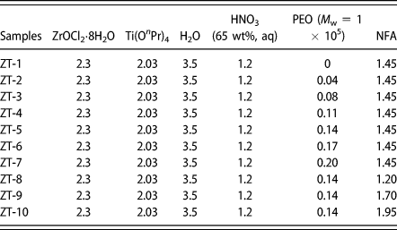
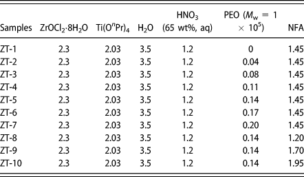
The starting compositions of the samples are listed in Table I. Zirconium and titanium cations are isovalent with different ionic radii, 0.72 Å for Zr4+ and 0.61 Å for Ti4+. These two cations can form different kinds of mixed oxides. However, ZrTiO4 is the only stoichiometric compound formed under atmospheric pressure [Reference Kim, Oh, Park and Hong30]. In this work, the molar ratio of these two elements in the precursors [ZrOCl2·8H2O and Ti(OnPr)4] was 1:1. To avoid the formation of precipitation or flocculation, acetic acid [Reference Backlund, Smatt, Rosenholm and Lindén24] and citric acid [Reference Guo, Zhu, Cai, Liu and Yang27] could be used as chelating agents to slow down the hydrolysis rate of transition metal alkoxides. The addition of strong acid such as nitric acid [Reference Konishi, Fujita, Oiwa, Nakanishi and Hirao25] and hydrochloric acid [Reference Konishi, Fujita, Nakanishi, Hirao, Morisato, Miyazaki and Ohira31] was another strategy. In this system, the hydrolysis rate of Ti(OnPr)4 was faster than that of ZrOCl2·8H2O. Nitric acid was chosen to reduce the reaction rate of Ti(OnPr)4, which made these two precursors hydrolyzed and polymerized together in the solvent to obtain transparent homogeneous solution.
Starting compositions of the samples (in grams).
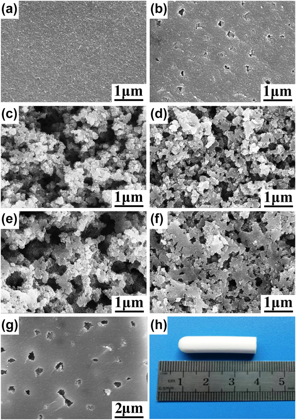
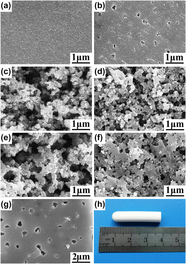
The addition of water-soluble polymer PEO could induce phase separation. Herein, the effect of PEO amount on the gel morphologies was investigated. Figure 1 shows the scanning electron microscope (SEM) images of dried gels prepared with different amounts of PEO. Without PEO, no obvious pore structure could be observed, phase separation almost does not take place, and transparent sample is obtained [Fig. 1(a)]. When the amount of PEO is 0.04 g, the obtained dried gel is translucent monolith with only isolated macropores in microstructure [Fig. 1(b)]. When PEO amount is up to 0.08 g, the gel skeleton is particle aggregates [Fig. 1(c)]. As the amount of PEO increases, the gel morphology in the micrometer range changes from co-continuous structure [Figs. 1(d)–1(f)] to isolated macroporous structure [Fig. 1(g)]. The obtained sample has no crack [Fig. 1(h)]. The change in the gel morphology is similar to that of the PEO-incorporated SiO2 system [Reference Nakanishi22]. There may be a critical degree of polymerization, where the complex of PEO and oxide oligomers is least stable and the phase separation occurs easily. In the SiO2 system, the phase separation tendency possesses a maximum at very low degree of polymerization (small amount of PEO). With the increase in PEO amount, the degree of polymerization is higher than the critical value and the phase separation tendency decreases [Reference Konishi, Fujita, Oiwa, Nakanishi and Hirao25]. In the present ZrO2–TiO2 system, taking ZT-5 as an example, only when the degree of polymerization is appropriate, the sol–gel transition occurs parallel to phase separation and the co-continuous structure induced by the phase separation could be frozen by the sol–gel transition. During the drying process, volume shrinks by about 30% because of the capillary force. The solid phase becomes the skeleton and the solution phase becomes the co-continuous macropores. After drying, the white circular cylindrical dried gel is obtained [Fig. 1(h)].
(a–g) SEM images of dried gels prepared with varied PEO amount: (a) ZT-1, (b) ZT-2, (c) ZT-3, (d) ZT-4, (e) ZT-5, (f) ZT-6, and (g) ZT-7. (h) Appearance of a dried gel ZT-5.
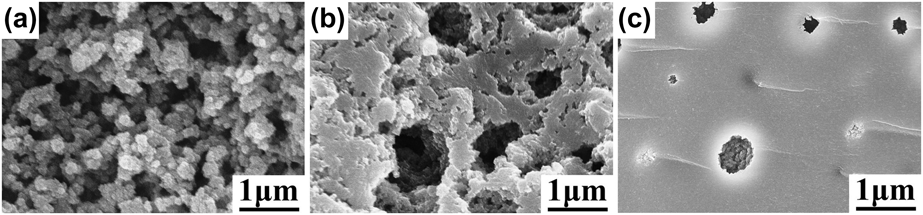
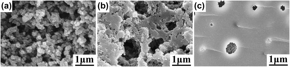
NFA was used as a gelation inducer to promote sol–gel transformation. Under the acidic and certain temperature conditions, NFA was gradually hydrolyzed to generate aqueous methylamine. Then, the pH of the sol rose slowly and uniformly. The polycondensation of the precursors was induced, which led to the gelation. The micro-morphologies of dried gels prepared with different NFA amounts are presented in Fig. 2. With the increase of NFA amounts, the micrometer-scale morphology of the gel gradually changes from particle aggregates [Fig. 2(a)], through co-continuous skeleton [Figs. 1(e) and 2(b)], to isolated macroporous structure [Fig. 2(c)]. The gelation time was reduced from 5 h (1.20 g of NFA) to 2 h (1.95 g of NFA). When the amount of NFA is small, the sol–gel transformation rate is slow and the phase separation trend is strong. When the amount of NFA is large, more methylamine is produced in the system and the sol–gel transformation trend is strong, which accelerate the gelation.
(a–c) SEM images of dried gels prepared with varied NFA amount: (a) ZT-8, (b) ZT-9, and (c) ZT-10.
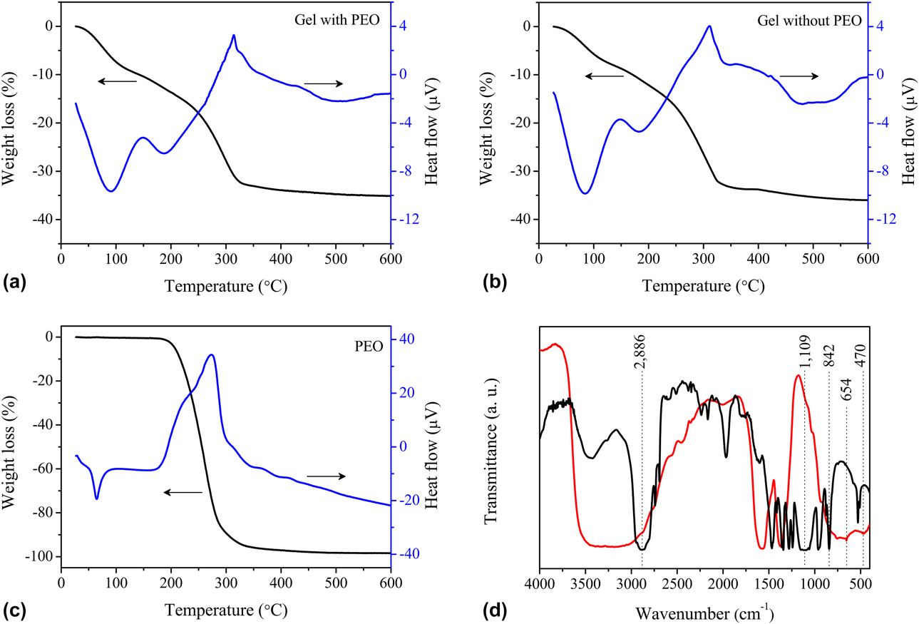
Thermal and infrared analyses were carried out to clarify the distribution of PEO between gel phase and liquid phase. As shown in Fig. 3, the thermogravimetry–differential thermal analysis (TG-DTA) curves of the gels prepared with PEO [Fig. 3(a)] and without PEO [Fig. 3(b)] are similar. The weight loss below 200 °C and the endothermic peak at around 90 and 190 °C are attributed to the evaporation of residual water and organic solvent components. The weight loss between 200 and 350 °C and the exothermic peak at around 320 °C belong to the decomposition of residual organic components. No exothermic peak belonging to the decomposition of PEO [at around 280 °C, Fig. 3(c)] can be found in the two samples, indicating that PEO is preferentially distributed into the liquid phase. This phenomenon is similar to the Al2O3 system [Reference Tokudome, Fujita, Nakanishi, Miura and Hirao23].
(a–c) TG-DTA curves. (d) FT-IR spectra of the dried gel ZT-5 (red line) and PEO (black line).
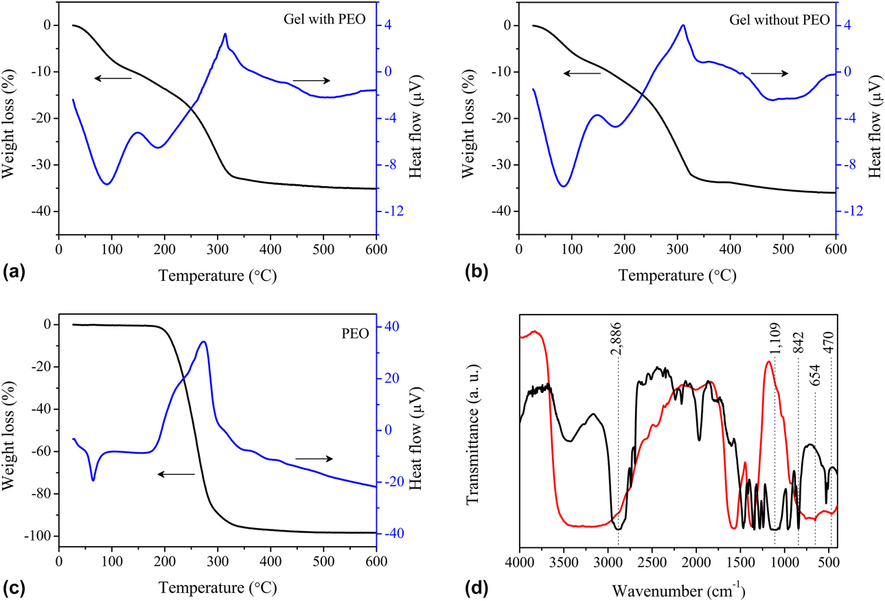
Figure 3(d) displays the Fourier transform infrared spectroscopy (FT-IR) spectra of the gel prepared with PEO (ZT-5) and PEO. The stretching vibration of C–C bonds (842 cm−1), C–O bonds (1109 cm−1), and C–H bonds (2886 cm−1) are derived from PEO. For ZT-5, no absorption peaks assigned to PEO can be found, indicating that PEO is preferentially distributed into the liquid phase. In the ZT-5 spectrum, the wide peak at around 3400 cm−1 and the absorption peaks at around 1600 cm−1 and 1340 cm−1 could be attributed to the hydroxyl stretching vibration peak of residual water, the deformation vibration of water contained in the gel framework and the bending vibration of the coordination hydroxyl group, respectively, which shows that the residual organics and other intermediates may be present in the solid phase. In addition, the bands at 654 cm−1 and 470 cm−1 belong to the vibration of Ti–O–Ti and Zr–O–Zr, respectively [Reference Zhu, Xu, Yu and Wang32].
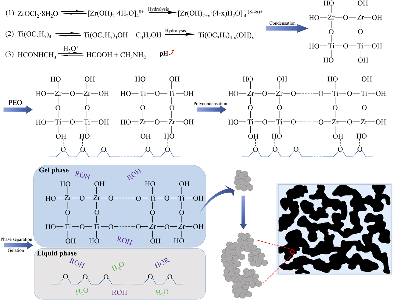
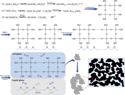
In the present ZrO2–TiO2 system, the preparation process and formation mechanism of the gel are proposed in Fig. 4. Zirconium oxychloride formed zirconium hydrate ions in the solution and existed as [Zr(OH)2·4H2O]48+. Initial hydrolysis of titanium(IV) n-propoxide also occurred. Then NFA was added. The methylamine produced by the NFA hydrolysis reaction increased the pH of the solution, which promoted the hydrolysis of the precursors and formed [Zr(OH)2+x·(4 − x)H2O]4(8−4x)+, Ti(OC3H7)4−x(OH)x. With the progress of condensation, oligomers Zr–O–Zr, Ti–O–Ti, and Zr–O–Ti were formed. The hydrolyzed oligomers were connected with each other and interacted with PEO chains through the hydrogen bond. However, this hydrogen bond was weak and easy to break or disappear. In addition, as the degree of polymerization increases, the compatibility between the polycondensation products and PEO chains was further reduced, resulting in phase separation, and forming the PEO-rich liquid phase and the polycondensation products-rich gel phase. During the reaction, the oligomers were continuously cross-linked to form the colloidal primary particles in their respective regions after phase separation. The colloidal particles were connected and assembled to form a three-dimensional gel framework. The solvent gradually lost its fluidity and filled in the gel framework and sol–gel transition occurred. When the sol–gel transition and the phase separation process were almost simultaneous, the co-continuous two-phase structure was fixed. After drying at a certain temperature, the solvent phase formed pores and the gel solids formed the final macro-mesoporous zirconium titanate monoliths.
The sol–gel process accompanied by phase separation of the ZrO2–TiO2 system.

Heat treatment of porous monoliths
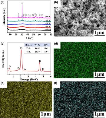
Sample ZT-5 has more obvious and uniform pores, which is selected as the object to study the effect of heat treatment on gel in the following. The dried gel ZT-5 was heat treated at 400–600 °C in air with the heating rate of 5 °C/min. Figure 5(a) shows the X-ray diffraction (XRD) patterns of the gels heat treated at various temperatures. No peaks can be observed for the gels dried and heat treated at 400 °C, indicating amorphous phase. Diffraction peaks appear at 450 °C due to the precipitation of nanocrystals. When heat treated at 500 °C, the diffraction peaks ascribed to ZrTiO4 (PDF No. 34-0415) appear and increasing the heat treatment temperature to 600 °C, a single phase of ZrTiO4 is maintained. Compared with other zirconium titanate preparation methods, such as coprecipitation or solid-state reactions [Reference Hu, Payzant, Booth, Rawn, Hunt and Allard13, Reference Gajović, Djerdj, Furić, Schlögl and Su14], the sol–gel accompanied by phase separation technique needs lower crystallization temperature, which may be due to the homogeneity on the molecular scale.
(a) XRD patterns of sample ZT-5 heat treated at different temperatures. (b) SEM image, (c) EDS, and (d–f) two-dimensional maps for Zr, Ti, and O, respectively, of ZT-5 heat treated at 500 °C.
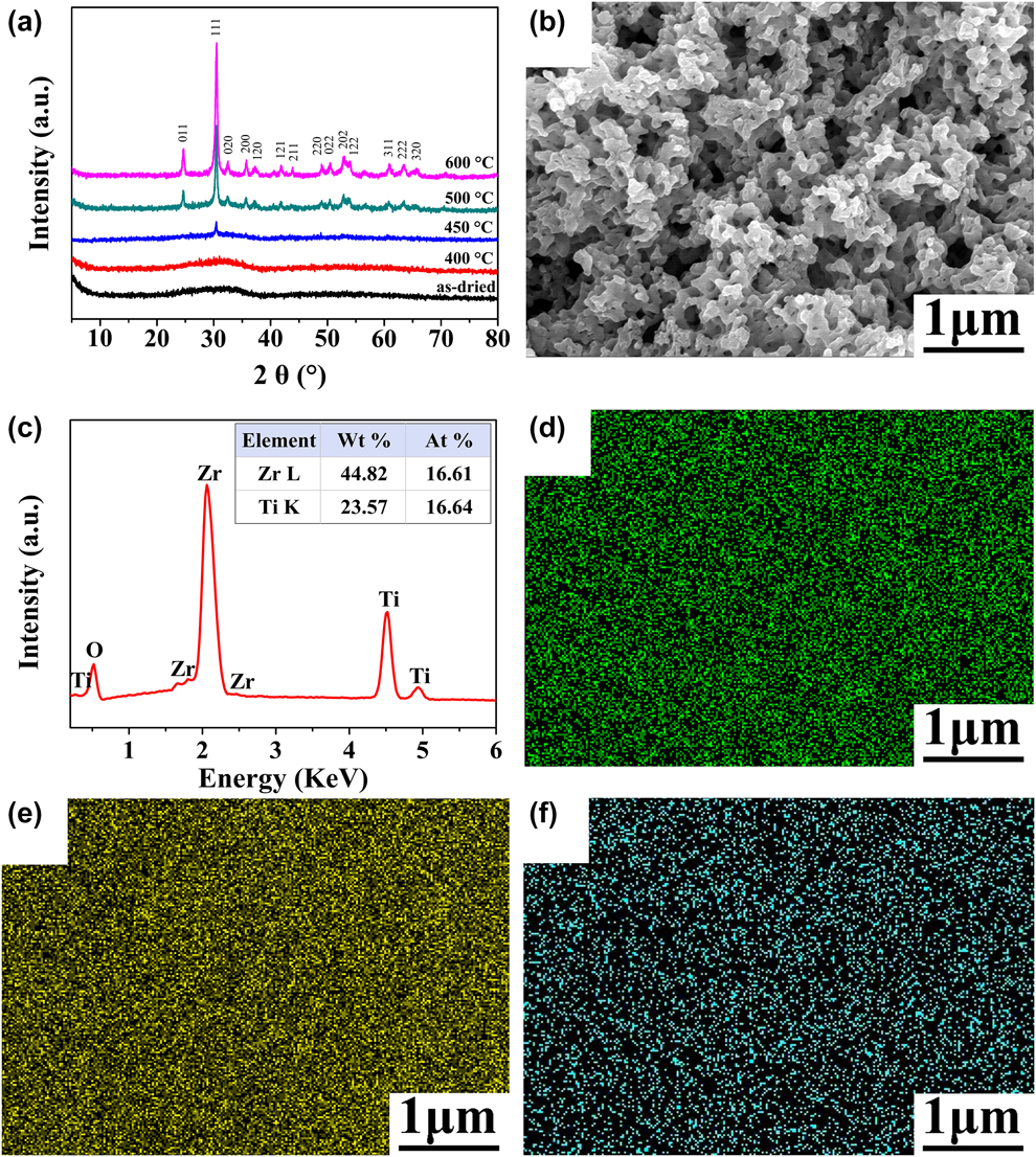
After heat treatment at 500 °C, ZT-5 dried gel shrinks by about 20%. The co-continuous macroporous structure is well maintained. Moreover, the macropore size becomes small and the surface of skeletons becomes smoother and thinner due to the crystallization and sintering [Fig. 5(b)]. Energy dispersive spectrometer (EDS) results confirm the expected elemental constituents of Zr, Ti, and O, and the Zr/Ti atom ratio is close to 1 [Fig. 5(c)]. Two-dimensional elemental analysis shows that Zr, Ti, and O distribute uniformly in the skeleton structure [Figs. 5(d)–5(f)].
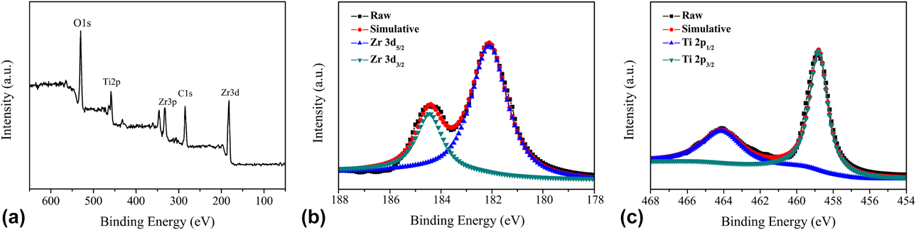
X-ray photoelectron spectroscopy (XPS) measurement was carried out to investigate the formation of chemical bonds of the sample ZT-5 heat treated at 500 °C. The XPS survey spectrum is shown in Fig. 6(a). Two peaks for zirconium (Zr 3p and Zr 3d) and one main peak for titanium (Ti 2p) are detected using the signal of carbon (C 1s) from ambient contamination as the calibration standard. Figures 6(b) and 6(c) show the detailed XPS spectra of Zr 3d and Ti 2p. In the Zr 3d spectrum, two peaks (182.1 and 184.4 eV) are ascribed to Zr 3d 5/2 and Zr 3d 3/2, respectively. For Ti 2p, the peaks Ti 2p 3/2 (458.8 eV) and Ti 2p 1/2 (464.1 eV) may testify the formation of the Ti–O–Zr bonds [Reference Naumkin, Kraut-Vass, Gaarenstroom and Powell33]. Such binding energies represent the fully oxidized zirconium ion Zr4+ and titanium ion Ti4+ [Reference Moulder, Stickle, Sobol and Bomben34, Reference Zhu, Zhang, Chen and Yin35], which could be attributed to ZrTiO4 [Reference Sham, Aranda, Farfan, Gottifredi, Martínez-Lara and Bruque17, Reference Ikawa, Yamada, Kojima and Matsumoto36].
(a) XPS survey spectrum and (b, c) detailed XPS spectra of Zr 3d and Ti 2p, respectively, of ZT-5 heat treated at 500 °C.
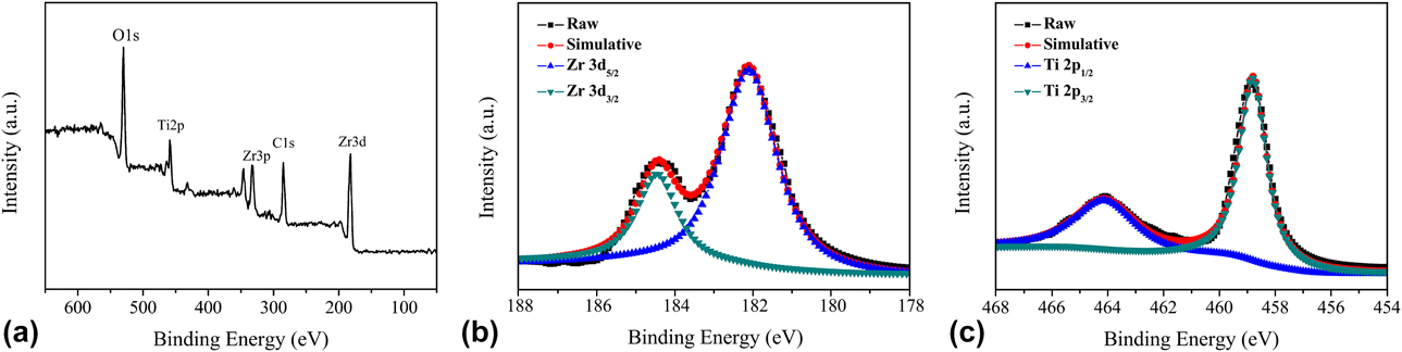
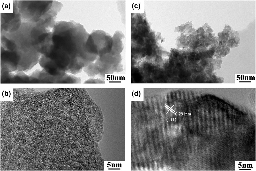
Figure 7 displays the transmission electron microscopy (TEM) images of sample ZT-5 before and after heat treatment at 500 °C. For the as-dried gel [Figs. 7(a) and 7(b)], fine and uniform primary particles assemble together to form large spherical particles with a diameter of 30–100 nm and then form an amorphous skeleton structure. The interstices among spherical particles contribute to the mesopores. After heat treatment at 500 °C [Figs. 7(c) and 7(d)], crystal particles with the size of 10–50 nm are formed due to sintering and aggregation, and mesopores also exist in the skeleton. In addition, the sample has good crystallinity and the lattice distance of 0.291 nm is consistent with (111) plane of ZrTiO4.
TEM and HRTEM images of ZT-5 before (a, b) and after (c, d) heat treatment at 500 °C.

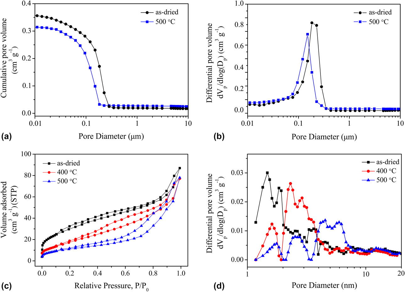
Figures 8(a) and 8(b) show the macropore diameter distributions of sample ZT-5 before and after heat treatment at 500 °C measured using mercury porosimetry. The macropore diameter of dried gel is concentrated around 0.18 μm and the macropore is derived from the spinodal decomposition. After heat treatment at 500 °C, the macropore diameter decreases slightly and is concentrated around 0.15 μm. Due to the decomposition of organic components, the porosity increases from 46 to 63%.
(a, b) Macropore size distributions, (c) nitrogen adsorption–desorption isotherms, and (d) DFT pore size distributions of sample ZT-5 heat treated at various temperatures.
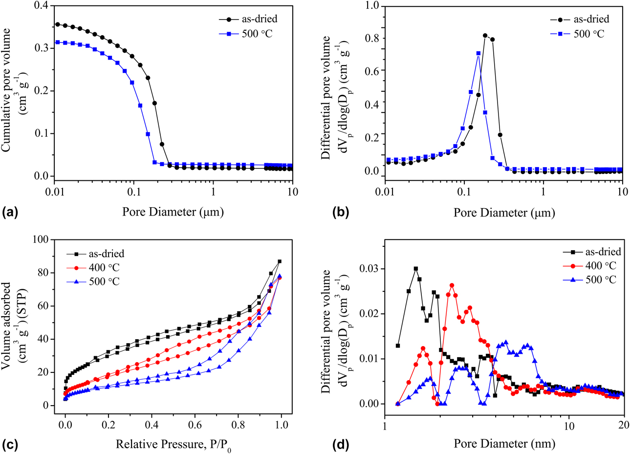
Nitrogen adsorption–desorption isotherms and the corresponding density functional theory (DFT) pore size distributions of ZT-5 as-dried and heat treated at various temperatures are shown in Figs. 8(c) and 8(d). The as-dried and heat treated gels exhibit type IV isotherms according to the IUPAC classification. The increased adsorption at higher relative pressure (P/P 0 > 0.90) indicates the existence of macropores. For the dried gel, the adsorption below P/P 0 = 0.05 indicates the existence of micropores originated from the interstices among primary particles. However, for the heat-treated gels, the lower adsorption below P/P 0 = 0.05 shows the decrease of micropores. As revealed from Fig. 8(c), the H3 type hysteresis loops indicate the existence of “slit-type” mesopores originated from the accumulation of globular or particulate gel structure.
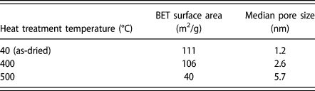
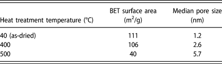
The pore properties of ZT-5 as-dried and heat treated at various temperatures are summarized in Table II. The dried gel exhibits the Brunauer–Emmett–Teller (BET) surface area of 111 m2/g with a median pore size of about 1.2 nm. When heat treated at 500 °C, the median pore size increases to 5.7 nm, which caused by the particle growth. The BET surface area decreases with the increase in temperature due to the decrease in micropores, phase transformation and the aggregation of nanoparticles induced by sintering.
Structural properties of as-dried and heat-treated ZT-5.
Conclusions
Zirconium titanate monoliths with macro-mesopores have been synthesized via a sol–gel process accompanied by phase separation. The pore size and the morphology could be controlled by changing PEO and NFA amount. Under suitable conditions, the obtained monoliths have co-continuous structure. The dried gel is amorphous. After heat treatment at 500 °C, the gel forms a pure ZrTiO4 phase, and the co-continuous macroporous structure is well maintained, the macropore size becomes small, and the surface of skeletons becomes smoother and thinner.
Experimental
Materials
Zirconium oxychloride (ZrOCl2·8H2O, 98%; Shanghai Macklin Biochemical Co., Ltd., Shanghai, China) and titanium(IV) n-propoxide [Ti(OnPr)4, 98%; Beijing HWRK Chemical Co., Ltd., Beijing, China] were used as zirconium and titanium sources, respectively. A mixture of distilled water (H2O) and nitric acid (HNO3, 65 wt%; Beijing Chemical Works, Beijing, China) was used as the solvent. PEO (Changchun Huagao Fine Chemicals Co., Changchun, China) with a number average molecular weight (M w) of 1 × 10⁵ was used to induce the phase separation and NFA (99%; Shanghai Macklin Biochemical Co., Ltd., Shanghai, China) to initiate gelation. Ethanol (EtOH, 99.9%; Beijing Chemical Works, Beijing, China) was used for solvent exchange. All the chemical reagents were used as received.
Preparation of zirconium titanate monoliths
In a typical synthesis, ZrOCl2·8H2O and PEO were dissolved in a mixture of H2O and HNO3. After dissolving completely, NFA was added into the solution. Then, Ti(OnPr)4 was slowly added with vigorous stirring under ice-cooled condition. After stirring for 10 min, the resultant homogeneous solution was sealed and kept at 60 °C for gelation. The gel formation occurred after about 4 h. The wet gels were aged for 24 h at 60 °C, then solvent-exchanged with ethanol two times (12 h for each time). After evaporation-dried at 40 °C, some of the dried gels were heat treated at various temperatures between 400 and 600 °C for 5 h with a heating rate of 5 °C/min in air.
Characterization
The morphology and microstructure of the dried and heat-treated gels were observed by an SEM (JSM-7500F; Hitachi Ltd., Tokyo, Japan) and a high-resolution TEM (HRTEM; Tecnai G2 F30, FEI Ltd., Oregon). Two-dimensional elemental analysis was performed using SEM equipped with EDS (SEM-EDS, HITACHI S-4800; Hitachi Ltd., Tokyo, Japan). TG-DTA (Thermo plus TG-DTA6200; Rigaku Corp., Tokyo, Japan) was performed at temperatures up to 600 °C at a heating rate of 10 °C/min with continuous supply of air at a rate of 50 mL/min. Infrared (IR) transmittance spectra were recorded on FT-IR spectrometer (NicoletiS5; Thermo Fisher Scientific Inc., New York) using the potassium bromide (KBr) pellet technique. The crystalline phase was confirmed by powder X-ray diffraction (Bruker D8 Advance; Bruker Science and Technology Ltd., Karlsruhe, Germany) using Cu Kα radiation (λ = 0.154 nm) as an incident beam in the 2θ range from 5° to 80°. XPS data were obtained with PHI Quantera II Spectrometer (Ulvac-Phi. Inc., Kanagawa, Japan) using 300 W Al Kα radiation in ultrahigh vacuum (approximately 3 × 10⁻⁹ mbar). Binding energies were calibrated with respect to the signal for adventitious carbon (binding energy = 284.6 eV). The macropore diameter of the samples was characterized using a mercury porosimeter (AutoPore IV 9520; Micromeritics Instruments Corp., Georgia). Nitrogen adsorption–desorption isotherms were obtained using a nitrogen adsorption–desorption apparatus (QUADRASORB SI; Quantachrome Instruments, Florida). Before the measurement, the samples were outgassed at 200 °C under vacuum for more than 4 h. The pore size distribution was calculated from the adsorption branch of the isotherm using DFT. The specific surface area was estimated by BET method.
Acknowledgments
We gratefully acknowledge the National Natural Science Foundation of China (20973022, 11472048) and the State Key Laboratory of Catalytic Materials and Reaction Engineering (33600000-14-ZC0607-0006) (RIPP, SINOPEC).