Introduction
In tropical and subtropical zones, climatic factors such as temperature and humidity favour the development of parasitic diseases (Satrija et al., Reference Satrija, Retnani, Ridwan and Tiuria2001). Intestinal helminthiasis is an important issue of public health in developing countries, the study of which has recently been neglected in favour of HIV/AIDS, malaria and tuberculosis. In tropical areas, gastrointestinal (GI) helminthiasis of domesticated animals is a major preoccupation of small-scale local farmers (Alawa et al., Reference Alawa, Adamu, Gefu, Ajanusi, Abdu, Chiezey, Alawa and Bowman2003). GI parasites cause high morbidity and often high mortality in livestock, resulting in significant economic losses (Ndao et al., Reference Ndao, Belot, Zinsstag and Pfister1995). In developed countries, GI parasites are controlled using commercial anthelmintics, whereas, in developing countries, traditional methods of helminth control remain largely dependent on medicinal plants. Because of increasing development of anthelmintic resistance within worm populations and, on the other hand, drug residues in animal products and the high cost of conventional anthelmintics, interest in alternative sources of anthelmintics is emerging (Sangster, Reference Sangster1999; Matthee et al., Reference Matthee, Dreyer, Hoffmann and Van Niekerk2002; Taylor et al., Reference Taylor, Hunt and Goodyear2002; Cirak et al., Reference Cirak, Crulegen and Baner2004; Wolstenholme et al., Reference Wolstenholme, Fairweather, Prichard, Von Samson-Himmelstjerna and Songster2004). In this work, we looked at Canthium mannii, used traditionally by the people of Dschang, Western Region of Cameroon (Cental Africa) as worm medicine. The traditional treatment consisted of eating a portion of the bark of the tree, sometimes with fried groundnuts, twice a day, for 3 days. This plant is commonly called ‘A koup memia in yemba’ (Wabo Poné, Reference Wabo Poné2001) or ‘kot missou in badjoue’ and has been listed as a plant used as an anthelmintic (Adjanohoun et al., Reference Adjanohoun, Aboubakar, Dramame, Ebat, Ekpére, Enoworock, Focho, Cibile, Kamanyi, Kamsu kom, Keita, Mbenkum, Mbi, Mbiele, Mbomé, Mubiri, Nancy, Kongmeneck, Satabie, Sofovora, Tamze and Wirmum1995). Heligmosomoides polygyrus, used in this study, is a useful experimental organism and has been widely used for the testing of anthelmintics (Smyth, Reference Smyth1996). The in vitro test is based on the hypothesis that a nematicidal activity observed would be indicative for potential in vivo activity. Medicinal plants may be effective, yet less expensive and less toxic for humans, animals and the environment compared to conventional anthelmintics (Lans & Brown, Reference Lans and Brown1998; Quilan et al., Reference Quilan, Quinlan and Nolan2002). Recent studies conducted in various parts of the world have revealed anthelmintic properties of extracts and/or essential oils of some medicinal plants (McGaw et al., Reference McGaw, Jager and Van Staden2000; Mpoame & Essomba, Reference Mpoame and Essomba2000; Noritaka et al., Reference Noritaka, Tavo and Viazulito2000; Assis et al., Reference Assis, Bevilaqua, Moraisa, Vieirab, Costar and Souzaa2003; Githiori et al., Reference Githiori, Hoglund, Waller and Baker2003a, Reference Githiori, Hoglund, Waller and Bakerb; Githiori, Reference Githiori2004; Wabo Poné et al., Reference Wabo Poné, Mpoame, Bilong Bilong and Kerboeuf2005, Reference Wabo Poné, Bilong Bilong, Mpoame, Fusi-Ngwa and Coles2006; Magdeleine et al., Reference Magdeleine, Hoste, Mathieu, Varo and Archimède2009; Oliveira et al., Reference Oliveira, Bevilaqua, Costa, Macedo, Barros, Rodrigues, Cumurça-Vasconcelos, Morais, Lima, Vieira and Navarro2009; Tariq et al., Reference Tariq, Chishti, Ahmad and Shawl2009). Taking the above facts into consideration, the present study aimed to evaluate the nematicidal properties of four extracts of C. manni (Rubiaceae) stem bark on different life-cycle stages of H. polygyrus (Nematoda, Heligmosomatidae), a parasite of rodents.
Materials and methods
Plant materials
Canthium mannii, belonging to the family Rubiaceae, is a climbing shrub up to 6 m high. The leaves are elliptical, cuneate at base, shortly acuminated at the apex; 8–15 cm long, 2.5–6 cm broad (Adjanohoun et al., Reference Adjanohoun, Aboubakar, Dramame, Ebat, Ekpére, Enoworock, Focho, Cibile, Kamanyi, Kamsu kom, Keita, Mbenkum, Mbi, Mbiele, Mbomé, Mubiri, Nancy, Kongmeneck, Satabie, Sofovora, Tamze and Wirmum1995). Species identification has been carried out at the National Herbarium of Cameroon (NHC). Fresh C. mannii stem bark, collected in Fondonera, a village situated 25 km from Dschang (Santchou subdivision, Menoua division, Western Region of Cameroon, Central Africa), was cut in small rectangles (2 cm × 5 cm), spread on cardboard and air dried for 3–4 h daily for 7 days, then ground and stored in airtight plastic bags for about 1 week at room temperature (24°C) and relative humidity (RH) of about 67% for subsequent use in the laboratory.
Preparation of extracts
Four types of extracts were prepared to compare the effects and to increase the chances of detecting anthelmintic activity: ethanol extract (ETE), hexane extract (HEE), cold water extract (CWE) and hot water extract (HWE).
Ethanol and hexane extracts
Four hundred grams of ground bark of C. manni were macerated in 5 litres of ethanol (95%), which removed more polar compounds, or hexane (100%) for semi-polar and apolar compounds (Balansard et al., Reference Balansard, Maillard and Vidal-Ollivier1991). The mixtures were stirred daily, and 72 h later the ethanol extract and hexane extracts were obtained (Ciulei, Reference Ciulei1982). This was followed by the dilution of 140 mg of the concentrated extract (using a rotary evaporator) with 0.2 ml of dimethyl sulphoxide (DMSO), a diluent which efficiently helped to dissolve the organic extracts. After 5–10 min, distilled water (DW) was added to obtain 20 ml, according to the method of Ibarra & Jenkins (Reference Ibarra and Jenkins1984). This resulted to a 7 mg/ml stock solution from which a series of dilutions were made to obtain solutions of different concentrations: 3.5, 3.0, 2.5, 2.0 and 1.5 mg/ml.
Cold and hot water extracts
Four hundred grams of ground bark of C. manni were macerated in 5 litres of DW. The mixture was stirred daily, and 48 h later (to avoid growth of fungi) a CWE was obtained. This solution was filtered through a filter paper of pore size 2.5 μm. A 250 ml aliquot of the filtrate was distributed between five beakers, which were then placed in an oven at 50°C for a week, allowing the extracts to dry. About 140 mg of the dried extract obtained were diluted with 20 ml of DW to get a stock solution which was serially diluted to obtain five different concentrations, as with the organic extracts.
For the HWE, a procedure similar to that of the CWE was applied except that the DW was boiled (100°C) and the infusion (400 g of powder in hot water) was allowed to stand at room temperature (24°C) for approximately 3 h to cool before serially diluting the stock solution.
Reference drug
The reference drug, mebendazole (MBZ), used in this study was bought from a local pharmacy. This drug was chosen due to its availability and also because some authors have used it for in vitro trials using different concentrations. The drug was diluted in DW to obtain a stock solution with a concentration of 7 mg/ml, which was serially diluted to obtain the same concentrations as with organic and water extracts. Controls used for the bioassay were 1% DMSO (maximum concentration of DMSO in test dishes) and distilled water.
Recovery of eggs and larvae
Heligmosomoides polygyrus L3 larvae, used in this study to infect mice orally, were of a laboratory-selected strain provided by Dr G.C. Coles, University of Bristol, UK. After 10 days, the fresh eggs were obtained from the faeces of experimentally infected mice (Michael et al., Reference Michael, Meinke and Shoop2001). Briefly, 3 g of faeces were collected, homogenized in a mortar, suspended in saturated sodium chloride solution (0.4% NaCl), and cleansed of organic debris by filtration through sieves (1 mm and 150 μm) in a 100 ml beaker. The contents of the latter were poured into eight tubes and centrifuged at 1000 g for 5 min. The supernatant containing eggs was poured through a 45 μm sieve. The retained material on the sieve, containing eggs, was washed with tap water to remove the salt solution. The sieve was then turned and the opposite side was washed with distilled water and eggs were collected in a 16-cm Petri dish (Euzeby, Reference Euzeby1981). At this stage eggs were not counted.
The egg solution obtained as above was cultured, using the technique described by Smyth (Reference Smyth1996). Briefly, 3 ml of the egg suspension was poured on filter paper covering the bottoms of three Petri dishes. The dishes were then covered to maintain a high relative humidity (65–67%) to prevent them from drying out, and were stored at 24°C. After 3, 4–5 and 6–7 days of incubation, L1, L2 and L3 larvae, respectively, were observable in Petri dishes. Larvae were differentiated using their morphological features and were concentrated with a Baermann apparatus (Cheesbrough, Reference Cheesbrough1987; Smyth, Reference Smyth1996).
In the larval development assay, a sufficient quantity of mice faeces was collected and a suspension made with 50 ml of DW. This suspension was filtered through a 150 μm sieve. The filtrate, collected in a 200 ml beaker, was resuspended in 100 ml water and centrifuged at 1000 g for 5 min. The supernatant was discarded, but 100 ml were saved for later addition to the final ova suspension to provide bacteria necessary for the foraging nematode larvae during their development (Michael et al., Reference Michael, Meinke and Shoop2001).
Evaluation of anthelmintic activity
The anthelmintic efficacy tests of the extracts on different lifecycle stages of H. polygyrus were performed, using four different procedures. To evaluate the effects of the various extracts on un-embryonated eggs, 1 ml of a diluted egg solution containing 30–40 eggs was distributed in each of the 12 Petri dishes (35 mm diameter × 10 mm) and mixed with the same volume of a specified extract or MBZ at different concentrations. The dishes were covered and the eggs incubated in at room temperature for 48 h. The number of L1 larvae and eggs per Petri dish was then counted using a microscope (at 4 × magnification). The percentage of hatched eggs (HE%) was determined using the relationship below (Hubert & Kerboeuf, Reference Hubert and Kerboeuf1992):

To assess the effect of various substances on embryonated eggs, the same numbers of un-embryonated eggs were distributed in the same number of Petri dishes and were allowed to stand at room temperature for about 24 h. When the L1 larvae became visible and started moving actively within the eggshell (>90% in the control Petri dish), 1 ml of each concentration of extracts or MBZ was added to each Petri dish, according to Dobson et al. (Reference Dobson, Donal, Waller and Snowdon1986). The dishes were then covered and incubated for a further 6 h at room temperature to allow hatching in the Petri dishes. When the eclodibility rates (E%) in DW and 1% DMSO controls were higher than 90%, 2–3 drops of Lugol's iodine (5%) were added to each Petri dish to stop egg hatching (Pessoa et al., Reference Pessoa, Movais, Bevilaqua and Luciano2002). Then, all the embryonated eggs and first-stage larvae (L1) were counted using a microscope (at 4 × magnification) and the percentage of eclodibility was calculated by the formula below (Wabo Poné et al., Reference Wabo Poné, Bilong Bilong, Mpoame, Fusi-Ngwa and Coles2006):

For the effect of C. mannii extracts on L1, L2 and L3 larvae of H. polygyrus, the same procedures described above were followed but 1 ml of solution containing 10–15 larvae was used. Death or immobilization of the different larval stages were assessed under a microscope (at 4 × magnification) after 2, 4, 6 and 24 h for rhabditoid larvae (Sinha et al., Reference Sinha, Sivastava, Singh, Sinha and Sinha1987), and after 24 and 48 h for filariform larvae (Boisvenue et al., 1983). Larvae that showed no movement after 5–10 s of continuous observation were recorded as dead. The mortality rate (M%) was assessed using the following formula, adopted from Hubert & Kerboeuf (Reference Hubert and Kerboeuf1992):

To assess the effect of the various extracts and MBZ on larval development, 1 ml suspensions containing 30–40 eggs and bacteria were distributed in each of the 12 Petri dishes (35 mm diameter × 10 mm). These dishes were covered and incubated in at room temperature for 48 h. After this period, the majority of nematode eggs had hatched to L1 larvae (Hubert & Kerboeuf, Reference Hubert and Kerboeuf1992). One ml of the previously prepared range of extracts or MBZ concentrations were added to each Petri dish. Five days later, the number of third-stage larvae (L3) was counted using a microscope (at 4 × magnification) by separating them from L1 and L2 larvae (Taylor, Reference Taylor1990; Hubert & Kerboeuf, Reference Hubert and Kerboeuf1992; Ademola, Reference Ademola2002). The percentage of development (D%) was assessed by the following formula adopted from Hubert & Kerboeuf (Reference Hubert and Kerboeuf1992):

All tests were repeated six times for each treatment and control.
Data analysis
The 50% ovicidal concentration (OC50), the 50% inhibitory concentration (IC50), and the 50% larvicidal concentration (LC50) were determined from the linear regression curve obtained between the mortality expressed in probit and the decimal logarithm of the concentrations (mg/ml). Comparisons were made using the chi-square test, results were regarded as significant at P < 0.05.
Results
Egg hatch assay
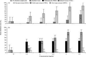
The effects of mebendazole, ethanol, hexane, cold and hot water extracts on fresh eggs of H. polygyrus are given in fig. 1. The mean hatching rates were 95.1 and 96.2% in 1% DMSO (negative control) and DW (placebo), respectively. Except for HEE, all values obtained with various concentrations of the extracts differed significantly from negative control and placebo (P < 0.05). Conversely, no statistical difference (P>0.05) was detected between 1% DMSO and DW. Eggs that failed to hatch were considered dead or damaged. Except for HEE, all extracts tested showed a linear relationship between the probit of egg hatch and the logarithm of the product concentration. The OC50 values obtained were 1.0, 1.1, 1.6, 1.8 and 6.3 mg/ml for MBZ, ETE, HWE, CWE and HEE, respectively. With embryonated eggs, it was shown that lower concentrations of CWE and HEE did not affect the egg hatching ( ≤ 10% inhibition). Conversely, HWE and ETE affected the eclodibility similarly, with 85 and 82% inhibition, respectively, at 3.5 mg/ml (fig. 2a and b). The percentage of inhibition due to MBZ was constant and closer to 45% for all concentrations tested. As for the fresh eggs, a linear relationship was observed between the probit of inhibition percentage of egg hatching and the logarithm of the concentration. The IC50 values were 1.2, 1.3, 1.6, 3.0, and 3.2 mg/ml for HWE, ETE, MBZ, CWE and HEE, respectively. In this assay, the effect of HWE was slightly greater than that of ETE.
Effects of different concentrations (mg/ml) of cold and hot water (a), ethanol and hexane (b) extracts of Canthium mannii on fresh eggs of Heligmosomoides polygyrus after 48 h of contact.

Effects of different concentrations (mg/ml) of cold and hot water (a), ethanol and hexane (b) extracts of Canthium mannii on embryonated eggs of Heligmosomoides polygyrus after 6 h of contact.

Larval development
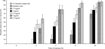
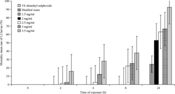
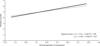
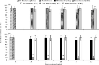
Only ETE significantly affected the survival of L1 and L2 larvae of H. polygyrus; in fact MBZ, CWE, HWE and HEE were inactive on these life-cycle stages. After incubation for 24 h, the highest mortalities of L1 and L2 due to C. mannii extracts tested were 97.2 and 92.7%, respectively, achieved at 3.5 mg/ml (figs 3 and 4). A dose (fig. 5) and time responses were also linear after log transformation of data. The LC50 values were low for both L1 (1.5 mg/ml) and L2 (1.0 mg/ml) after 6 and 24 h, respectively. For filariform larvae (L3), all the substances at all concentrations tested were inactive and all infective sheathed larvae were still very mobile after 48 h of contact. Only ETE reduced significantly the development of rhabditiform larvae (fig. 6a and b). Furthermore, the concentrations 3.0 and 3.5 mg/ml had a similar L3 production rate of 6%. There was a significant difference (P < 0.05) between the production rate of different extracts. The LC50 after 5 days of contact was relatively low (0.7 mg/ml) for ETE and higher for the other extracts ( ≥ 8.9 mg/ml). A comparison between the egg-hatching test and the larvicidal activity demonstrated that the eggs were much more susceptible than the larvae to the extracts.
Mortality mean rate of L1 larvae of Heligmosomoides polygyrus according to concentrations (mg/ml) of ethanol extract (ETE) and exposure time.

Mortality mean rate of L2 larvae of Heligmosomoides polygyrus according to concentrations (mg/ml) of ethanol extract (ETE) and exposure time.

Mortality (in probit) of L1 and L2 larvae of Heligmosomoides polygyrus according to the decimal logarithm of concentrations (μg/ml) of ethanol extract after 6 h (L1, solid line) and 24 h (L2, dashed line) of exposure.

Larval development rate (L1 to L3) of Heligmosomoides polygyrus according to the different concentrations (mg/ml) of cold and hot water (a), ethanol and hexane (b) extracts after 5 days of contact.

Discussion
Extracts of the shrub, C. mannii, examined in the present study, had detectable in vitro effects on life-cycle stages of the nematode H. polygyrus. Concentrations active against parasitic stages of H. polygyrus were higher than those ( ≥ 0.125 mg/ml) effective against the same stages of Ancylostoma caninum (Wabo Poné et al., Reference Wabo Poné, Mpoame, Bilong Bilong and Kerboeuf2005, Reference Wabo Poné, Bilong Bilong, Mpoame, Fusi-Ngwa and Coles2006). The high concentrations required against the different stages of H. polygyrus could be due to the multi-resistance features of this rodent parasite strain. The dose- and time-dependent responses observed with extracts of the plant were similar to those obtained by Dobson et al. (Reference Dobson, Donal, Waller and Snowdon1986), Pessoa et al. (Reference Pessoa, Movais, Bevilaqua and Luciano2002) and Wabo Poné et al. (Reference Wabo Poné, Mpoame, Bilong Bilong and Kerboeuf2005, Reference Wabo Poné, Bilong Bilong, Mpoame, Fusi-Ngwa and Coles2006). The ETE appeared more active on the fresh eggs, whereas HWE was slightly more active on embryonated eggs (IC50 = 1.2 mg/ml for HWE vs. IC50 = 1.3 mg/ml for ETE). These findings could be explained by the fact that different solvents extract different compounds (Ciulei, Reference Ciulei1982). Hot water could be extracting compounds such as heterosides and iridoids (Balansard et al., Reference Balansard, Maillard and Vidal-Ollivier1991), affecting egg hatching, while the better activity of ETE could be attributed to the greater concentration of alcohol-soluble active anthelmintic compounds, such as terpenoids and tannins, affecting embryonation. Egg hatching could have been inhibited by some saponins, present in the extracts. These molecules are known to stop the egg-hatch mechanism. Also, the percentage of inhibition due to MBZ was constant. Since MBZ is a purified drug, the active compound after dilution would have had the same concentration in each Petri dish.
The mode of action of C. mannii extracts on H. polygyrus eggs is not completely understood. Some compounds might act by penetrating the eggshell; these could prevent development of eggs in C. manni solution, as with MBZ (Dupouy-Camet, Reference Dupouy-Camet2000). Other compounds in the extracts could paralyse the first-stage larvae within the eggs in C. manni solution, as seen with levamisole (Dobson et al., Reference Dobson, Donal, Waller and Snowdon1986). Hence, the decrease in the rate of hatching observed in the present bioassay can be attributed to the action of the extracts, since the proportion of eggs hatched was high in the control (>95%). The fact that eggs were more susceptible to extracts could be due to a soluble active anthelmintic compound present in the extract, which affects both embryonation and hatching. The similar eclodibility activity of HWE and ETE may be due to the fact that hot water and ethanol may help to concentrate more anthelmintic compounds that affect the hatching mechanism.
The lower activity of HWE, HEE, CWE on larval development may be due to a low concentration of larvicidal compound in these extracts. The ETE effect on rhaditiform larvae may be due to ingestion through the oral route during larval feeding or by diffusion through the cuticle (Dobson et al., Reference Dobson, Donal, Waller and Snowdon1986). These two routes of action could be used simultaneously.
These results suggested that the extracts of C. mannii could contain a large spectrum of compounds with different modes of action. This could explain the multiple target activity (on egg hatching and larval development) of the extracts. None of the extracts or MBZ (the reference drug) affected the filariform larvae of H. polygyrus. This inactivity could be the result of the inability of H. polygyrus infective larvae to feed, thus not consuming the drug, and/or the non-permeability of the sheath of the L3 larvae which protects them from chemical substances (Smyth, Reference Smyth1996).
The mean larval development, i.e. from L1 to L3, was 88.4% in the control dishes. In this assay, about 40 eggs/ml were incubated; this low density could justify the relative high rate of larval development. As Hubert & Kerboeuf (Reference Hubert and Kerboeuf1992) indicated, too many eggs per culture are incompatible with satisfactory larval development and the percentage of infective larvae decreases sharply. The fact that ETE, compared to other extracts, drastically reduced the production of third-stage larvae is probably due on the one hand, to the greater concentration of alcohol-soluble active anthelmintic compound(s) and, on the other hand, to a more transcuticular and/or oral absorption of the extract into the body of rhabditiform larvae. The nematicidal activity of ETE shown in this work could be due to the fact that the stem bark of C. mannii contained secondary metabolites such as alkaloids, flavonoids, polyphenols and saponins, which were identified after phytochemical screening of ETE (Wabo Poné, Reference Wabo Poné2007). In related Canthium sp., glycosides such as iridoide diglycosides and flavonol, which are hydrophilic molecules, have been isolated by Kanchanapoom et al. (Reference Kanchanapoom, Kasai and Yamasaki2002) and Gunasegaran et al. (Reference Gunasegaran, Subramani, Azantha Parimala, Ramachandra Nair, Rodriguez and Souzaa2003), respectively. The stem and bark of plants represent a site of the biosynthesis and storage of secondary metabolites responsible for the biological properties of medicinal plant extracts (Busindou, Reference Busindou1997). In all the assays, MBZ had a low activity compared to ETE and HWE; these results could be explained by the multi-resistant feature of the parasite strain and/or by the fact that benzimidazoles are weakly soluble in water (Boisvenue et al., 1983; Elard, Reference Elard2000).
In conclusion, C. mannii stem bark extracts exhibited in vitro negative effects on eggs, rhabditoid larvae, and on larval development of H. polygyrus, a parasite of mice. The ETE showed the highest nematicidal activity on the different parasite stages, with dose- and time-dependent responses. These results suggest that ETE, and to a lesser extent HWE, can regulate the risk of H. polygyrus and other nematode host infections. The ETE in vitro activity has to be confirmed in vivo during treatment of infected animals, by measuring the total worm counts and faecal egg counts. In addition, toxicological investigations are necessary.
Acknowledgements
The authors express their sincere thanks to Fondonera's community, and particularly to Mr Innocent Tafadji Mekuikeu and Mrs Albertine Djitia-Fongou for helping in the collection of the plant specimens, and to Dr G.C. Coles for providing the parasite used in this study.