Introduction
Intranasal teeth are rare. Trauma is a documented cause;Reference Johnson1 however, cases with this aetiology have been rarely reported.
Identification and treatment of intranasal teeth is important because of potential morbidity, for example due to pain, infection, bleeding or congestion.Reference Kirmeier, Truschnegg, Payer, Malyk, Daghighi and Jakse2–Reference Smith, Gordon, De Luchi and Intranasal7 However, cases may be diagnosed late, as was the presented case, due to the absence (or variability) of symptoms and the timing of presentation.
This article presents a case of trauma causing displacement of a tooth into the nasal cavity in a child.
Case report
A fit and healthy six-year-old girl was presented to our accident and emergency department due to a two-month history of unilateral, right-sided nasal congestion and laboured breathing at night. Her parents reported a tendency for her to repeatedly blow her nose. She did not have any other symptoms. Her medical history showed that, at two years of age, she had fallen from a high chair and had caught her mouth on a window ledge, resulting in trauma to her upper right deciduous central incisor. Her parents noticed the tooth to be missing following the trauma, but did not follow this up with any specific medical or dental investigations.
On examination, a hard, white mass was noted in the child's right nasal cavity. The nature of the foreign body was unclear, and an attempt was made to remove it. This resulted in bleeding; thus, further attempts to remove the foreign body were abandoned.
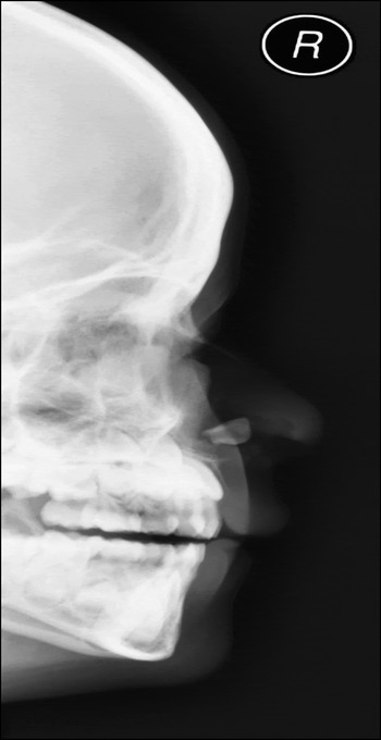
A lateral cephalogram radiograph was requested, which revealed an opaque, calcified structure in the nostril (Figure 1).
Lateral radiograph showing a calcified structure (the tooth) in the nostril. R = right

The child was directly referred to the ENT department for further investigation. There, it was concluded that the object in her right nostril was most likely to be of dental origin, i.e. a tooth.
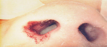
Extraction of the intranasal tooth was performed under direct vision by the oral surgeon, with an approximate operating time of 10 minutes (Figure 2). Post-extraction, the nasal mucosa appeared intact, with no obvious oro-nasal communication.
Surgical photograph showing the examination findings in the operating theatre.

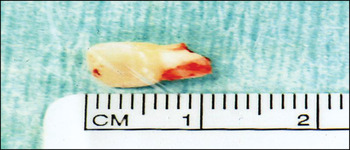
The extracted tooth appeared to be a deciduous incisor with its apex missing (Figure 3).
The extracted tooth, a deciduous upper right central incisor.

In view of this post-operative finding, and the child's earlier history involving trauma to an incisor tooth, it was thought likely that the trauma had caused intrusion of the tooth into the nasal cavity.
At the follow-up appointment a week later, the oral surgeons noted a very good recovery. An upper standard occlusal radiograph confirmed the presence of both upper central permanent incisors.
Discussion
Intranasal teeth are rare, and most commonly are due to supernumerary teeth. However, they can also arise from ectopic deciduous or permanent teeth.Reference Smith, Gordon, De Luchi and Intranasal7, Reference Lee8 Ectopic eruptions may be found in all areas of the maxillofacial skeleton, for example, the maxillary sinus, palate, mandibular condyle, coronoid process, orbits, nasal cavity, and in patients with cleft lip and palate.Reference Murty, Hazarika and Hebbar5, Reference Smith, Gordon, De Luchi and Intranasal7 The majority of intranasal teeth are unilateral and are discovered before adulthood.Reference Lin, Chung-Feng, Su, Kao and Peng4
The aetiology of these intranasal teeth eruptions is not clear but is thought to be multifactorial, namely, infection, trauma, anatomical malformation (cleft lip and palate) or hereditary factors. Of those reported in patients with cleft lip and palate, most result from the surgery that is required to correct the malformation.Reference Andrade, Marchionni, de Oliveira and Heitz9 Trauma as a cause of intranasal teeth eruption has rarely been reported.
The symptomatology can be variable, ranging from complete absence of symptoms to problems such as nasal discharge,Reference Kirmeier, Truschnegg, Payer, Malyk, Daghighi and Jakse2, Reference Pracy, Williams, Montgomery and Nasal6 nasal obstruction,Reference Kohli and Verma3–Reference Murty, Hazarika and Hebbar5, Reference Smith, Gordon, De Luchi and Intranasal7 epistaxis,Reference Lin, Chung-Feng, Su, Kao and Peng4 headacheReference Murty, Hazarika and Hebbar5 and facial pain.Reference Pracy, Williams, Montgomery and Nasal6, Reference Andrade, Marchionni, de Oliveira and Heitz9 Complications include chronic ulceration or infection,Reference Kohli and Verma3, Reference Moghaddam, Hyde and Williamson10 paranasal sinusitis,Reference Lin, Chung-Feng, Su, Kao and Peng4, Reference Pracy, Williams, Montgomery and Nasal6 oro-nasal fistulaeReference Kohli and Verma3, Reference Coonar, Crean and Bennett11 and rhinolith formation.Reference Coonar, Crean and Bennett11 In the presented case, the patient had nasal congestion which presented some time after the initial traumatic insult.
• Intranasal teeth are rare and usually present in children
• Trauma is a very rare cause, and can directly displace the tooth into the nasal cavity
• Symptoms may be delayed or absent
• Extraction is required to treat symptoms and prevent subsequent morbidity
Diagnosis is based upon the findings of clinical examination and radiological investigation, including plain radiographs or computed tomography. Once a diagnosis has been established, early treatment should be pursued, in the form of surgical extraction, to prevent significant morbidity if the condition is left untreated. In our patient, the tooth was extracted under direct vision, a relatively simple procedure with no complications. Endoscopic extraction of intranasal teeth is another useful method and enables good visualisation and precise dissection.Reference Kirmeier, Truschnegg, Payer, Malyk, Daghighi and Jakse2, Reference Lin, Chung-Feng, Su, Kao and Peng4, Reference Lee8, Reference Moghaddam, Hyde and Williamson10
Conclusion
Trauma is a rare cause of an intranasal tooth eruption. Diagnosis may be difficult due to an absence of symptoms or a delay in presentation. Accurate diagnosis and early treatment are fundamental to prevent morbidity. Treatment by extraction of the tooth leads to a successful outcome. The presented case highlights the importance of a detailed history, which proved to be vital for the final diagnosis. Our patient's parents were advised to ensure regular follow-up visits with her general dental practitioner to closely monitor the development of permanent dentition.