Introduction
A 66-year-old woman presented with dysphagia secondary to a multinodular lingual goitre. A previously silent lingual thyroid had undergone multinodular change to cause dysphagia and eventual airway compromise. The goitre was excised via a midline, mandible-splitting approach.
Case report
A 66-year-old woman was seen in the ENT clinic with progressive dysphagia, choking episodes and a sensation of airway obstruction. She had been referred to our department by an anaesthetist who had experienced difficulty intubating the patient for a routine, elective, laparoscopic cholecystectomy. The patient had noticed an increasing swelling in the submental region for over 30 years. Ten years prior to the onset of swelling, she had undergone an operation in Turkey to remove a presumed thyroglossal cyst, following which she was placed on thyroid hormone replacement to suppress any recurrence of the cyst pathology.
Neck examination showed a 5 × 5 cm swelling in the submental region, which was firm in consistency and situated above the old surgical incision for the thyroglossal cyst.
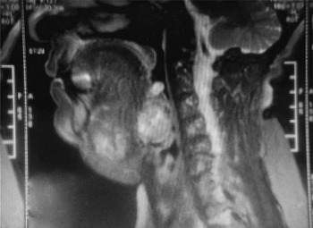
Fibre-optic laryngoscopy revealed a large mass in the tongue base. Magnetic resonance imaging of the neck showed two masses. The first, a multinodular swelling situated slightly to the left of the midline, measured 52 × 34 × 41 mm, and extended from the floor of the mouth to the root of the tongue, displacing the mylohyoid muscle inferiorly (Figure 1). The second multinodular swelling, measuring 26 × 26 × 26 mm, was closely related to the first and lay in the base of the tongue. Superior to the second swelling was a small nodule which was thought to be part of the same complex. There was no thyroid tissue in the anatomically conventional, para-tracheal position.
Sagittal magnetic resonance imaging scan of the swelling arising in the region of the tongue base.

The patient underwent a radioiodine scan, which showed vivid iodine uptake at the site of the two main lesions. The normal anatomical position of the thyroid did not show any radioiodine uptake. Thyroid function tests, performed after stopping the patient's thyroxine medication, were normal. On stopping her thyroxine, there was no rebound growth of the patient's ectopic, multinodular goitre.
The patient was scheduled for elective surgical excision of the ectopic thyroid, in view of its compressive symptoms and size.
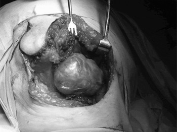
A surgical tracheotomy was performed for tracheotomy intubation. Exploration of the prelaryngeal region revealed the parathyroid glands in the para-tracheal space; no thyroid tissue could be seen in the prelaryngeal area. The ectopic thyroid was approached via a midline, lip-splitting incision with midline mandibulotomy (Figure 2). The left submandibular gland was excised, after identifying and preserving the lingual and hypoglossal nerves. The floor of the mouth was divided and the two lesions dissected out in toto.
Intra-operative photograph of the smaller lobe of the multinodular lingual goitre. In this inferior view, with the patient's head tilted to the observer's left, the goitre is clearly visible wrapped around the posterior edge of the mylohyoid muscle. The mandible was later split to gain access to the rest of the lesion.

The patient recovered well post-operatively, and was decannulated on the 10th post-operative day.
Post-operative histopathological analysis confirmed the tissue to be of thyroid origin (Figures 3 and 4).
Photomicrograph of the lingual thyroid, showing multiple colloid follicles (H&E 100×).

Photomicrograph of the lingual thyroid, showing multiple follicles (H&E 200×).

On follow up, the patient had normal tongue function, and her dysphagia and choking sensation had abated.
Discussion
The human thyroid gland develops from two primordial structures, the thyroid diverticulum and the ultimobranchial bodies.Reference De Felice and Di Lauro1 The thyroid diverticulum arises from the posterior midline of the embryonic mouth, between the first and second pharyngeal pouches. This diverticulum deepens into the mesenchyme. The ultimobranchial bodies are a pair of epithelial bodies found on the posterior part of the fourth branchial pouches. The C-cell precursors migrate from the neural crest bilaterally to become localised in the ultimobranchial bodies in the fourth pharyngeal pouch. The thyroid diverticulum fuses with the ultimobranchial bodies, differentiating into thyroid follicles and C-cells. It is widely believed that the thyroid follicles develop from the diverticulum and the C-cells from the ultimobranchial bodies. The thyroid gland then migrates from the foramen caecum to the thyroid bed in front of the trachea. The hyoid develops later; thus, thyroglossal cysts can be located either above, below or within the hyoid bone.
Alt et al. analysed the development of thyroid vessel architecture in zebrafish and mouse embryos.Reference Alt, Elsalini, Schrumpf, Haufs, Nathan and Schwabe2 They concluded that defects or changes in pharyngeal and cervical blood vessels correlated with abnormal thyroid development in both species. Based on their study, they concluded that the carotids were the vessels most likely to define morphogenesis of the thyroid gland in mammals.
An ectopic thyroid can occur at any location along the path of migration. The commonest type of ectopic thyroid is a lingual thyroid, lying between the circumvalate papillae and the epiglottis. This type has an incidence of one in 3000 to 10 000.Reference Williams, Sclafani, Slupchinskij and Douge3 Other locations include sublingual, suprahyoid, infrahyoid, at the level of the hyoid bone, within the trachea, submandibular, intracardiac, and associated with the duodenum, gall bladder and porta hepatis.Reference Brandwein, Som and Urken4–Reference Ghanem, Bley, Altehoefer, Hogerle and Langer10 A dual ectopic location of the thyroid gland has also been reported, wherein the gland occurs in two different ectopic sites simultaneously.Reference Ulug, Ulubil and Alagol11–Reference Long, McChesney and Beggs13
The first published case of lingual thyroid was reported by Hickman in 1869.Reference Hickman14 The case involved a lingual thyroid in a 16-hour-old infant who died from asphyxiation caused by the lingual goitre. Lingual thyroid has subsequently been extensively reported in the literature, but virtually exclusively in children and young adults.
The various presenting symptoms of lingual thyroids include a feeling of a lump in the throat, foreign body sensation, change in voice, swallowing difficulties, dyspnoea, orthopnoea, obstructive sleep apnoea and intubation difficulties.Reference Taibah, Ahmed, Baessa, Saleem, Rifai and Al-Arifi15 Clinical evaluation should include a thorough head and neck examination, along with fibre-optic laryngoscopic examination to visualise the region posterior to the circumvalate papillae and to assess the gland size and the airway. Magnetic resonance imaging is usually performed to evaluate the anatomy of the ectopic thyroid tissue, as well as to identify the presence of any normal thyroid tissue in the thyroid bed. Analysis of tri-iodothyronine, thyroxine and thyroid-stimulating hormone (TSH) concentrations are required in order to assess thyroid gland function.
In our patient's case, there was no histological proof of her original ‘thyroglossal cyst’. It is possible that she had two separate ectopic sites each containing thyroid tissue. As the bulk of the thyroid was in the lingual site, a descending tract containing a thyroglossal cyst would seem unlikely. A coincidental dermoid cyst would be another explanation.
There is little consensus on the management of ectopic thyroids, due to their rarity.
Medical management consists of therapy with exogenous thyroxine to suppress endogenous TSH production and thus remove any stimulus for ectopic thyroid enlargement. The effect of this on multinodular goitres in the lingual site is likely to be comparable to that on multinodular goitres in the usual anatomical site. In our patient's case, such therapy, at best, slowed the goitre's growth.
• This paper describes a case of multinodular goitre developing in a lingual thyroid
• The presenting symptoms of a lingual thyroid include a feeling of a lump in the throat, foreign body sensation, change in voice, swallowing difficulties, dyspnoea, orthopnoea, obstructive sleep apnoea and intubation difficulties
Radioiodine ablation therapy may be of use for patients unfit for surgery or those who refuse surgery. Again, its effectiveness in ectopic multinodular goitres can be presumed to be similar to that in conventionally sited multinodular goitres.
Surgical removal is offered to patients with compressive symptoms or an unsatisfactory response to suppressive therapy with thyroxine. There are various surgical approaches to the lingual thyroid: transoral, transhyoid pharyngotomy, lateral pharyngotomy and, as in our case, midline mandibulotomy.Reference Ward, Cantrell and Allan19 Endoscopic CO2 laser excision of the lingual thyroid gland has been advocated in view of the associated shorter post-operative stay and reduced morbidity.Reference Hafidh, Sheahan, Khan, Colreavy and Timon16 However, subtotal removal of a multinodular goitre, whether in the neck or an ectopic site, must be undertaken with the knowledge that the remaining tissue is likely to continue to grow, and may need further intervention.
Excision of the lingual thyroid does not risk hypoparathyroidism, as these glands are present in the normal cervical position.
In the absence of a normal thyroid, the patient will need lifelong thyroxine replacement. For this reason, there have been successful attempts to re-implant the ectopic thyroid tissue either in the rectus sheath or on a transoral pedicle into the oral cavity.Reference Minuto, Fazzuoli, Rollandi, Derchi and Biassoni17, Reference Wu, Zheng, Dong, Li, Zhang and Zhao18 Due to the size of the multinodular goitre, this was not attempted in our patient.
To the best of our knowledge, our patient represents the first reported case of a multinodular goitre developing in a lingual thyroid.
Acknowledgements
We thank Mandy Esprit and Julie John, ENT Administrators, Ealing General Hospital, for their help during the preparation of the manuscript.