Introduction
Cynodontia, the therapsid group including mammals as its extant subclade (Kemp, Reference Kemp1982; Rubidge and Sidor, Reference Rubidge and Sidor2001; Botha et al., Reference Botha, Abdala and Smith2007), first appeared during the late Permian and formed a key component of the Triassic tetrapod fauna (Kemp, Reference Kemp1982; Battail, Reference Battail2001; Abdala and Ribeiro, Reference Abdala and Ribeiro2010). The non-mammalian cynodonts (sensu Abdala et al., Reference Abdala, Gaetano, Martinelli, Soares, Hancox and Rubidge2020) acquired diverse modifications in dentition, jaw articulation, and basicranium prior to development of the extant mammalian clade (Kemp, Reference Kemp1982, Reference Kemp2005; Kielan-Jaworowska et al., Reference Kielan-Jaworowska, Cifelli and Luo2004). The evolution of mammal-like features in non-mammalian cynodonts and early mammals is debated extensively (Zhou et al., Reference Zhou, Wu, Martin and Luo2013; Świło et al., Reference Świło, Niedźwiedzki and Sulej2014; O'Meara and Asher, Reference O'Meara and Asher2016; Luo et al., Reference Luo, Meng, Grossnickle, Liu, Zhang, Neander and Ji2017; Huttenlocker et al., Reference Huttenlocker, Grossnickle, Kirkland, Schultz and Luo2018; Krause et al., Reference Krause, Hoffmann, Hu, Wible, Rougier, Kirk, Groenke, Rogers, Rossie, Schultz, Evans, von Koenigswald and Rahantarisoa2020). One of the key features was the subdivision of the single-rooted postcanines into double-rooted premolars and multi-rooted molars (Rowe, Reference Rowe1988; Luo, Reference Luo, Fraser and Sues1994; Lucas et al., Reference Lucas, Heckert, Harris, Seegis and Wild2001; Shapiro and Jenkins, Reference Shapiro and Jenkins2001; Sulej et al., Reference Sulej, Niedźwiedzki, Tałanda, Dróżdż and Hara2020). Cynodonts experienced their first phase of diversification just before the end of the Permian. Following a decline after the Permo-Triassic Extinction, this group recovered to a peak diversity during the Middle Triassic (Anisian; Abdala and Ribeiro, Reference Abdala and Ribeiro2010; Abdala and Gaetano, Reference Abdala, Gaetano and Tanner2018; Lukic-Walther et al., Reference Lukic-Walther, Brocklehurst, Kammerer and Fröbisch2019). The earliest records of cynodonts are from the late Permian (Wuchiapingian) Tropidostoma Assemblage Zone of South Africa, these include Charassognathus (Botha et al., Reference Botha, Abdala and Smith2007) and Abdalodon (Kammerer, Reference Kammerer2016). Other Gondwanan records are from southern Brazil (Martinelli et al., Reference Martinelli, Bonaparte, Schultz and Rubert2005, Reference Martinelli, Soares and Schwanke2016, Reference Martinelli, Eltink, Da-Rosa and Langer2017; Soares et al., Reference Soares, Martinelli and De Oliveira2014), Argentina (Martinelli et al., Reference Martinelli, Soares and Schwanke2016; Fiorelli et al., Reference Fiorelli, Rocher, Martinelli, Ezcurra, Hechenleitner and Ezpeleta2018), South Africa (Sidor and Hancox, Reference Sidor and Hancox2006), and India (Chatterjee, Reference Chatterjee1982; Datta et al., Reference Datta, Das and Luo2004; Ray, Reference Ray2015). Non-mammalian cynodonts are also known from the Late Triassic of Laurasian landmasses, which in today's geography include such regions and countries as Greenland (Shapiro and Jenkins, Reference Shapiro and Jenkins2001), the United States (Lucas and Oakes, Reference Lucas and Oakes1988; Sues et al., Reference Sues, Olsen, Kroehler, Fraser and Sues1994; Liu and Olsen, Reference Liu and Olsen2010; Liu and Sues, Reference Liu and Sues2010; Heckert et al., Reference Heckert, Mitchell, Schneider and Olsen2012), Germany (Lucas et al., Reference Lucas, Heckert, Harris, Seegis and Wild2001), Poland (Świło et al., Reference Świło, Niedźwiedzki and Sulej2014; Sulej et al., Reference Sulej, Niedźwiedzki, Tałanda, Dróżdż and Hara2020), and several continental European countries.
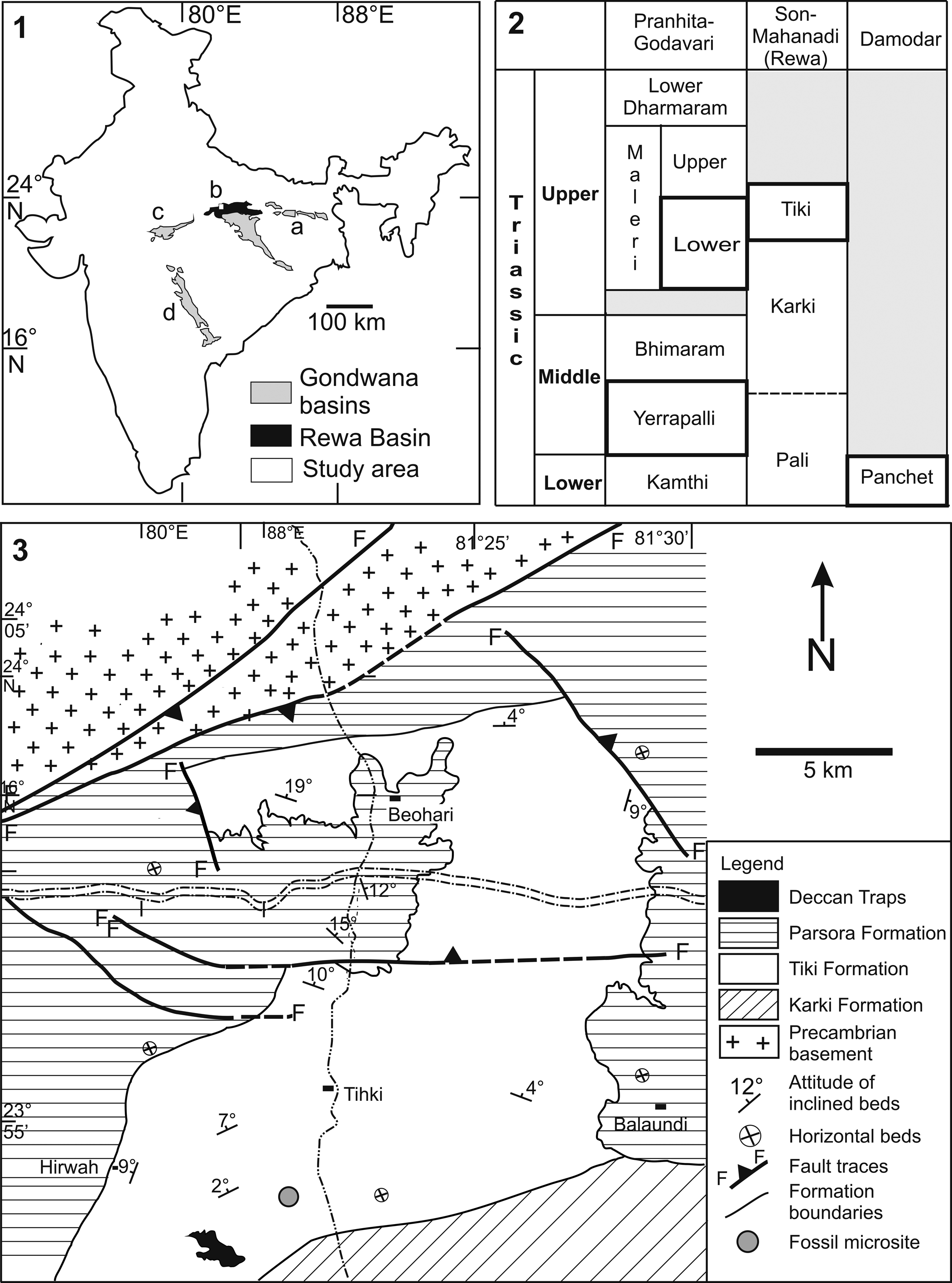
Non-mammalian cynodonts are known from the Triassic sediments of the various Gondwana basins of India (Fig. 1.1, 1.2). These included Thrinaxodon bengalensis Satsangi, Reference Satsangi1987 (Bandyopadhyay, Reference Bandyopadhyay, Calvo, Porfiri, Gonzalez Riga and Santos2011) and Panchetocynodon damodarensis Das and Gupta, Reference Das and Gupta2012 from the Lower Triassic Panchet Formation of the Damodar Basin, and indeterminate trirachodontid teeth from the Middle Triassic Yerrapalli Formation of the Pranhita-Godavari Basin (Bandyopadhyay and Sengupta, Reference Bandyopadhyay and Sengupta1999, Reference Bandyopadhyay, Sengupta, Harris, Lucas, Spielmann, Lockley, Milner and Kirkland2006; Bandyopadhyay and Ray, Reference Bandyopadhyay and Ray2020). The Late Triassic is represented by the traversodontid Exaeretodon statisticae Chatterjee, Reference Chatterjee1982 and isolated postcanine teeth of the chiniquodontid Deccanodon maleriensis Nath and Yadagiri, Reference Nath and Yadagiri2007 from the lower part of the Maleri Formation (Chatterjee, Reference Chatterjee1982; Nath and Yadagiri, Reference Nath and Yadagiri2007), and sectorial teeth of the dromatheriid Rewaconodon tikiensis Datta, Das, and Luo, Reference Datta, Das and Luo2004 from the Tiki Formation of the Rewa Basin (Datta et al., Reference Datta, Das and Luo2004). Ray (Reference Ray2015) described a large traversodontid Ruberodon roychowdhurii from the Tiki Formation based on an ontogenetic series of five nearly complete and partial lower jaws. In addition, the Tiki Formation has yielded isolated teeth of two mammaliaforms. The first is Gondwanadon tapani Datta and Das, Reference Datta and Das1996, which has a triconodont-like crown in its lower molar, and has been assigned to the Morganucodonta (Datta and Das, Reference Datta and Das1996). The second is Tikitherium, which has a transversely expanded upper molar (Datta, Reference Datta2005). By its similarity to the upper molar of docodonts, Tikitherium has been hypothesized to be more closely related to the Docodonta (Luo and Martin, Reference Luo and Martin2007; Abdala and Gaetano, Reference Abdala, Gaetano and Tanner2018; Panciroli et al., Reference Panciroli, Benson and Luo2019; Zhou et al., Reference Zhou, Bhullar, Neander, Martin and Luo2019) than to other mammaliaform clades. Assuming this hypothesis, Tikitherium would be a significant data point for the earliest possible time for the split of mammaliaform groups.
(1) Major Gondwana basins of peninsular India—Damodar Basin (a), Son-Mahanadi Basin (b), Satpura Basin (c), Pranhita-Godavari Basin (d). (2) Generalized Triassic stratigraphy of the Gondwana basins (after Bandyopadhyay and Ray, Reference Bandyopadhyay and Ray2020); cynodont-bearing horizons are marked in bold. (3) Geological map of the study area showing the fossil microsite (after Bhat et al., Reference Bhat, Ray and Datta2018a).

The Tiki Formation of India, which is exceptionally rich in the Late Triassic vertebrate fossils (Ray et al., Reference Ray, Bhat, Mukherjee and Datta2016), is a fluvial succession (~400 m in thickness) and comprises red-dominated mudstones with color mottles, paleosols, sheet and channel-fill sandstones, and caliche-derived, cross-stratified peloidal calcirudite horizons. The depositional environment of the Tiki Formation is interpreted to be an extensive floodplain drained by large river systems (Mukherjee et al., Reference Mukherjee, Ray, Chandra, Pal and Bandyopadhyay2012). Based on the fossil fauna and flora, the Tiki Formation is correlated with several Late Triassic horizons, such as the Ischigualasto Formation of Argentina, the upper part of the Santa Maria Formation and the overlying lower Caturrita Formation of Brazil, and the Isalo II Beds of Madagascar, along with the lower Tecovas Formation of the Texas. The Tiki Formation ranges in age from late Carnian to early/middle Norian (Datta et al., Reference Datta, Ray and Bandyopadhyay2019a).
In an integrated study of the Tiki vertebrates, a rich fossil locality was recovered near the village of Tihki (23°56′N, 81°22′58″E; Fig. 1.3), which is the type locality of the Tiki Formation (Mukherjee et al., Reference Mukherjee, Ray, Chandra, Pal and Bandyopadhyay2012). The locality has a highly diverse bonebed of multiple fossil taxa, dominated by metoposaurids. This fauna has yielded not only large fossils but also numerous micro-vertebrates in isolated teeth and skeletal elements (Rakshit and Ray, Reference Rakshit and Ray2020). The bonebed resulted from two distinct biostratinomic modes. The first biostratinomic mode is of terrestrial and semi-aquatic animals of large body size that lived in the surrounding floodplain; this mode consisted of natural death, decomposition, and differential preburial modification of the animals. The second biostratinomic mode is by mass-death assemblage, formed from breaching of banks during flooding, and burial of fishes and unionid bivalves in high-energy deposition of sediments. The macrofossils included disarticulated skeletal remains of an undescribed metoposaurid (Rakshit and Ray, Reference Rakshit and Ray2020), a derived phytosaur (Datta et al., Reference Datta, Kumar and Ray2019b), the rhynchosaur Hyperodapedon tikiensis Mukherjee and Ray, Reference Mukherjee and Ray2014, along with numerous, large unionid bivalves, and the scroll coprolite Tikicopros triassicus Rakshit et al., Reference Rakshit, Bhat, Mukherjee and Ray2019. The vertebrate microfossils, on the other hand, included isolated teeth and skeletal remains of hybodontids, xenacanthids, dipnoans, actinopterygians, various archosauriforms including basal saurischians, and non-mammalian cynodonts (Ray et al., Reference Ray, Bhat, Mukherjee and Datta2016, Reference Ray, Bhat and Datta2019; Bhat, Reference Bhat2018; Bhat et al., Reference Bhat, Ray and Datta2018a, Reference Bhat, Ray and Dattab; Rakshit et al., Reference Rakshit, Bhat, Ray and Datta2018; Bhat and Ray, Reference Bhat and Ray2020). This assemblage has yielded several isolated teeth of the dromatheriid Rewaconodon tikiensis and well-preserved teeth and jaw remains of three new cynodont taxa, which are the focus of the present study. These new taxa are described and compared with other cynodonts recovered from coeval horizons, and their significance in terms of cynodont dental evolution and paleobiogeography is discussed.
Material and methods
Material
The collection comprises 10 well-preserved, isolated teeth and a partial dentary (IITKGPR583D). All the specimens are housed in the Vertebrate Paleontology Laboratory of the Department of Geology and Geophysics, Indian Institute of Technology, Kharagpur, India. The Tiki specimens are compared with the teeth of other valid non-mammalian cynodonts such as Dromatherium (Emmons, Reference Emmons1857), Microconodon (Osborn, Reference Osborn1886; Simpson, Reference Simpson1926a; Sues, Reference Sues2001; Heckert et al., Reference Heckert, Mitchell, Schneider and Olsen2012), Pseudotriconodon (Hahn et al., Reference Hahn, Lepage and Wouters1984), Gaumia (Hahn et al., Reference Hahn, Wild and Wouters1987), Tricuspes (Godefroit and Battail, Reference Godefroit and Battail1997), Meurthodon (Sigogneau-Russell and Hahn, Reference Sigogneau-Russell, Hahn, Fraser and Sues1994), Hahnia (Godefroit and Battail, Reference Godefroit and Battail1997), and Rewaconodon (Datta et al., Reference Datta, Das and Luo2004), and the Late Triassic and Early Jurassic morganucodontids Eozostrodon (Parrington, Reference Parrington1941), Morganucodon (Kühne, Reference Kühne1949), Erythrotherium (Crompton, Reference Crompton1964), Helvetiodon (Clemens, Reference Clemens1980), Hallautherium (Clemens, Reference Clemens1980), Gondwanadon (Datta and Das, Reference Datta and Das1996), Rosierodon (Debuysschere et al., Reference Debuysschere, Gheerbrant and Allain2015), and Polonodon (Sulej et al., Reference Sulej, Niedźwiedzki, Tałanda, Dróżdż and Hara2020).
Field techniques
The sites of microfossils are demarcated in the field by surface indicators of fossils such as fragmentary remains of vertebrates and abundant unionid shells. Bulk-sampling of nearly 8.6 tons of red mudstone was carried out following the procedures outlined by Cifelli et al. (Reference Cifelli, Madsen and Larson1996) and Bhat (Reference Bhat2017), screen-washed, and then hand-picked using fine brushes in a picking tray under stereomicroscope. A rich microfossil assemblage comprising ~1,800 isolated teeth and skeletal remains of varied vertebrates was recovered, which included the cynodont teeth under this study.
Scanning Electron Microscopy
SEM imaging of the specimens followed the standard procedures outlined by Reed (Reference Reed2005). The specimens were washed with distilled water and immersed in acetone. The isolated teeth were mounted on small aluminum metallic stubs with the help of carbon tape to reduce the charging effect. SEM investigation was carried out under Field Emission (FE)-SEM (ZEISS) AURIGA COMPACT at the Central Research Facility at the Indian Institute of Technology, Kharagpur, India.
Tooth morphology, orientation, and nomenclature
The terminology used to describe tooth and mandibular morphology follows Crompton (Reference Crompton1974), Kermack and Kermack (Reference Kermack and Kermack1984), Godefroit and Battail (Reference Godefroit and Battail1997), and Debuysschere et al. (Reference Debuysschere, Gheerbrant and Allain2015). The mesial or anterior side is towards the jaw symphysis, tip of the root is the apex of the tooth, with the gum-line marking the junction between root and crown (Kermack and Kermack, Reference Kermack and Kermack1984), whereas the dorsal side of the crown is the occlusal side. Nomenclature for details of tooth morphology follows Crompton and Jenkins (Reference Crompton and Jenkins1968), Pacey (Reference Pacey1978), Godefroit and Battail (Reference Godefroit and Battail1997), Clemens (Reference Clemens2011), Debuysschere et al. (Reference Debuysschere, Gheerbrant and Allain2015), Hendrickx et al. (Reference Hendrickx, Abdala and Choiniere2019). For the upper and lower jaw teeth, the capital and lower-case letters designate the cusps, respectively. The major cusp, which is usually aligned medially in the main row, is termed as A/a, whereas there are two cusps, B/b and C/c, on either side of the major cusp. Cusp B/b is on the mesial and cusp C/c is on the distal side of the cusp A/a. The distal cingular cusps are termed as D/d because these are positioned distally to C/c. The mesial cingular cusps are termed E/e, and these are the most mesial cusps on the lingual cingulum. The distolingual cingular cusp in relation to E/e is E′/e′, whereas E″/e″ is the distolingual cingular cusp in relation to E′/e′. F/f is the mesial cusp on the labial cingulum, whereas g is the basal lingual cingular cusp at the base of a and c. The latter is also known as the kühnecone (Parrington, Reference Parrington1967). I/i is the mesiolingual cingular cusp in relation to D/d. An unusual cingular cusp, i′, which is at the same height as i, is identified on the distal margin just below the cusp d.
All the examined teeth are postcanines because the crowns have three distinct linearly arranged cusps (sensu Rose, Reference Rose2006). However, the regional differentiation of the teeth into premolars and molars is not possible because these teeth were found isolated. The crowns with more major and cingular cusps are considered posterior in position as in other carnivorous or insectivorous non-mammalian cynodonts where morphological complexity increases posteriorly (Osborn and Crompton, Reference Osborn and Crompton1973). The isolated teeth are oriented as suggested by Godefroit and Battail (Reference Godefroit and Battail1997), Datta et al. (Reference Datta, Das and Luo2004), and Sulej et al. (Reference Sulej, Niedźwiedzki, Tałanda, Dróżdż and Hara2020), where the more convex side is regarded as the labial side for both upper and lower jaw teeth. The upper postcanine teeth have a continuous cingulum around the crown, whereas in the lower tooth, the labial cingulum is absent (Kielan-Jaworowska et al., Reference Kielan-Jaworowska, Cifelli and Luo2004). Inclination of the cusps is towards the distal/posterior side. In addition, accessory cusps are numerous or better developed on the distal side of the crown. The number of major, accessory, and cingular cusps was determined by comparisons with those in the lower jaw of Alemoatherium (Martinelli et al., Reference Martinelli, Eltink, Da-Rosa and Langer2017, p. 406, fig. 3A). The relative positions of the postcanine teeth are determined based on complete jaws of Microconodon tenurostris Osborn, Reference Osborn1886 (Sues, Reference Sues2001, p. 39, fig. 1), Brasilitherium riograndensis Bonaparte et al., Reference Bonaparte, Martinelli, Schultz and Rubert2003 (Bonaparte et al., Reference Bonaparte, Martinelli, Schultz and Rubert2003, fig. 15) and Alemoatherium huebneri Martinelli et al., Reference Martinelli, Eltink, Da-Rosa and Langer2017 (Martinelli et al., Reference Martinelli, Eltink, Da-Rosa and Langer2017, fig. 3A). Wear facets developed on the lingual side of the upper tooth and labial side of the lower tooth, and are usually found in the adults or in individuals that have stopped tooth replacement (Bonaparte et al., Reference Bonaparte, Martinelli, Schultz and Rubert2003, Reference Bonaparte, Martinelli and Schultz2005).
Measured parameters
CH, crown height; CL, crown length; CW, crown width along the major cusp a; RH, root height.
Repository and institutional abbreviation
IITKGP, Indian Institute of Technology, Kharagpur, India.
Systematic paleontology
Order Therapsida Broom, Reference Broom1905
Suborder Cynodontia Owen, Reference Owen1861
Infraorder Eucynodontia Kemp, Reference Kemp1982
Family Dromatheriidae Gill, Reference Gill1872
Genus Rewaconodon Datta, Das, and Luo, Reference Datta, Das and Luo2004
Type species
Rewaconodon tikiensis Datta, Das, and Luo, Reference Datta, Das and Luo2004 from the Upper Triassic Tiki Formation of the Rewa Gondwana Basin, India.
Generic diagnosis
Labiolingually compressed, triconodont-like crown, three to four cusps aligned in a longitudinal row, incipient division of the root by a shallow groove on both the lingual and labial sides, occlusal wear between the main cusp and the two accessory cusps (after Datta et al., Reference Datta, Das and Luo2004).
Rewaconodon indicus new species
Figures 2, 3
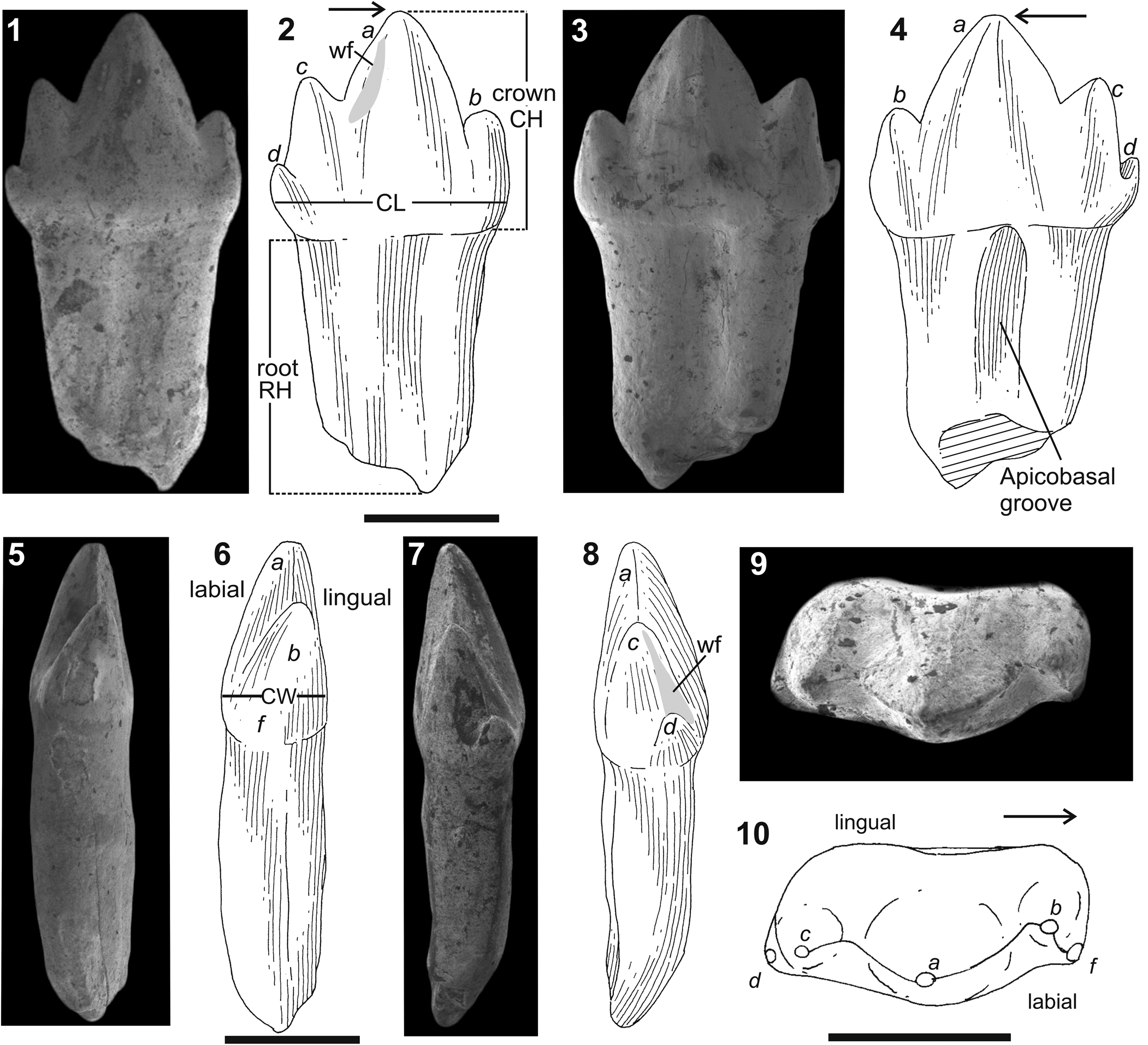
Rewaconodon indicus n. sp. Holotype, IITKGPR583C, a right lower postcanine tooth in (1, 2), labial, (3, 4), lingual, (5, 6), mesial, (7, 8), distal, (9, 10), occlusal views. Letters in italics correspond to cusp nomenclature. Abbreviations: CH, crown height; CL, crown length; CW, crown width; RH, root length; wf, wear facet. Arrows indicate anterior/mesial direction. Scale bars represent 0.5 mm.

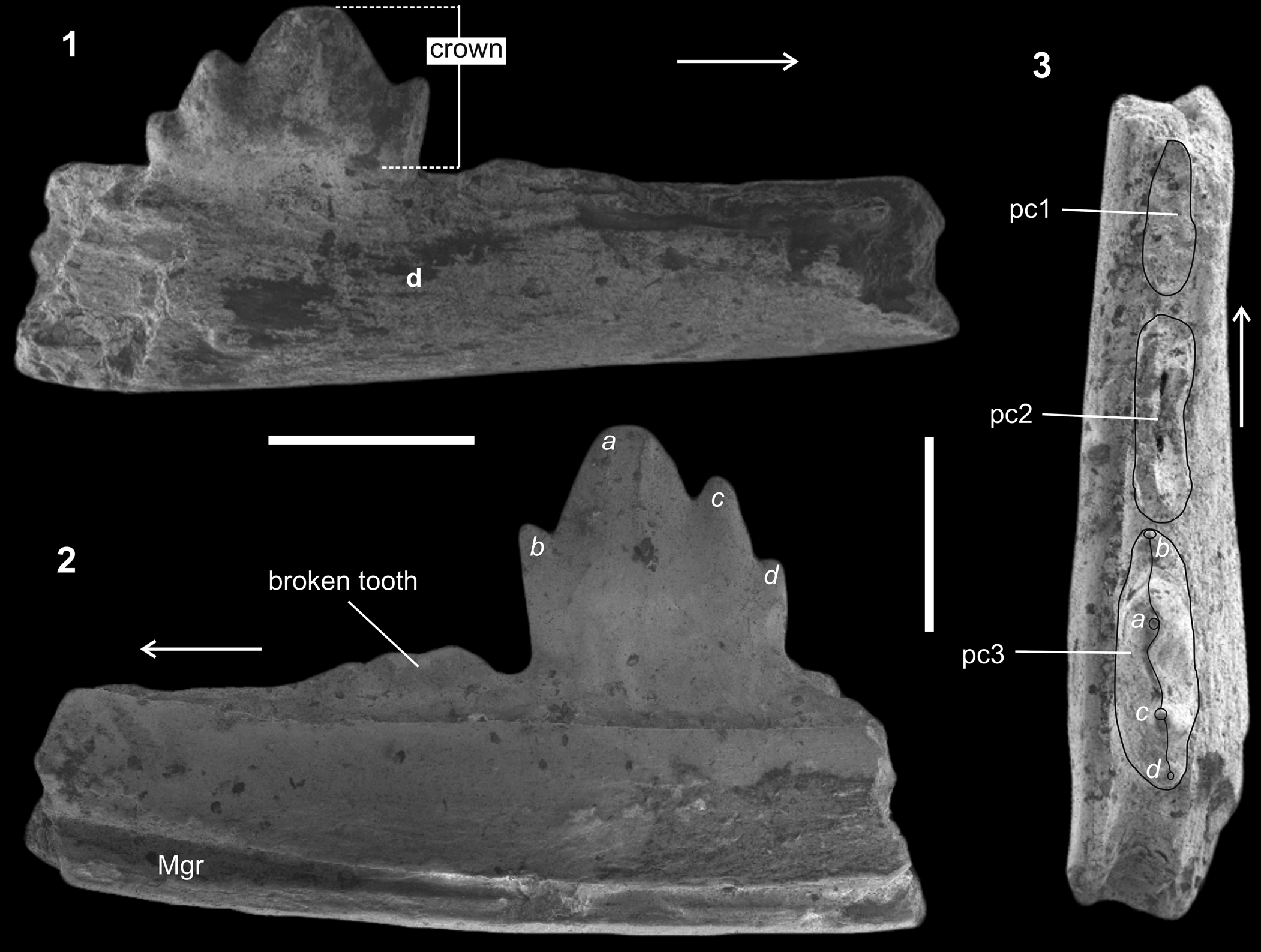
Rewaconodon indicus n. sp. IITKGPR583D, a partial right dentary in (1) lateral, (2) medial, and (3) occlusal views. Letters in italics correspond to cusp nomenclature. Abbreviation: d, dentary; Mgr, Meckelian groove; pc, postcanine tooth. Arrows indicate anterior/mesial direction. Scale bars represent 0.5 mm.

Holotype
IITKGPR583C, a right lower postcanine tooth.
Differential diagnosis
Tri- or tetra-cuspid asymmetrical crown; long mesial edge of major cusp a; asymmetrical mesial and distal edges; a > c > b > d; a small conical cingular cusp f; marked constriction at crown-root junction; crown-root length ratio <1. These teeth are differentiated from those of R. tikiensis in showing a more distinctive constriction between the crown and root, presence of an accessory cusp d placed distally with respect to c, and a mesial cingular cusp f at the crown-root junction.
Occurrence
Tiki Formation of the Rewa Gondwana Basin (Late Triassic); near the village of Tihki (23°56′N, 81°22′58″E), Shahdol District, Madhya Pradesh, India.
Description
The species is described based on three postcanine teeth, including the holotype, IITKGPR583C (Fig. 2.1–2.10), and a partial right dentary (Fig. 3.1–3.3). The holotype has a long (CH/CL = 1.12) and asymmetrical crown, which is tetracuspid with a principal cusp (a), three accessory cusps (b, c, d), and a cingular cusp (f) aligned mesiodistally (Fig. 2.1, 2.2). The principal cusp (a) and accessory cusps (b, c, d) are pointed, straight, and upright (Fig. 2.1, 2.2). The main cusp (a) is asymmetrical in labial/lingual views, with its anterior/mesial margin much longer than the posterior/distal margin (Fig. 2.1, 2.2). Distally, the crown bears two cusps, c and d (Fig. 2.2, 2.4), of which cusp d is smaller than cusp b. Cusp d is positioned in the distolabial corner of the crown and slightly offset from the mesiodistal axis (Fig. 2.7, 2.8). Cingular cusp f is present on the labial margin just below cusp b (Fig. 2.5, 2.6) and is smaller than cusp d (Fig. 2.9, 2.10). Labially, the crown bears a distinctly vertical, U-shaped wear facet on the distal face of major cusp a (Fig. 2.2), whereas lingually, small v-shaped depressions are visible on the mesial edges of cusps c and d (Fig. 2.4). The latter appears to be an artifact and not a wear facet. The overall arrangement of the main cusps is a > c > b and that of the cingular cusps is d > f. The occlusal surface of the tooth is arched labially (Fig. 2.9, 2.10) and results in a sharp cutting edge. There is a marked constriction between the crown and root on both labial and lingual surfaces (Fig. 2.1–2.8). The root is long in comparison to the crown, where the crown-root ratio is 0.93:1. Both the labial and lingual views (Fig. 2.1–2.4) show a wide central longitudinal groove on the root, which is more pronounced on the lingual face (Fig. 2.3, 2.4) and suggests incipient root bifurcation. In other specimens examined (IITKGPR581A, IITKGPR582C), there are four mesiodistally aligned cusps with a slight displacement of the principal cusp a labially. These are more posterior in position with respect to the holotype (IITKGPR583C) as the crown complexity increases posteriorly.
The taxon also includes a partial right dentary (IITKGPR583D; Fig. 3.1–3.3) bearing three teeth with a triconodont pattern. The dentary is robust, and its labial surface is convex and deep (Fig. 3.1), and the deepest part of the dentary is just below pc3 (Fig. 3.2). The lingual surface of the dentary shows the Meckelian groove (Fig. 3.2), which extends horizontally along the body of the mandible, parallel and close to the ventral margin of the dentary. The margin of the Meckelian groove is well defined and the groove as a whole is very distinct. The preserved teeth are aligned mesiodistally, and show no offset with respect to one another. The postcanine (pc3) tooth has four cusps aligned horizontally along the length of the bone (Fig. 3.1–3.3). Cusp a is larger and slightly tilted distally. The mesial margin of cusp a is long and twice the height of the distal margin. Cusp b is lower than cusp c, which is followed by a small cusp d. The overall arrangement of the cusps is a > c > b, as on the isolated holotype tooth, IITKGPR583C (Fig. 2).
Etymology
The species is named after India.
Materials
Two isolated partial lower left postcanine teeth (IITKGPR581A, IITKGPR582C), and a partial right dentary (IITKGPR583D) with three postcanine teeth, of which one tooth is complete.
Remarks
Datta et al. (Reference Datta, Das and Luo2004) reported several postcanine teeth characterized by labiolingually compressed triconodont-like crowns with three or four cusps, no cingulum or constriction at the crown-root junction, and incipient root subdivision. These teeth were assigned to the new dromatheriid taxon Rewaconodon tikiensis (Datta et al., Reference Datta, Das and Luo2004). In the current study, three teeth with triconodont-like asymmetrical crowns are described, which are characterized by four distinct cusps, where the main cusp a has a long mesial edge, and are assigned to the genus Rewaconodon. These teeth are differentiated from those of R. tikiensis in showing a more distinctive constriction between the crown and root, presence of an accessory cusp d placed distally with respect to c, and a mesial cingular cusp f at the crown-root junction. Hence, these Tiki specimens are assigned to a new species of Rewaconodon, R. indicus n. sp.
Genus Inditherium new genus
Type species
Inditherium floris n. gen. n. sp., from the Upper Triassic Tiki Formation of the Rewa Gondwana Basin, Shahdol District, Madhya Pradesh, India.
Diagnosis
As for the type and only species.
Etymology
The genus name is after India and the Latin word ‘therium’ derived from Greek word ‘therion’ meaning beast.
Holotype
IITKGPR584A, a right lower postcanine tooth.
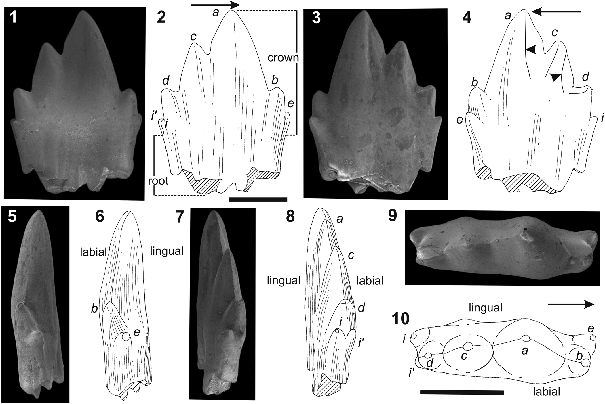
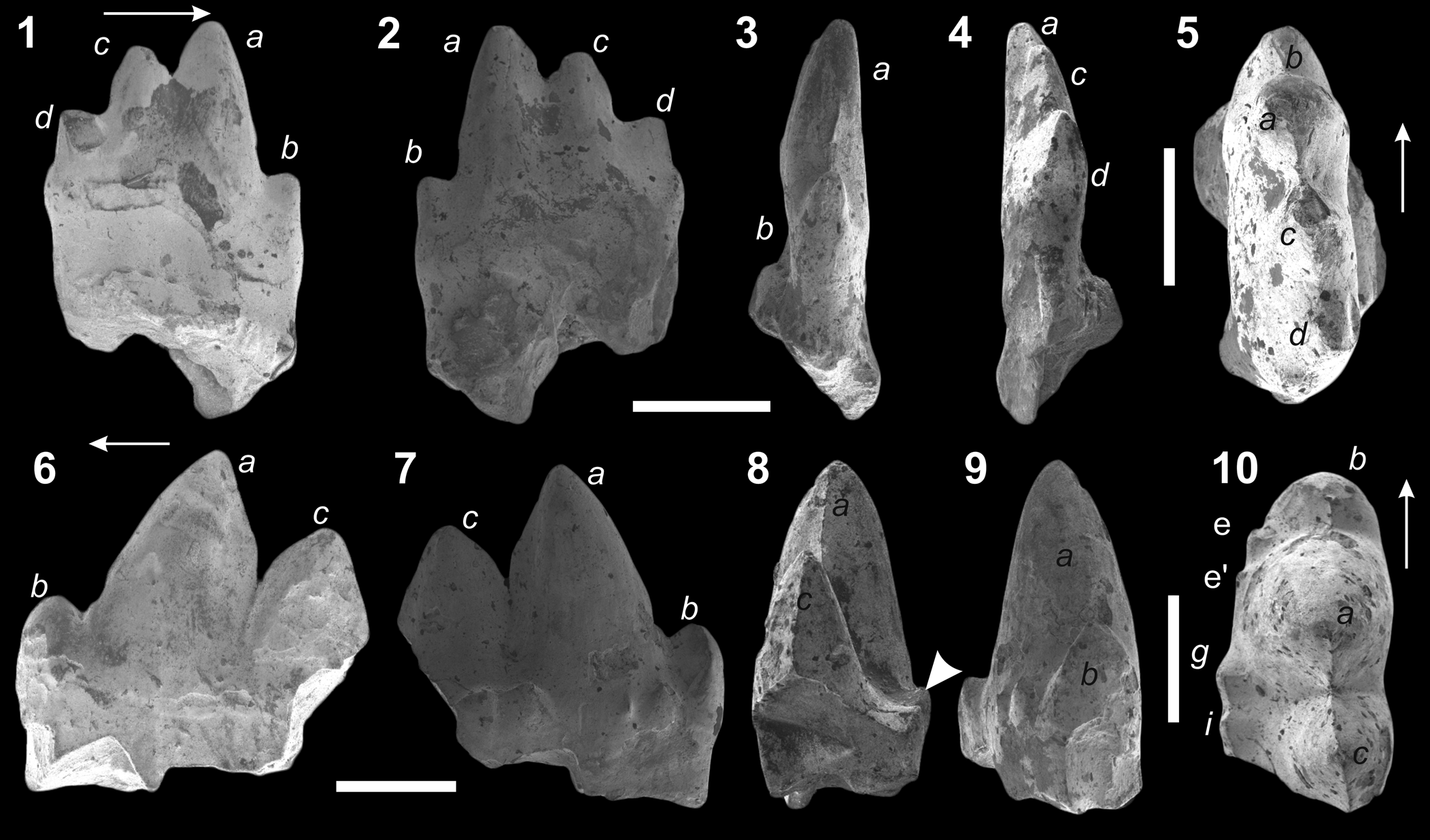
Inditherium floris n. gen. n. sp. Holotype, IITKGPR584A, a right lower postcanine tooth in (1, 2), labial, (3, 4), lingual, (5, 6), mesial, (7, 8), distal, (9, 10), occlusal views. Letters in italics correspond to cusp nomenclature. Arrows and arrowheads indicate anterior/mesial direction and a deep wedge between the cusps (a and c, c and d, respectively). Scale bars represent 0.5 mm.

Inditherium floris n. gen. n. sp. Partial lower postcanine teeth in (1, 2), labial, (3, 4), lingual, (5, 6), mesial, (7, 8), distal, (9, 10), occlusal views. (1–5) IITKGPR584B, right lower; (6–10) IITKGPR584C, left lower. Letters in italics correspond to cusp nomenclature. Arrows and an arrowhead indicate anterior/mesial direction and a distinct lingual cingulum, respectively. Scale bars represent 0.5 mm.

Diagnosis
Broad crown (CL/CH = 1); flower-shaped; tetra- or pentacuspid; diverging cusps; concave lingual crown face; long mesial edge of major cusp a; a >c >> b > d > e; small cingular cusps g and i; unusual cingular cusp i′; faint wear facets between cusps; weak lingual cingulum.
Occurrence
Tiki Formation of the Rewa Gondwana Basin (Late Triassic); near the village of Tihki (23°56′N, 81°22′58″E), Shahdol District, Madhya Pradesh, India.
Description
The description is based on three specimens (IITKGPR584A–C), including the holotype (IITKGPR548A; Fig. 4.1–4.10), where the crowns are well preserved but the roots are broken. The crowns are broad (CH/CL = 1), flower-shaped, with diverging and pointed cusps. The major cusp a is the highest and reaches twice the height of cusp b. The mesiodistal length of the primary cusp a is almost half the crown length. In the holotype (IITKGPR584A), cusps b and d are nearly at the same level and positioned on the extreme mesial and distal margins, respectively (Fig. 4.1, 4.2). Two cingular cusps, e and i (Pacey, Reference Pacey1978), are present on the distal margin at the same height (Fig. 4.3, 4.4). A small unusual cingular cusp i′ (Fig. 4.1, 4.2, 4.7–4.10) is present labially at the same height as cusp i (Fig. 4.1–4.4). A long vertical groove visible on the cusp a lingually appears to be an artifact and not a wear facet because it is not visible on the occlusal side (Fig. 4.9, 4.10). In occlusal view (Fig. 4.9, 4.10), the crowns are labiolingually compressed (CL/CW = 3.25) and cusps a, c, and d are aligned in a straight line, although cusp a is slightly offset and aligned lingually. In addition, cusp b is offset from the main crest and positioned more labially (Fig. 4.9, 4.10).
Another specimen (IITKGPR584B; Fig. 5.1–5.5) is a right lower postcanine tooth with the crown dominated by four diverging cusps a–d. As in the holotype, the crown is labiolingually compressed. In occlusal view, cusps a, c, and d are aligned in a straight line, with cusp b being slightly offset labially (Fig. 5.5). The crown has a partially preserved root with a fragment of alveolar bone attached labially (Fig. 5.3, 5.4). There is another partial tooth in the collection, IITKGPR584C (Fig. 5.6–5.10), which exhibits three prominent cusps, a, b, c; the portion containing d is missing. The major cusp a is distally inclined and, as in other specimens, has a long anterior/mesial edge (Fig. 5.6, 5.7). Triangular, mesiodistally oriented wear facets are present on the mesial edge of cusp b (Fig. 5.10). The tooth is characterized by a prominent lingual cingulum that bears multiple cingular cusps (Fig. 5.8–5.10). The cingular cusp e and e′ are present mesiolingually just below cusp b (Fig. 5.10), whereas the other two cingular cusps g (‘Kühnecone’, sensu Parrington, Reference Parrington1967) and i are positioned between the bases of cusp a and c (Fig. 5.10). In occlusal view, the major and cingular cusps are separately aligned (Fig. 5.10), which results in a distinct valley between them. The root is partially present in IITKGPR584B (Fig. 5.1, 5.2), and shows a weak constriction between the root and the crown.
Etymology
Species name after the flower-shaped crown of the holotype specimen.
Materials
IITKGPR584B–IITKGPR584D, three partial postcanine teeth, without roots.
Remarks
The tooth crown of the new taxon is broad and bears four or five diverging cusps, giving it a distinct flower-like shape. Such a crown is not present in the two species of Rewaconodon (R. tikiensis Datta et al., Reference Datta, Das and Luo2004 and R. indicus n. sp.), the crowns of which are essentially triconodont with upright, non-divergent cusps. Moreover, specimens IITKGPR584A and IITKGPR584C are characterized by a distinct lingual cingulum, which is absent in the two species of Rewaconodon, although R. indicus n. sp. shares a marked constriction between the crown and root. In addition, multiple cingular cusps (e, e′, g, i, i′) are evident in the examined specimens. Hence, based on the crown characteristics, which included crown proportions, diverging cusps, multiple cingular cusps, and a lingual cingulum, specimens IITKGPR584A–C differ from those of Rewaconodon and are assigned to a new taxon, namely Inditherium floris n. gen. n. sp.
Mammaliamorpha Rowe, Reference Rowe1988
Family incertae sedis
Genus Tikiodon new genus
Type species
Tikiodon cromptoni n. gen. n. sp., from the Upper Triassic Tiki Formation of the Rewa Gondwana Basin, Shahdol District, Madhya Pradesh, India.
Diagnosis
As for type and only species.
Etymology
The genus is named after Tiki Formation of India and Greek word ‘odon’ meaning tooth.
Holotype and only known specimen
IITKGPM1, a left lower postcanine tooth.
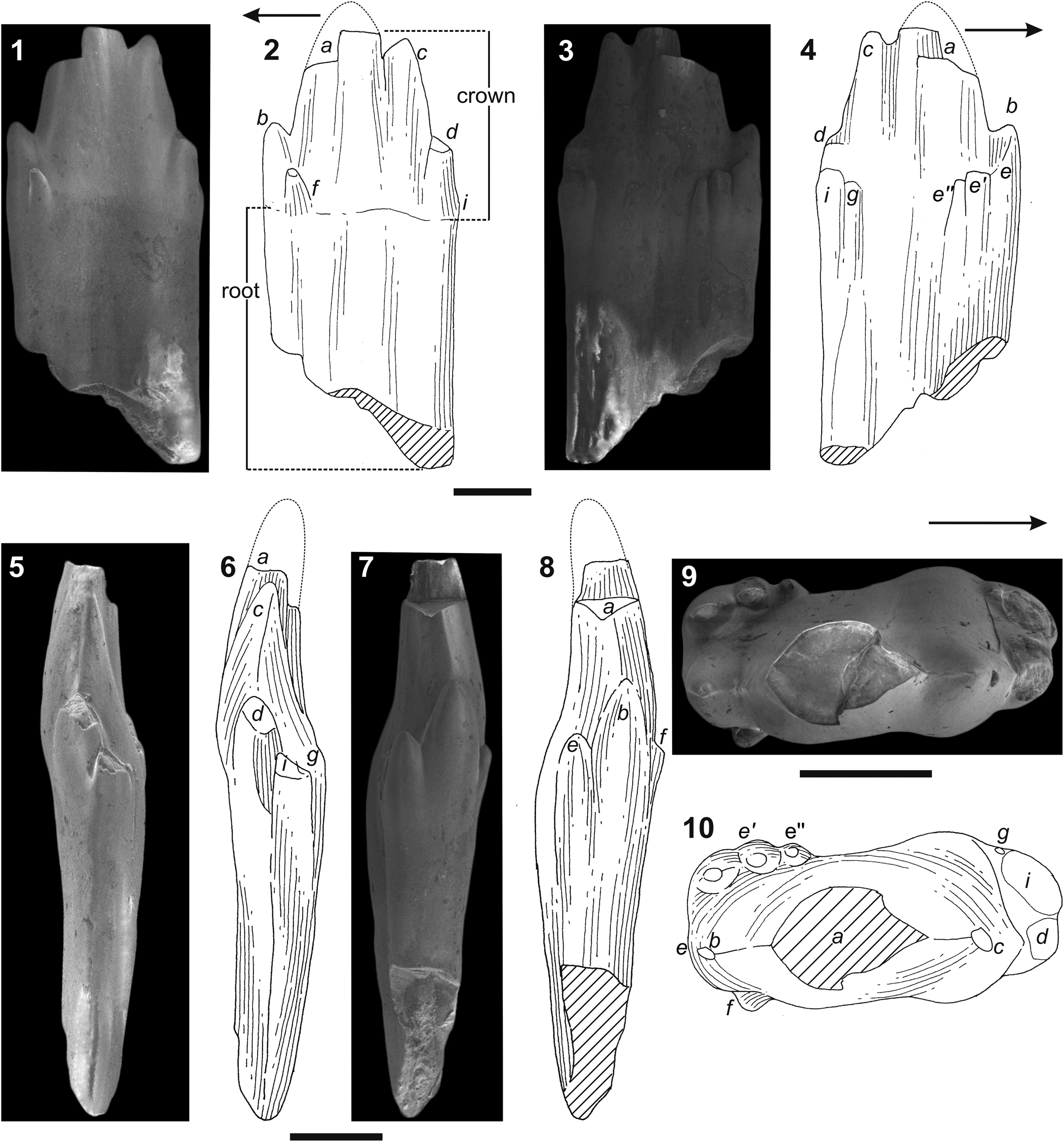
Tikiodon cromptoni n. gen. n. sp. Holotype, IITKGPM1, a lower left postcanine tooth in (1, 2), buccal/labial, (3, 4), lingual, (5, 6), distal, (7, 8), mesial, (9, 10), occlusal views. Letters in italics correspond to cusp nomenclature. Arrows indicate anterior/mesial direction. Scale bar represents 0.5 mm.

Diagnosis
A small mammaliamorph characterized by a unique combination of following features: shovel-shaped crown; multicuspid with closely spaced cusps; quadrangular in occlusal view; cusp formula a > c >> b, small distal cingular cusp d in line with principal and accessory cusps, weakly developed labial and lingual cingula; three cingular cusps e, e′, and e″ arranged mesiodistally, cingular cusp f present mesially, cingular cusp g at the base of cusp c, cingular cusp i present distolingually, crown-root ratio <1; incipient root bifurcation with a deep, wide, and median groove in labial and lingual views.
Occurrence
Tiki Formation of the Rewa Gondwana Basin (Late Triassic); near the village of Tihki (23°56′N, 81°22′58″E), Shahdol District, Madhya Pradesh, India.
Description
The taxon is described based on the only specimen, the holotype, IITKGPM1 (Fig. 6.1–6.10). It has three main closely spaced, upright cusps (a, b, and c) and a cingular cusp d, aligned along a straight line mesiodistally (Fig. 6.1–6.4). The apices of these cusps are slightly tilted lingually, resulting in a shovel-like crown (Fig. 6.5–6.8). Cusp a, albeit broken, is large and wider than cusp c, which is the second cusp in length and width (Fig. 6.1–6.4). The mesial length of cusp a is longer than the distal length (Fig. 6.1–6.4). In occlusal view, the crown is sub-quadrangular, wide (CL/CW = 2.5), with the apex of major cusp a centrally positioned (Fig. 6.9, 6.10), and the smallest cusp b and the cingular cusp d are at the same level (Fig. 6.1–6.4). On the lingual cingulum, three small cingular cusps e, e′, and e″, are arranged mesiodistally (Fig. 6.3, 6.4) and are of the same height as cusp g (‘Kühnecone’; Fig. 6.3, 6.4), which is positioned distally at the base of primary cusp c. Cusp i (Pacey, Reference Pacey1978) is at the extreme position of the distal margin (Fig. 6.5, 6.6) in relation to cusp d. On the labial face (Fig. 6.1, 6.2), a small and conical cusp f is positioned at the base of cusp a and b (Fig. 6.9, 6.10). The apices of the major and accessory cusps are abraded (Fig. 6.5–6.10). On the primary cusp a, the wear facet is present on the distalolabial margin, and in the accessory cusps c and d, there are mesially oriented, faint wear facets. The slopes of the mesial and distal margins are vertical and straight (Fig. 6.5–6.8). The root of the crown is flattened labiolingually and twice the height of major cusp a. The root of the holotype (IITKGPM1) has a wide and deep sulcus on labial and lingual views, but is not fully bifurcated (Fig. 6.1–6.8).
Etymology
Species name is after A.W. Crompton for his contribution to the study of early mammals.
Remarks
The specimen has a shovel-liked crown with weak but distinct labial and lingual cingula bearing multiple cingular cusps. This derived advanced crown morphology is combined with a long, undivided root (CH/RH = 0.88), the latter being characterized by a distinct median apicobasal groove in the labial and lingual views. Such a feature is also noted in a postcanine tooth reported from the Upper Triassic of Germany (Lucas et al., Reference Lucas, Heckert, Harris, Seegis and Wild2001). Any further loss of the thin layer between mesial and distal margin would transform these teeth into true ‘morganucodontid’ morphology (Kielan-Jaworowska et al., Reference Kielan-Jaworowska, Cifelli and Luo2004). A combination of derived crown and basal root morphology makes it difficult to assign the Tiki specimen to a particular clade. Compared to other Late Triassic forms, such as Rewaconodon tikiensis (Datta et al., Reference Datta, Das and Luo2004, p. 76, fig. 2A–E), Rewaconodon indicus n. sp. (current study), Alemoatherium huebneri (Martinelli et al., Reference Martinelli, Eltink, Da-Rosa and Langer2017, p. 410, fig. 6A–O), and Polonodon woznikiensis Sulej et al., Reference Sulej, Niedźwiedzki, Tałanda, Dróżdż and Hara2020 (Sulej et al., Reference Sulej, Niedźwiedzki, Tałanda, Dróżdż and Hara2020, p. 85, fig. 4K–M,), the Tiki specimen (IITKGPM1) has a multi-cuspid shovel-shaped crown. As a result, the specimen is assigned to Tikiodon cromptoni n. gen. n. sp., and placed within Mammaliamorpha (Rowe, Reference Rowe1988).
Discussion
The Indian dromatheriids and other taxa
The dromatheriids are small- to medium-sized carnivorous, Late Triassic sectorial-toothed non-mammalian cynodonts with triconodont-like postcanines (Abdala, Reference Abdala2019) and are known from Carnian–Norian strata of the eastern United States (Emmons, Reference Emmons1857; Sues et al., Reference Sues, Olsen, Kroehler, Fraser and Sues1994; Sues, Reference Sues2001), central and western Europe (Godefroit and Battail, Reference Godefroit and Battail1997; Kielan-Jaworowska et al., Reference Kielan-Jaworowska, Cifelli and Luo2004), and India (Datta et al., Reference Datta, Das and Luo2004). The dromatheriids are mostly represented by isolated teeth and partial jaw fragments. Although their phylogenetic position is problematic, they are considered derived cynodonts (Hopson and Kitching, Reference Hopson and Kitching1972; Sues, Reference Sues2001), and are currently represented by the genera Dromatherium, Microconodon, Pseudotriconodon, Tricuspes, Meurthodon, Rewaconodon, and Polonodon (Emmons, Reference Emmons1857; Hahn et al., Reference Hahn, Lepage and Wouters1984; Sues et al., Reference Sues, Olsen, Kroehler, Fraser and Sues1994; Godefroit and Battail, Reference Godefroit and Battail1997; Datta et al., Reference Datta, Das and Luo2004; Heckert et al., Reference Heckert, Mitchell, Schneider and Olsen2012; Sulej et al., Reference Sulej, Niedźwiedzki, Tałanda, Dróżdż and Hara2020).
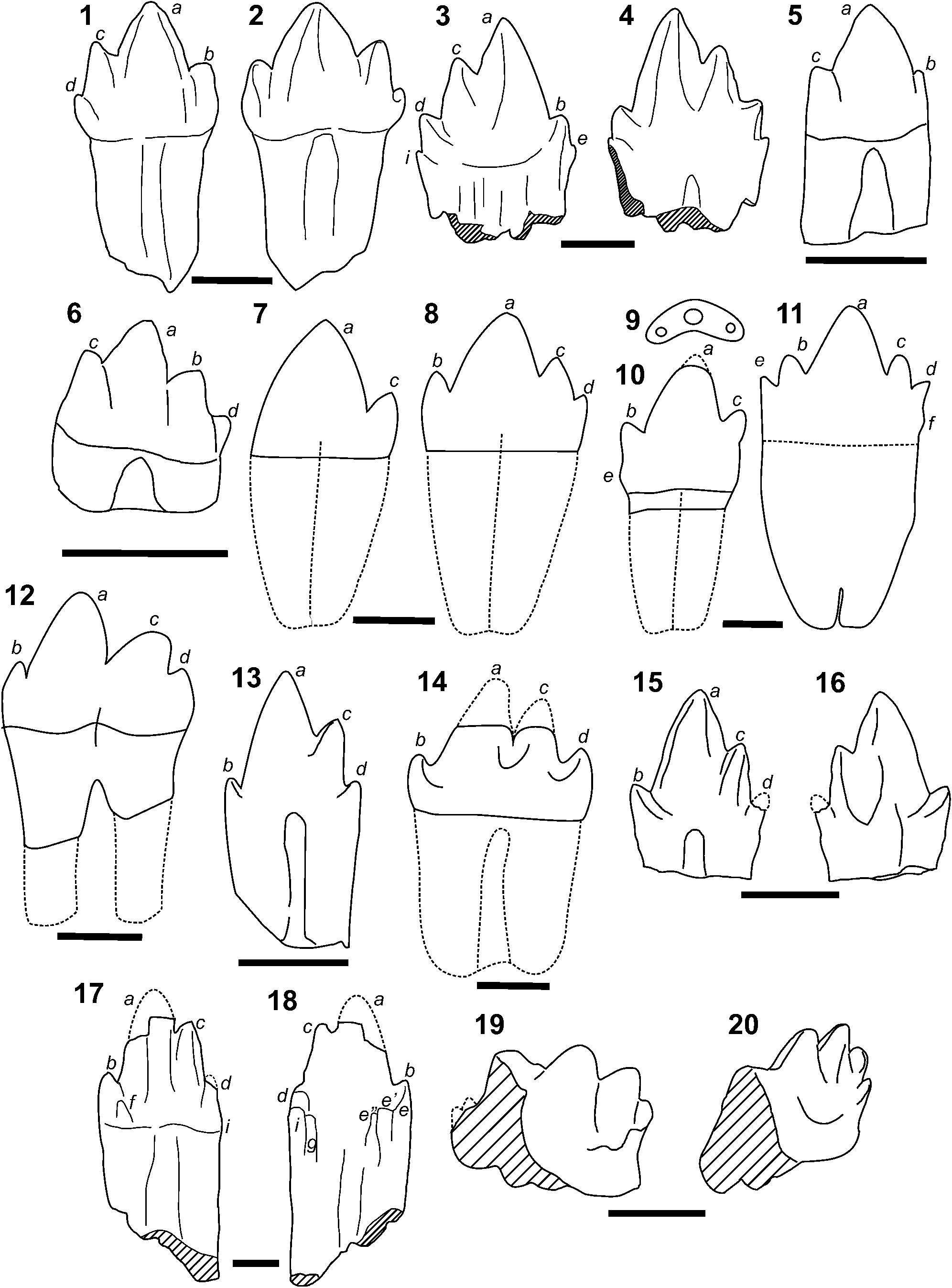
The specimens of the new taxa Rewaconodon indicus n. sp. (IITKGPR581A, IITKGPR582C, IITKGPR583C–D; Fig. 7.1, 7.2) and Inditherium floris n. gen. n. sp. (IITKGPR584A–D, Fig. 7.3, 7.4) are considered dromatheriids based on sharp-edged, tetra- and penta-cuspid crowns with the cusps arranged linearly and an undivided root. Specimens IITKGPR581A, IITKGPR582C, and IITKGPR583C–D are assigned to the genus Rewaconodon (R. tikiensis Datta et al., Reference Datta, Das and Luo2004) based on morphological similarity in terms of labiolingually compressed crown, mesiodistal alignment of the three main cusps, and incipient root bifurcation (Fig. 7.5, 7.6). However, these new specimens (IITKGPR581A, IITKGPR582C, IITKGPR583C–D) are distinct from R. tikiensis Datta et al., Reference Datta, Das and Luo2004 in having a broad, tetracuspid crown (Fig. 2.1–2.10), distal cusp c higher than mesial cusp b, cusp formula of a > c > b rather than a > b > c of R. tikiensis Datta et al., Reference Datta, Das and Luo2004, and a marked constriction between the crown and the root. Based on these features, the specimens are assigned to a new species of Rewaconodon, R. indicus n. sp. It is to be noted that the new species possibly developed tetra- or pentacuspid crowns, suggesting positional variations of the teeth (Osborn and Crompton, Reference Osborn and Crompton1973), as seen in Pseudotriconodon and Microconodon (Godefroit and Battail, Reference Godefroit and Battail1997; Sues, Reference Sues2001).
Lower postcanine tooth of different Triassic cynodont genera for comparison. (1, 2) Rewaconodon indicus n. sp.; (3, 4) Inditherium floris n. gen. n. sp.; (5, 6) R. tikiensis; (7) Dromatherium; (8) Microconodon; (9, 10) Tricuspes; (11) Pseudotriconodon; (12) Meurthodon; (13) Polonodon; (14) Therioherpeton; (15, 16) Alemoatherium; (17, 18) Tikiodon cromptoni n. gen. n. sp.; (19, 20) Gondwanadon tapani. Sources of information: Osborn (Reference Osborn1886, Reference Osborn1887); Bonaparte and Barberena (Reference Bonaparte and Barberena1975); Lucas and Oakes (Reference Lucas and Oakes1988); Sigogneau-Russell and Hahn (Reference Sigogneau-Russell, Hahn, Fraser and Sues1994); Datta and Das (Reference Datta and Das1996); Godefroit and Battail (Reference Godefroit and Battail1997); Sues (Reference Sues2001); Datta et al. (Reference Datta, Das and Luo2004); Cuny (Reference Cuny2004); Martinelli et al. (Reference Martinelli, Eltink, Da-Rosa and Langer2017); Sulej et al. (Reference Sulej, Niedźwiedzki, Tałanda, Dróżdż and Hara2020). Letters in italics correspond to cusp nomenclature. All teeth have been brought to the same crown height for comparative purpose. Scale bars represent 1 mm.

The preserved mandibular ramus of R. indicus n. sp. is robust and low, as in Dromatherium (Emmons, Reference Emmons1857) and Microconodon (Sues, Reference Sues2001, p. 40, fig. 3). The teeth of Rewaconodon indicus n. sp. resemble the postcanines of Dromatherium (Emmons, Reference Emmons1857), Microconodon (Osborn, Reference Osborn1886; Sues, Reference Sues2001; Heckert et al., Reference Heckert, Mitchell, Schneider and Olsen2012), Tricuspes (Godefroit and Battail, Reference Godefroit and Battail1997), Pseudotriconodon (Hahn et al., Reference Hahn, Lepage and Wouters1984), Meurthodon (Sigogneau-Russell and Hahn, Reference Sigogneau-Russell, Hahn, Fraser and Sues1994), and Polonodon (Sulej et al., Reference Sulej, Niedźwiedzki, Tałanda, Dróżdż and Hara2020) in: (1) triconodont-like crown, (2) three main cusps situated in a mesodistally elongated line, (3) narrow crown (CH ≥ CL) with more or less parallel sides, and (4) incipient division of root labially and lingually.
However, Rewaconodon indicus n. sp. is markedly different from these taxa based on crown morphology and cusp proportions. The postcanine teeth of Dromatherium (Emmons, Reference Emmons1857; Fig. 7.7) have a highly pointed bicuspid crown, where a > > c and cuspules may be present. The teeth of Microconodon (Fig. 7.8) have shallow sulci extending the entire apicobasal height of the root (Osborn, Reference Osborn1886; Simpson, Reference Simpson1926b; Sues, Reference Sues2001; Heckert et al., Reference Heckert, Mitchell, Schneider and Olsen2012), but lack the more pronounced constriction on the Tiki teeth. In addition, the tooth crowns of Microconodon are compressed labiolingually, nearly symmetrical with b and c at more or less equal height. On the other hand, the cusps of Tricuspes (Fig. 7.9, 7.10) are arranged in a marked V in occlusal view, on a nearly symmetrical crown where a > b = c. There is a marked constriction at the crown-root junction, although the rest of the root is broken (Godefroit and Battail, Reference Godefroit and Battail1997, p. 576, fig. 6B). The teeth of Pseudotriconodon (Fig. 7.11) from Saint-Nicolas-de-Port France (Godefroit and Battail, Reference Godefroit and Battail1997) are characterized by an incipient root bifurcation, and linear arrangement of the principal and accessory cusps (Cuny, Reference Cuny2004) distinguishing them from the Tiki specimens, which have accessory cusps offset from the midline (Figs. 2.9, 2.10, 4.9, 4.10). On the other hand, the crowns of Hahnia and Meurthodon (Godefroit and Battail, Reference Godefroit and Battail1997) are curved either labially or distally, which are otherwise straight in the Tiki specimens. Meurthodon has a completely bifurcated root (Fig. 7.12). The postcanines of Rewaconodon indicus n. sp. (IITKGPR583C) differs from those of Polonodon woznikiensis of the middle to late Carnian Woźniki claypit of Silesia, Poland (Sulej et al., Reference Sulej, Niedźwiedzki, Tałanda, Dróżdż and Hara2020, p. 85, fig. 4K–M) in having a relatively long mesial edge of the major cusp a and the cusps b and d more or less at the same height (Fig. 7.13).
Small-sized probainognathians, such as Therioherpeton and Alemoatherium, also have triconodont teeth (Fig. 7.14–7.16). The postcanine tooth of Therioherpeton cargnini Bonaparte and Barberena, Reference Bonaparte and Barberena1975 (Fig. 7.14) bears four cusps in a straight line, has an incipient root division, and lacks cingula (Bonaparte and Barberena, Reference Bonaparte and Barberena1975; Oliveira, Reference Oliveira2006), which is different from Rewaconodon indicus n. sp. The sectorial postcanine teeth of Alemoatherium huebneri have four main cusps (Fig. 7.15, 7.16), with a cusp formula of a > c > > b > > d (Martinelli et al., Reference Martinelli, Eltink, Da-Rosa and Langer2017). However, these teeth differ from R. indicus n. sp. in the labial positioning of cusp b with respect to cusps c and d, and lacking a cingular cusp.
It is evident that the new dromatheriid taxon Inditherium floris n. gen. n. sp. (IITKGPR584A–D) is characterized by a flower-shaped, pentacuspid crown with diverging cusps, development of a weak lingual cingulum and cingular cusps (Fig. 7.3, 7.4), differing from other dromatheriids including the two species of Rewaconodon. Such development of a lingual cingulum was either related to ontogeny or shows evolutionary advancement (Bonaparte et al., Reference Bonaparte, Martinelli, Schultz and Rubert2003; Kielan-Jaworowska et al., Reference Kielan-Jaworowska, Cifelli and Luo2004). However, the teeth of Polonodon woznikiensis Sulej et al., Reference Sulej, Niedźwiedzki, Tałanda, Dróżdż and Hara2020 are markedly similar to Inditherium floris n. gen. n. sp. in having mesiodistally aligned cusps with incipient root division, but differ in the number and orientation of cingular cusps.
Comparison of the new mammaliamorph Tikiodon cromptoni n. gen. n. sp. with other taxa
Mammaliamorpha (Rowe, Reference Rowe1988, Reference Rowe, Szalay, Novacek and Mckenna1993), comprising the last ancestor shared by Tritylodontidae and Mammalia, is diagnosed by several cranial and postcranial synapomorphies (Rowe, Reference Rowe1988; Luo et al., Reference Luo, Ji, Wible and Yuan2003; Gurovich, Reference Gurovich2005). The lineage includes Brasilodontidae, Tritheledontidae, Tritylodontidae, and Mammaliaformes (Bonaparte et al., Reference Bonaparte, Martinelli, Schultz and Rubert2003, Reference Bonaparte, Martinelli and Schultz2005; Luo, Reference Luo2007; Huttenlocker et al., Reference Huttenlocker, Grossnickle, Kirkland, Schultz and Luo2018; Abdala, Reference Abdala2019; Wallace et al., Reference Wallace, Martínez and Rowe2019; Abdala et al., Reference Abdala, Gaetano, Martinelli, Soares, Hancox and Rubidge2020). Wallace et al. (Reference Wallace, Martínez and Rowe2019) added the probainognathian cynodont Pseudotherium to the mammaliamorph clade, which has incipient root division similar to Tikiodon n. gen. Root division is of particular interest in mammaliamorphs because the basal-most mammaliamorph tritylodonts are characterized by multiple and fully divided roots (Huttenlocker et al., Reference Huttenlocker, Grossnickle, Kirkland, Schultz and Luo2018; Wallace et al., Reference Wallace, Martínez and Rowe2019). They even show within-clade variation in tooth root(s), although some can have a single root in the lower postcanines (Luo, Reference Luo, Fraser and Sues1994). Undivided or incipient roots developed in several other taxa, such as Microconodon, Therioherpeton, Prozostrodon, Pachygenelus, Riograndia, Irajatherium, Botucaraitherium, Brasilodon, Brasilitherium, Pseudotherium (Bonaparte and Barberena, Reference Bonaparte and Barberena1975; Shubin et al., Reference Shubin, Crompton, Sues and Olsen1991; Sues, Reference Sues2001; Bonaparte et al., Reference Bonaparte, Martinelli, Schultz and Rubert2003, Reference Bonaparte, Martinelli and Schultz2005; Martinelli et al., Reference Martinelli, Bonaparte, Schultz and Rubert2005; Soares et al., Reference Soares, Schultz and Horn2011, Reference Soares, Martinelli and De Oliveira2014; Wallace et al., Reference Wallace, Martínez and Rowe2019), and Tikiodon n. gen. (current study). Recent studies show that the divided roots can be even secondarily coalesced into a compound root, suggesting evolutionary reversals of root division, as found in Vilevolodon (Luo et al., Reference Luo, Meng, Grossnickle, Liu, Zhang, Neander and Ji2017) and Borealestes (Panciroli et al., Reference Panciroli, Benson and Luo2019). However, the mammaliamorph lacks differentiation of the molars and premolars similar to the eucynodonts (Kielan-Jaworowska et al., Reference Kielan-Jaworowska, Cifelli and Luo2004).
The Tiki specimen, IITKGPM1, a lower postcanine tooth, is characterized by a shovel-shaped, long crown with closely spaced cusps, multiple cingular cusps and a distinct lingual cingulum (Fig. 6). It has long but incipient root bifurcation, but joined by a deep, wide, and median groove, which is more distinct and narrow in comparison to the dromatheriids. Moreover, no dromatheriid tooth has a pronounced lingual cingulum (Lucas et al., Reference Lucas, Heckert, Harris, Seegis and Wild2001) as seen in the crown of IITKGPM1 (Fig. 7.17, 7.18). Hence, the tooth of Tikiodon n. gen. is markedly different from the dromatheriid genera Pseudotriconodon (Hahn et al., Reference Hahn, Lepage and Wouters1984), Tricuspes and Hania (Godefroit and Battail, Reference Godefroit and Battail1997), Gaumia (Hahn et al., Reference Hahn, Wild and Wouters1987), Meurthodon (Sigogneau-Russell and Hahn, Reference Sigogneau-Russell, Hahn, Fraser and Sues1994), Polonodon (Sulej et al., Reference Sulej, Niedźwiedzki, Tałanda, Dróżdż and Hara2020), and Rawaconodon species. Moreover, the postcanines of dromatheriid genera are symmetrical between the labial and lingual aspects of the crowns. In contrast, Tikiodon n. gen. has a more concave lingual side and a slightly convex labial side resulting in a shovel-like tooth crown. Its long crown has a more strongly developed mesiolingual cingulum with multiple cingular cusps (Fig. 6).
Because of the derived characters of the tooth crown, the Tiki specimen (IITKGPM1) also can be compared with the basal mammaliaform family Morganucodontidae. The family is represented by Gondwanadon, Morganucodon, Eozostrodon, Helvetiodon, Erythrotherium, Rosierodon, and Hallautherium (Parrington, Reference Parrington1941; Kühne, Reference Kühne1949; Crompton, Reference Crompton1964; Clemens, Reference Clemens1980; Datta and Das, Reference Datta and Das1996; Debuysschere et al., Reference Debuysschere, Gheerbrant and Allain2015). Of these, the type genus, Morganucodon (Kühne, Reference Kühne1949), is the best known and represented by skulls, isolated teeth, jaws, and cranial and postcranial elements from the Upper Triassic of France, India, and Switzerland (Clemens, Reference Clemens1980; Cuny, Reference Cuny, Lucas and Morales1993; Sigogneau-Russell and Hahn, Reference Sigogneau-Russell, Hahn, Fraser and Sues1994; Datta and Das, Reference Datta and Das1996; Godefroit, Reference Godefroit1997; Debuysschere et al., Reference Debuysschere, Gheerbrant and Allain2015), the Lower Jurassic of England, China, and Arizona, United States (Rigney, Reference Rigney1963; Parrington, Reference Parrington1971; Kermack et al., Reference Kermack, Mussett and Rigney1973, Reference Kermack, Mussett and Rigney1981; Jenkins and Parrington, Reference Jenkins and Parrington1976; Young, Reference Young1978; Jenkins et al., Reference Jenkins, Crompton and Downs1983; Luo and Wu, Reference Luo, Wu, Fraser and Sues1994; Butler and Sigogneau-Russell, Reference Butler and Sigogneau-Russell2016), and possibly from the Lower Cretaceous of England (Butler et al., Reference Butler, Sigogneau-Russell and Ensom2012).
The lower molariform teeth of Morganucodon peyeri Clemens, Reference Clemens1980 are smaller than those of Morganucodon oehleri Rigney, Reference Rigney1963 (Debuysschere et al., Reference Debuysschere, Gheerbrant and Allain2015), but have principal and accessory cusps of similar sizes as in M. watsoni Kühne, Reference Kühne1949. Morganucodon peyeri has well-demarcated principal and accessory cusps (Debuysschere et al., Reference Debuysschere, Gheerbrant and Allain2015), although the principal cusp is slightly recurved and joined to the accessory cusps by a wide and deep notch (Clemens, Reference Clemens1980, p. 78, pl. 11, figs. 1, 2). This crown morphology is not similar to that of the Tiki specimens, which have high, upright, and stout principal and accessory cusps. Hallautherium schalchi Clemens, Reference Clemens1980, from the same bed as M. peyeri, differs from the Tiki specimens in lacking g (‘Kühnecone,’ Parrington, Reference Parrington1967). The Tiki specimen cannot be compared with Helvetiodon schutzi Clemens, Reference Clemens1980 because it is mostly represented by either upper molars or premolars. Rosierodon anceps Debuysschere, Geerbrant, and Allain, Reference Debuysschere, Gheerbrant and Allain2015 from Saint-Nicolas de Port, France, is based on specimens showing morphological features seen both in the morganucodonts and kuehneotheriids. These differ from the Tiki material (IITKGPM1) in having a very high principal cusp a flanked on either side by two subequal accessory cusps, strongly convex labial margin, and an occlusal outline forming a half-circle (Debuysschere et al., Reference Debuysschere, Gheerbrant and Allain2015, p. 840, fig. 11C). Erythrotherium parringtoni Crompton, Reference Crompton1964, reported from the Upper Elliot Formation of South Africa, is represented by dentary fragments. Its postcanine teeth (Crompton, Reference Crompton1964, figs. 1, 2) show similarities with the Tiki specimen (IITKGPM1) in having well-developed cusps, three cingular cusps on the lingual face, and progressive increase in the size of the cusps posteriorly. But Erythrotherium parringtoni differs from the Tiki specimens in having a central cingular cusp (c.c.c., sensu Crompton, Reference Crompton1964) at the junction of the principal and first posterior cusps. This feature is present at the base of cusp c in the Tiki specimens and identified as cusp g (Fig. 6.3, 6.4). Such a cingular cusp g is also seen on the lower postcanine teeth of Brasilitherium riograndensis (Bonaparte et al., Reference Bonaparte, Martinelli, Schultz and Rubert2003, p. 14, fig. 15C) and is regarded as a morganucodontid type tooth morphology (sensu Bonaparte et al., Reference Bonaparte, Martinelli, Schultz and Rubert2003). The lower molar of Eozostrodon parvus Parrington, Reference Parrington1941 has posteriorly reclined cusps a and c along with a reduced cingular cusp g or ‘Kühnecone’ (Kielan-Jaworowska et al., Reference Kielan-Jaworowska, Cifelli and Luo2004), which otherwise is well developed in the Tiki specimens.
Moreover, the new Tiki specimen differs from Gondwanadon tapani (Fig. 7.19, 7.20) recovered from the same horizon (Datta and Das, Reference Datta and Das1996) in having stout and upright cusps and cuspules, which otherwise are slightly tilted distally in the latter. The crown of G. tapani, dominated by three cusps (Datta and Das, Reference Datta and Das1996, p. 220, fig. 2), is similar to the currently described specimen of Tikiodon n. gen., but differs in crown formula (a > b > c > d). The cingular cusp g (‘Kühnecone’) of IITKGPM1 is slightly shifted towards the base of cusp c, in contrast to that of G. tapani. Tikiodon n. gen. has a small but distinctive cusp i (Pacey, Reference Pacey1978) at the extreme position of the distal margin in relation to cusp d, which is not present in G. tapani. Although Tikitherium copei Datta, Reference Datta2005 is known from the same horizon, it is omitted from comparison because it is only known from a single left, transversely expanded, upper molar (Datta, Reference Datta2005). Hence, Tikiodon n. gen. is distinguishable from the non-mammalian cynodonts and from more-derived mammaliaforms such as morganucodontids in several derived features: more cingular cusps (e, e′, and e″) aligned mesiodistally; singular cusps are at the same height as the more distally positioned cusp g (‘Kühnecone’), and cusp i (sensu Pacey, Reference Pacey1978) at the distal extreme of the crown, behind cusp d. Its root is plesiomorphic and not yet divided. Tikiodon n. gen. is here assigned to Mammaliamorpha, although its affinity to other mammaliamorph sub-clades is still uncertain.
Cynodonts from India and evolution of dental morphology
The non-mammalian cynodonts, a group of paraphyletic taxa (Abdala et al., Reference Abdala, Gaetano, Martinelli, Soares, Hancox and Rubidge2020), are characterized by three dental patterns: sectorial (aligned series of cusps with/without cingulum; Abdala, Reference Abdala2019), gomphodont (labiolingually expanded crowns allowing crown-to-crown occlusion; Crompton, Reference Crompton, Joysey and Kemp1972; Hendrickx et al., Reference Hendrickx, Abdala and Choiniere2019), and expanded crown (parallel series of cusps separated by furrows; Abdala, Reference Abdala2019), the latter having aligned rows of cusps disposed in parallel series, and resembling crowns of haramiyids. The non-mammalian cynodonts continuously replaced their teeth (Abdala et al., Reference Abdala, Jasinoski and Fernandez2013). In contrast, the mammaliaforms have a derived tooth replacement pattern of only two generations (also known as diphyodonty; Luo et al., Reference Luo, Kielan-Jaworowska and Cifelli2004). Postcanines of these cynodonts are not differentiated into premolars and molars, as seen in mammaliaforms (Kermack and Kermack, Reference Kermack and Kermack1984; Crompton and Luo, Reference Crompton, Luo, Szalay, Novacek and McKenna1993). In India, the record of cynodonts is totally lacking in the late Permian, and very poor in the Triassic (Bandyopadhyay and Ray, Reference Bandyopadhyay and Ray2020). This gap in cynodont record is conspicuous in comparison to the high diversity and abundance of Triassic cynodonts elsewhere (Abdala, Reference Abdala2019).
The Indian cynodonts are mostly represented by partial crania and isolated teeth recovered from different Triassic horizons of several Gondwana basins (Bandyopadhyay, Reference Bandyopadhyay1999). The Lower Triassic Panchet Formation of India is similar to the Lystrosaurus Assemblage Zone of South Africa in terms of synapsid composition (Bandyopadhyay and Ray, Reference Bandyopadhyay and Ray2020), and has yielded a partial mandible of Panchetocynodon damodarensis Das and Gupta, Reference Das and Gupta2012 and an undescribed skull of Thrinaxodon bengalensis Satsangi, Reference Satsangi1987. Thrinaxodon, the dominant non-mammalian cynodont from the Lystrosaurus Assemblage Zone of South Africa, is also known from the Lower Fremouw Formation of Antarctica, suggesting their widespread dispersal across Gondwana (Colbert and Kitching, Reference Colbert and Kitching1977). The teeth of Panchetocynodon display a cusp morphology similar to that of Probainognathus, which comprises lingually oriented cingular cusps (Das and Gupta, Reference Das and Gupta2012, p. 178, figs 3–5). Both genera have a large central cusp a flanked on either side by two small accessory cusps b and c, which are of nearly equal heights. Thrinaxodon is characterized by postcanine crowns with prominent cusp a flanked by a small accessory cusp with a poorly developed lingual cingulum (Osborn and Crompton, Reference Osborn and Crompton1973).
Middle Triassic cynodonts were specialized for carnivorous and herbivorous diets. For example, the teeth of traversodontids are characterized by transverse crests guarding the deep occlusal basins that were able to masticate food (Abdala and Ribeiro, Reference Abdala and Ribeiro2010; Ray, Reference Ray2015). Similar labiolingually expanded postcanines were also present in therocephalians and procolophonids of same period. However, India lacks Middle Triassic forms, except for the undescribed trirachodontid teeth (Bandyopadhyay and Sengupta, Reference Bandyopadhyay and Sengupta1999, Reference Bandyopadhyay, Sengupta, Harris, Lucas, Spielmann, Lockley, Milner and Kirkland2006).
The Late Triassic was a pivotal time of great diversification of cynodonts and the first appearance of the Mammaliaformes (Abdala and Gaetano, Reference Abdala, Gaetano and Tanner2018), which later diversified with key innovations, including increasing complexity of postcanine teeth for specialized dietary niches (Constantino et al., Reference Constantino, Bush, Barani and Lawn2016). There are rich Late Triassic vertebrate-bearing stratigraphic horizons in India, namely the Maleri and the lower part of the overlying Dharmaram formations of the Pranhita-Godavari Basin and the Tiki Formation of the Rewa Basin (Bandyopadhyay and Ray, Reference Bandyopadhyay and Ray2020). Of these, the Maleri and Tiki formations have yielded a relatively rich record of non-mammalian cynodonts and early mammals, which are represented by well-preserved cranial and mandibular remains (Datta and Das, Reference Datta and Das1996; Datta et al., Reference Datta, Das and Luo2004; Bandyopadhyay, Reference Bandyopadhyay, Calvo, Porfiri, Gonzalez Riga and Santos2011; Ray, Reference Ray2015; Bandyopadhyay and Ray, Reference Bandyopadhyay and Ray2020). The lower Maleri Formation has yielded a partial skull, a nearly complete lower jaw, and several remains of the traversodontid Exaeretodon statisticae Chatterjee, Reference Chatterjee1982, and five isolated sectorial teeth, of which one was assigned to the genus Deccanodon (Nath and Yadagiri, Reference Nath and Yadagiri2007). Although lower postcanine teeth are not preserved in E. statisticae, the upper postcanines are transversely ovate and twice as broad and long in occlusal view and composed of lingual and labial lobes with a valley in between (Chatterjee, Reference Chatterjee1982, p. 207, fig. 3). The crown morphology of the sectorial teeth of the chiniquodontid Deccanodon is similar to that of Microconodon (Nath and Yadagiri, Reference Nath and Yadagiri2007, p. 59, fig. 2), and is characterized by six laterally compressed cusps with mesial cusp b being the highest and cusp d the smallest.
Cynodonts and mammaliaforms of the Tiki Formation show a wide disparity of teeth. Sectorial, essentially triconodont teeth of the dromatheriids are represented by the two genera, Rewaconodon (R. tikiensis Datta et al., Reference Datta, Das and Luo2004, and R. indicus n. sp.) and Inditherium floris n. gen. n. sp., whereas gomphodont teeth are seen in a large traversodontid Ruberodon roychowdhurii Ray, Reference Ray2015. The latter has sub-quadrangular crowns with a deep central occlusal basin and an anteriorly placed transverse ridge supporting two cusps with crown-to-crown occlusion (Ray, Reference Ray2015), which was interpreted as an adaptation for herbivory (Hopson, Reference Hopson1984; Reisz and Sues, Reference Reisz, Sues and Sues2000). Hence, the Late Triassic non-mammalian cynodonts of the Tiki Formation show a variety of postcanine tooth morphologies, which included the gomphodont teeth of the large herbivorous traversodontid (Ray, Reference Ray2015) and the sectorial teeth of the small carnivorous Rewaconodon (Datta et al., Reference Datta, Das and Luo2004) and Inditherium n. gen. (current study). Datta et al. (Reference Datta, Das and Luo2004) suggested that incipient root division in the dromatheriids implies a tooth morphology more derived than that of other non-mammalian cynodonts such as thrinaxodontids, probainognathians, chiniquodontids (sensu stricto), and gomphodonts (sensu stricto).
The most-derived mammaliaforms are the morganucodontid Gondwanadon tapani Datta and Das, Reference Datta and Das1996 (Kielan-Jaworowska et al., Reference Kielan-Jaworowska, Cifelli and Luo2004) and Tikitherium copei Datta, Reference Datta2005, which has been hypothesized to be closely related to docodontans (Luo and Martin, Reference Luo and Martin2007; Abdala and Gaetano, Reference Abdala, Gaetano and Tanner2018; Panciroli et al., Reference Panciroli, Benson and Luo2019). In Gondwanadon tapani, the right lower molariform tooth is double rooted, with the principal cusps (a > b > c > d) arranged in a linear fashion (Fig. 7.19, 7.20), and a weak lingual cingulum with a few cuspules (Datta and Das, Reference Datta and Das1996). On the other hand, Tikitherium copei is represented by an upper molar that has a trapezoidal occlusal outline and is tricuspid, with the cusps arranged in a triangle, a pronounced labial cingulum, and two roots (Datta, Reference Datta2005, fig. 4). The transition between the non-mammalian cynodonts and crown mammals had many known intermediate stages (Rose, Reference Rose2006; Luo, Reference Luo2007), including diphyodont tooth replacement, precise occlusion of upper and lower molars for mastication, and the dentary-squamosal jaw hinge. Tikiodon cromptoni n. gen. n. sp. represents a combination of derived crown and basal-root morphology, where the lower postcanine tooth has a shovel-shaped crown with closely spaced cusps, multiple lingual cingular cusps, and a pronounced lingual cingulum. Hence, the current study highlights the highly diverse Tiki cynodont fauna, and adds to a growing body of data on the Indian Late Triassic vertebrate fauna.
Paleobiogeographic implications
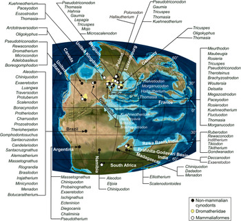
Eighty cynodont genera, including basal mammaliaforms (Abdala and Gaetano, Reference Abdala, Gaetano and Tanner2018; current work), have been reported from different Late Triassic horizons around the worldwide (Fig. 8). The Late Triassic basal mammaliamorphs are mostly known from Laurasia (Fig. 8), which includes North America (Osborn, Reference Osborn1887; Lucas and Oakes, Reference Lucas and Oakes1988; Lucas and Luo, Reference Lucas and Luo1993; Sues et al., Reference Sues, Olsen, Kroehler, Fraser and Sues1994), England (Fraser et al., Reference Fraser, Walkden and Stewart1985), Germany (Clemens, Reference Clemens1980), Switzerland (Clemens, Reference Clemens1980), France (Cuny, Reference Cuny, Lucas and Morales1993; Godefroit and Battail, Reference Godefroit and Battail1997; Debuysschere et al., Reference Debuysschere, Gheerbrant and Allain2015; Debuysschere, Reference Debuysschere2016), and Belgium (Godefroit, Reference Godefroit1997). In India, the Late Triassic basal mammaliaforms are known from only one stratigraphic horizon, the Tiki Formation, which has yielded the morganucodontid Gondwanodon, and the mammaliamorphs Tikitherium (Datta and Das, Reference Datta and Das1996; Datta, Reference Datta2005) and Tikiodon n. gen. (current work).
Paleogeographic map of the Late Triassic Pangaea (Scotese Reference Scotese2001) showing distribution of the cynodont taxa. Sources of information: Osborn (Reference Osborn1887); Rigney (Reference Rigney1963); Crompton (Reference Crompton1964); Clemens (Reference Clemens1980); Chatterjee (Reference Chatterjee1982); Fraser et al. (Reference Fraser, Walkden and Stewart1985); Lucas and Oakes (Reference Lucas and Oakes1988); Cuny (Reference Cuny, Lucas and Morales1993); Lucas and Luo (Reference Lucas and Luo1993); Sues et al. (Reference Sues, Olsen, Kroehler, Fraser and Sues1994); Datta and Das (Reference Datta and Das1996); Godefroit and Battail (Reference Godefroit and Battail1997); Godefroit et al. (Reference Godefroit, Cuny, Delsate and Roche1998); Godefroit (Reference Godefroit1999); Lucas et al. (Reference Lucas, Heckert, Harris, Seegis and Wild2001); Shapiro and Jenkins (Reference Shapiro and Jenkins2001); Cuny (Reference Cuny2004); Heckert (Reference Heckert2004); Datta et al. (Reference Datta, Das and Luo2004); Datta (Reference Datta2005); Martinelli et al. (Reference Martinelli, Bonaparte, Schultz and Rubert2005, Reference Martinelli, Soares and Schwanke2016, Reference Martinelli, Eltink, Da-Rosa and Langer2017); Sidor and Hancox, (Reference Sidor and Hancox2006); Kammerer et al. (Reference Kammerer, Flynn, Ranivoharimanana and Wyss2008); Abdala and Smith (Reference Abdala and Smith2009); Abdala and Ribeiro (Reference Abdala and Ribeiro2010); Heckert et al. (Reference Heckert, Mitchell, Schneider and Olsen2012); Soares et al. (Reference Soares, Martinelli and De Oliveira2014); Świło et al. (Reference Świło, Niedźwiedzki and Sulej2014); Ray, (Reference Ray2015); Abdala and Gaetano (Reference Abdala, Gaetano and Tanner2018); Fiorelli et al. (Reference Fiorelli, Rocher, Martinelli, Ezcurra, Hechenleitner and Ezpeleta2018); Wallace et al. (Reference Wallace, Martínez and Rowe2019); Abdala et al., Reference Abdala, Gaetano, Martinelli, Soares, Hancox and Rubidge2020; Hendrickx et al., Reference Hendrickx, Gaetano, Choiniere, Mocke and Abdala2020; Mocke et al. (Reference Mocke, Gaetano and Abdala2020); Sulej et al. (Reference Sulej, Niedźwiedzki, Tałanda, Dróżdż and Hara2020); current study.

Distributional patterns of Late Triassic cynodonts show distinct belts along which these concentrates. The cynodonts have only been found within a narrow belt ranging from the Equator to 30°N in the northern paleolatitudes, whereas in the southern hemisphere, they occur at relatively higher latitudes, ranging from 40°S to 60°S paleolatitude. It is possible this latitudinal distribution is due to insufficient sampling and/or lack of Late Triassic rock exposures (Close et al., Reference Close, Benson, Upchurch and Butler2017). However, this distributional pattern of the Late Triassic cynodonts parallels that of pseudosuchians (Mannion et al., Reference Mannion, Benson, Carrano, Tennant, Judd and Butler2015; Tolchard et al., Reference Tolchard, Nesbitt, Desojo, Viglietti, Butler and Choiniere2019). Moreover, most cynodonts show area-specific distribution, where the cynodont taxa are restricted either to the northern or southern hemisphere, except for the dromatheriid genus Rewaconodon, which is known from the lower Kalgary locality, Tecovas Formation of Texas, United States (Heckert et al., Reference Heckert2004) and the Tiki Formation of India (Datta et al., Reference Datta, Das and Luo2004). Ezcurra (Reference Ezcurra2010) grouped tetrapod assemblages from India and Europe with the South American assemblages during the Late Triassic, whereas Chatterjee et al. (Reference Chatterjee, Scotese and Bajpai2017) suggested both Gondwanan and Laurasian affinities for the Indian assemblages. This possibly suggests a biotic interchange between India and Laurasian regions and/or similarity in paleoclimatic conditions, but this requires further study.
Acknowledgments
We are grateful to D. Mukherjee, D. Datta, and N. Rakshit for active participation in the field during the fossil excavation. We acknowledge F. Abdala for his comments on the partial right dentary. We sincerely thank the reviewers, H.-D. Sues (National Museum of Natural History, Smithsonian Institution), Z.-X. Luo (University of Chicago), and the editor for constructive comments and corrections. We acknowledge N. Bhowmik for facilitating Scanning Electron Microscopy at CRF, IIT Kharagpur. Financial assistance and infrastructural facilities were provided by the Department of Science and Technology, India (SERB/SR/S4/ES-604/2012) and the Indian Institute of Technology, Kharagpur, India, respectively.