INTRODUCTION
Neglected tropical diseases, primarily affecting the poorest individuals living in developing countries, account for up to 90% of the global disease burden but get less than 10% of the research funds for new drug development approaches by both pharmaceutical industry and the public sector (Nour et al. Reference Nour, Khalid, Kaiser, Brun, Abdallah and Schmidt2009). Chagas disease (CD) is the sixth most important tropical infection faced by many Latin American countries (Martins-Melo et al. Reference Martins-Melo, Ramos, Alencar and Heukelbach2012). This parasitic disease caused by the flagellated protozoan organism, Trypanosoma cruzi still demands safer and more selective medicines since the current drugs (benznidazole and nifurtimox) present several undesirable characteristics such as long term of administration (30–60 days), high levels of adverse effects and limited efficacy especially during the later chronic phase (Soeiro et al. Reference Soeiro, Daliry, Silva, de Souza, Oliveira, Salomão, Menna Barreto, Castro, Méndez-Vilas and Díaz2010). In the last few years of experimental chemotherapy investigation, some natural products like sesquiterpene lactones (SL) (Schmidt et al. Reference Schmidt, Khalid, Romanha, Alves, Biavatti, Brun, Da Costa, de Castro, Ferreira, de Lacerda, Lago, Leon, Lopes, das Neves Amorim, Niehues, Ogungbe, Pohlit, Scotti, Setzer, Soeiro, Steindel and Tempone2012a , Reference Schmidt, Khalid, Romanha, Alves, Biavatti, Brun, Da Costa, de Castro, Ferreira, de Lacerda, Lago, Leon, Lopes, das Neves Amorim, Niehues, Ogungbe, Pohlit, Scotti, Setzer, Soeiro, Steindel and Tempone b ) and synthetic agents like arylimidamides (Soeiro et al. Reference Soeiro, Werbovetz, Boykin, Wilson, Wang and Hemphill2013a ) and inhibitors of fungal sterol 14α-demethylase (CYP51) have been reported as promising trypanocidal agents (Urbina, Reference Urbina2009; Hargrove et al. Reference Hargrove, Kim, de Nazaré Correia Soeiro, da Silva, Batista, Batista, Yazlovitskaya, Waterman, Sulikowski and Lepesheva2012). Among the last class of compounds, antifungal CYP51 inhibitors posaconazole and ravuconazole are finally in clinical trials for CD but the development of cheaper and more efficient anti-T. cruzi chemotherapies is still needed (Soeiro et al. Reference Soeiro, de Souza, da Silva, da Gama Jaen Batista, Batista, Pavão, Araújo, Aiub, da Silva, Lionel, Britto, Kim, Sulikowski, Hargrove, Waterman and Lepesheva2013b ). In this context, our present aim was to investigate through in vitro and in vivo studies the biological effect of the chloroaryl-substituted imidazole viniconazole with the goal of identifying novel anti-T. cruzi leads for possible future development into alternative therapies for CD.
MATERIALS AND METHODS
Compounds
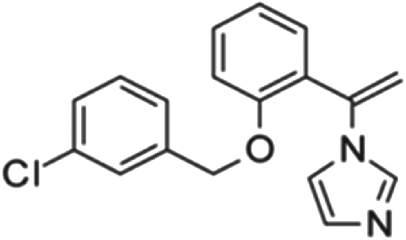
Viniconazole (Fig. 1) was kindly received from Dr M. Witschel, BASF (Ludwigshafen, Germany). Benznidazole (Bz, Laboratório Farmacêutico do Estado de Pernambuco – LAFEPE, Brazil) was used as the reference drug (Batista et al. Reference Batista, Batista, de Oliveira, do Amaral, Lannes-Vieira, Britto, Junqueira, Lima, Romanha, Sales, Stephens, Boykin and Soeiro2010). Stock solutions of the compounds (3·3 mm) were prepared in dimethyl sulfoxide (DMSO). Fresh final solvent concentration in the assays never exceeded 0·6%, which is not toxic for both parasites and mammalian cells. For in vivo studies, a stock solution of each compound was first prepared in DMSO and then diluted using sterile distilled water. The final concentration of DMSO never exceeded 10%, a concentration which is well tolerated by mice (Da Silva et al. Reference da Silva, Batista, Oliveira, de Souza, Hammer, da Silva, Daliry, Araujo, Britto, Rodrigues, Liu, Farahat, Kumar, Boykin and Soeiro2012).
Structure of viniconazole.

Cell cultures
Toxicity and infection assays: primary cultures of cardiac cells (CM) were obtained from mouse embryos as reported (Meirelles et al. Reference Meirelles, Araujo-Jorge, Miranda, De Souza and Barbosa1986). The cultures were maintained in Dulbecco's modified Eagle's medium (DMEM) supplemented with 10% horse serum, 5% fetal bovine serum (FBS), 2·5 mm CaCl2, 1 mm L-glutamine and 2% chicken embryo extract. Cell cultures were maintained at 37 °C in an atmosphere of 5% CO2 in air, and assays were run at least three times in duplicates.
Parasites
The Y strain of T. cruzi was used throughout the experiments. Bloodstream trypomastigotes (BT) were harvested by heart puncture from T. cruzi-infected Swiss mice at the day of parasitaemia peak (Meirelles et al. Reference Meirelles, Araujo-Jorge, Miranda, De Souza and Barbosa1986). Intracellular amastigotes lodged within cardiac cell cultures were employed as reported (Da Silva et al. Reference da Silva, Batista, Oliveira, de Souza, Hammer, da Silva, Daliry, Araujo, Britto, Rodrigues, Liu, Farahat, Kumar, Boykin and Soeiro2012).
In vitro cytotoxicity assays
Uninfected cardiac cultures were incubated for 24 and 48 h at 37 °C in the presence or absence of each compound diluted in DMEM. The cardiac cell morphology and spontaneous contractibility were evaluated by light microscopy. The cell death rates were measured by MTT (3-(4,5-dimethyl-2-thiazolyl)-2,5-diphenyl-2H-tetrazolium bromide – Sigma Aldrich) colorimetric assay, for the determination of EC50 (compound concentration that reduces 50% of cellular viability) as reported (Batista et al. Reference Batista, Batista, de Oliveira, Britto, Rodrigues, Stephens, Boykin and Soeiro2011).
Trypanocidal analysis
The BT were incubated at 37 °C for 24 h in the presence of increasing non-toxic concentrations of the tested compounds diluted in RPMI 1640 medium (Roswell Park Memorial Institute, Sigma Aldrich, USA) supplemented with 5% FBS. Death rates were determined by light microscopy through direct quantification of the number of live parasites using a Neubauer chamber, and the EC50 (compound concentration that reduces 50% of the number of the treated parasites) calculated. For the analysis of the effect against intracellular amastigotes, after 24 h of parasite–host cell interaction (10:1 parasite: CM ratio), the infected cultures were washed to remove free parasites and then incubated for another 48 h with increasing but non-toxic doses of the compound. The CM were maintained at 37 °C in an atmosphere of 5% CO2 in air, and the medium was replaced every 24 h. Then, untreated and treated infected CM were fixed and stained with Giemsa solution and the mean number of infected host cells and of parasites per infected cells scored as reported (Da Silva et al. Reference da Silva, Batista, Oliveira, de Souza, Hammer, da Silva, Daliry, Araujo, Britto, Rodrigues, Liu, Farahat, Kumar, Boykin and Soeiro2012). As light microscopy of untreated T. cruzi-infected cell cultures allows the identification of parasites through their typical nuclei and kDNA structures, only those intracellular forms that display these characteristic cellular elements were considered as live/viable parasites (Da Silva et al. Reference da Silva, Batista, Oliveira, de Souza, Hammer, da Silva, Daliry, Araujo, Britto, Rodrigues, Liu, Farahat, Kumar, Boykin and Soeiro2012). Control samples included parasites and infected cell cultures incubated solely with vehicle solution (DMSO not exceeding 0·6%). The activity of viniconazole was estimated by calculating the infection index (percentage of infected cells times the average number of intracellular amastigotes per infected host cell). All in vitro assays were run at least twice in duplicates.
Transmission electron microscopy (TEM) analysis: BT treated or not for 2 h at 37 °C with the corresponding EC50 value of the tested compound were fixed for 60 min at 4 °C with 2·5% glutaraldehyde and 2·5 mm CaCl2 in 0·1 m cacodylate buffer, pH 7·2 and post-fixed for 1 h at 4 °C with 1% OsO4, 0·8% potassium ferricyanide and 2·5 mm CaCl2 using this same buffer. Samples were routinely processed for TEM and examined in a Zeiss EM10C electron microscope (Oberkochen, Germany) (Da Silva et al. Reference da Silva, Batista, Batista, de Souza, da Silva, de Oliveira, Meuser, Shareef, Boykin and Soeiro2008).
Acute toxicity for mice: Maximum tolerated dose (MTD) was determined using female Swiss Webster mice (20–23 g). On day 1, mice were treated with the tested compound by i.p. or oral (p.o.) route at doses ranging from 25–400 mg kg−1 (Batista et al. Reference Batista, Batista, de Oliveira, do Amaral, Lannes-Vieira, Britto, Junqueira, Lima, Romanha, Sales, Stephens, Boykin and Soeiro2010). The animals were then continuously inspected for toxic and sub-toxic symptoms according to OECD (Organization for Economic Co-operation and Development) guidelines. Forty-eight hours after compound injection, the MTD values were determined based on animal survival rates and behaviour alterations (Da Silva et al. Reference da Silva, Batista, Oliveira, de Souza, Hammer, da Silva, Daliry, Araujo, Britto, Rodrigues, Liu, Farahat, Kumar, Boykin and Soeiro2012).
Mice infection and treatment schemes
Male Swiss mice were obtained from the Fundação Oswaldo Cruz (FIOCRUZ) animal facilities (Rio de Janeiro, Brazil). Mice were housed at 5 animals per cage and kept in a conventional room at 20–24 °C under a 12/12 h light/dark cycle. The animals were provided with sterilized water and chow ad libitum. Infection was performed by i.p. injection of 104 (Y strain) BT (Batista et al. Reference Batista, Batista, de Oliveira, do Amaral, Lannes-Vieira, Britto, Junqueira, Lima, Romanha, Sales, Stephens, Boykin and Soeiro2010). The animals (18–21 g) were divided into the following groups (five mice per group): uninfected (non-infected and untreated); untreated (infected with T. cruzi but treated only with vehicle); and treated (infected and treated – i.p. and orally (p.o.) up to 100 mg kg−1 day−1 viniconazole or 100 mg kg−1 day−1 benznidazole). Mice received two doses (0·1 mL via i.p.): the first one at 5 and the second one at 8 days post-infection (dpi) according to a previously standardized protocol designed for evaluating the efficacy of a novel trypanocidal compound in an experimental mouse model of acute T. cruzi infection, running in parallel with the reference drug benznidazole (Bz) (Batista et al. Reference Batista, Batista, de Oliveira, do Amaral, Lannes-Vieira, Britto, Junqueira, Lima, Romanha, Sales, Stephens, Boykin and Soeiro2010). For Bz treatment, infected mice received 0·2 mL oral dose (gavage) following the same therapeutic scheme as described above. Only those mice that presented positive parasitaemia were used in these studies.
Parasitaemia, mortality rates and ponderal curve analysis
Parasitaemia was individually checked by direct microscopic counting of parasites in 5 μL of blood, as described (Brener, Reference Brener1962). At different times, the body weight of each animal was recorded and the different mouse groups compared. Mortality was checked daily until 30 days post treatment and expressed as percentage of cumulative mortality (% CM) (Batista et al. Reference Batista, Batista, de Oliveira, do Amaral, Lannes-Vieira, Britto, Junqueira, Lima, Romanha, Sales, Stephens, Boykin and Soeiro2010).
Histopathological analysis
For acute toxicity analysis, tissues (heart, spleen, liver and kidneys) were removed after 48 h of viniconazole administration (25–400 mg kg−1), longitudinally cut, rinsed in ice-cold PBS, and fixed in Millonig–Rosman solution (10% formaldehyde in PBS). The tissues were dehydrated and embedded in paraffin. Sections (3 μm) were stained by routine haematoxylin–eosin and analysed by light microscopy as reported (Batista et al. Reference Batista, Batista, de Oliveira, do Amaral, Lannes-Vieira, Britto, Junqueira, Lima, Romanha, Sales, Stephens, Boykin and Soeiro2010).
Ethics
All procedures were carried out in accordance with the guidelines established by the FIOCRUZ Committee of Ethics for the Use of Animals (CEUA 0028/09).
RESULTS
The trypanocidal in vitro activity of the chloroaryl-substituted imidazole, viniconazole, screened against BT showed a time and dose-dependent effect, exhibiting EC50 values of 16 μ m after treatment for 24 h at 37 °C (Fig. 2A). This compound was also active against intracellular parasites, showing EC50 = 24 μ m, after 48 h of treatment (Fig. 2B), while the incubation with cardiac cell cultures resulted in EC50 = 90 μ m (data not shown).
Effect of viniconazole on bloodstream trypomastigotes and amastigotes of Trypanosoma cruzi (Y strain) in vitro. The activity was evaluated against the two parasite forms of T. cruzi: (A) bloodstream forms incubated at 37 °C for 2 and 24 h followed by the determination of the percentage of dead parasites, and (B) intracellular amastigote forms incubated at 37 °C for 48 h followed by the determination of the infection index (II).

Aiming to identify the main cellular targets of viniconazole, BT were exposed to this chloroaryl-substituted imidazole and processed for routine TEM. The ultrastructural studies showed that untreated parasites exhibited normal morphology such as typical plasma membrane, mitochondrion, nucleus and Golgi complex (Fig. 3A and B). When BTs were treated, we observed an intense intracellular vacuolization displaying vesicles of different sizes and electron densities (Fig. 3C, H and I), myelin-like figures (Fig. 3I, arrowhead) and endoplasmic reticulum profiles surrounding intracellular components (Fig. 3H, arrow). A great number of membrane blebs and shedding events were present, discharging cytoplasmic and membrane components (Fig. 3D, E, G). Also, alterations in the membrane profile were found in the nuclei of the treated cells (Fig. 3E, inset), besides a frequent and intense swelling of Golgi elements (Fig. 3F and E).
Transmission electron microscopy analysis (A–C) of trypomastigotes exposed to viniconazole: (A,B) untreated and (C–I) treated with EC50/24 h. Untreated parasites display typical morphology, while viniconazole-treated parasites show mitochondrial swelling (Fig. 2C, H, G and I), intense intracellular vacuolization with a great number of membrane blebs and with shedding of intracellular contents (Fig. 2D, E and G), with the occurrence of large multivesicular profiles (Fig. 2C, D and H), swelling Golgi complex (Fig. 2 F and E), cytoplasmic myelin-like figures (Fig. 2I), and alterations in the nuclear membrane (Fig. 2E inset).

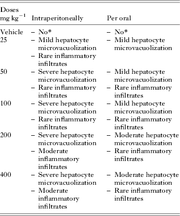
Our first in vivo step was to evaluate aspects of acute toxicity of viniconazole aiming to determine its MTD value in order to set and follow efficacy studies. Thus, Swiss female mice received viniconazole via the i.p. and p.o. route (25 mg kg−1 up to 400 mg kg−1) and were followed for non-invasive side effects up to 48 h after injection. The administration of the compound at all tested doses with both routes of application did not: (a) cause alteration on typical behaviour, (b) result in mortality, (c) induce visible neurological disorders or (d) alter the body weight gain (data not shown). However, histopathology of liver, spleen, heart and kidney showed major alterations in liver samples (Table 1). A dose-dependent reactivity related to hepatic microvacuolization and mononuclear inflammation was observed mainly at doses >50 mg kg−1 via the i.p. route (Table 1, Fig. 4).
Histopathological analysis of representative samples from untreated (A) and treated female mice with 25 mg kg−1 (B) and 400 mg kg−1 (C,D) doses of viniconazole. Liver sections stained with haematoxylin-eosin (HE) of untreated animals showing characteristic morphology (A). Liver sections of treated mice exhibit hepatocyte vacuolization (star) (B,D). 400×.

Analysis of hematoxilin and eosin stained liver samples collected from mice treated with viniconazole

* No alteration.
Next, the efficacy of viniconazole was tested in Swiss male mice inoculated with 104 bloodstream parasites using a therapeutic scheme employing doses administered once a day, at 5 and 8 dpi (corresponding to the onset and parasitaemia peak, respectively in this animal model). Bz treatment was run in parallel using a standard protocol (100 mg kg−1 via p.o.), following the same therapeutic scheme as described above.
As expected for this experimental mouse model of T. cruzi acute infection, infected and untreated animals (untreated group) presented high parasitaemia and no animal survival (Fig. 5). When viniconazole was administrated via the i.p. route, all doses failed to reduce parasitaemia or protect against mortality (all treated animals died even earlier than the untreated group). Although gavage administration of viniconazole gave a mild reduction in parasitaemia peak levels (about 35%), all mice died (Fig. 5). As expected, Bz completely suppressed the parasitaemia and gave 100% survival (Fig. 5).
In vivo effect of viniconazole on acute mouse model of Trypanosoma cruzi infection, using Y strain: The effects of viniconazole (via i.p. – 25–100 mg kg−1 day−1 and via p.o route – 100 mg kg−1 day−1) and benznidazole (100 mg kg−1 day−1 by p.o.) on parasitaemia (A) and mortality rates (B) are presented.

DISCUSSION
Our present aim was to investigate the potential efficacy and selectivity of a chloroaryl-substituted imidazole named viniconazole against T. cruzi. In fact, azoles are considered a primary target for clinical and agricultural antifungal agents and are also well known as effective inhibitors of T. cruzi infection and development in vitro and in vivo due to their ability to inhibit CYP51, an enzyme catalysing ergosterol production (Urbina, Reference Urbina2009). Recent studies regarding the direct inhibition of CYP51 activity across phylogeny, including the human pathogens Trypanosoma brucei, T. cruzi and Leishmania infantum, identified novel protozoa-specific inhibitory scaffolds that are easy to synthesize and nontoxic, and among them, the VNI molecule ((R)-N-(1-(2,4-dichlorophenyl)-2-(1H-imidazol-1-yl)ethyl)-4-(5-phenyl-1,3,4-oxadi-azol-2-yl)benzamide) presented high antiprotozoan activity, killing T. cruzi amastigotes at low nanomolar concentration, being able to suppress parasitaemia and improve the survival rates of T. cruzi-infected mice (Soeiro et al. Reference Soeiro, de Souza, da Silva, da Gama Jaen Batista, Batista, Pavão, Araújo, Aiub, da Silva, Lionel, Britto, Kim, Sulikowski, Hargrove, Waterman and Lepesheva2013b ; Villalta et al. Reference Villalta, Dobish, Nde, Kleshchenko, Hargrove, Johnson, Waterman, Johnston and Lepesheva2013). Thus, in view of the promising trypanocidal activity of azoles, presently the efficacy in vitro and in vivo of a novel chloro-imidazole compound was investigated. Our findings showed that viniconazole displayed a trypanocidal in vitro effect against both BT (EC50 = 16 μ m) and intracellular amastigotes (EC50 = 24 μ m) localized within cardiac host cells, but presented a considerable toxicity towards mammalian cell cultures, leading to quite low selectivity indices (4 to 6). Although in acute toxicity studies no mortality and no major side effects were observed through non-invasive methods followed up to 48 h after viniconazole injection, histopathology analysis demonstrated liver reactivity especially at doses >50 mg kg−1 by the i.p. route. Also, it failed to reduce parasite burden and protect against animal mortality using a highly stringent mouse model of T. cruzi infection using the Y strain. The administration of viniconazole for only 2 days, by both i.p. and p.o. routes, using doses of up to 100 mg kg−1 day−1 led to similar results as those found in the vehicle-treated group while Bz 100 mg kg−1 (p.o.) under the same therapeutic scheme suppressed parasitaemia and gave 100% of animal survival.
Transmission electron microscopy was also performed aiming to show the most frequent alteration triggered by viniconazole in BT. Our present data showed intense damage to the membranes of the treated parasites, especially plasma membrane (blebs and shedding events) and Golgi (swelling aspects). Also, a high number of vacuoles could be found in treated bloodstream forms which associated to the previous alterations suggesting an autophagic process, although other biochemical approaches are needed to confirm these data. Autophagy induced by pharmacological agents has been reported in trypanosomatids. Helenalin and cynaropicrin, two SLs, induce a strong vacuolization in Leishmania mexicana and in T. cruzi that was associated with autophagic processes (Barrera et al. Reference Barrera, Jimenez-Ortiz, Tonn, Giordano, Galanti and Sosa2008; Da Silva et al. Reference da Silva, Batista, Oliveira, de Souza, Hammer, da Silva, Daliry, Araujo, Britto, Rodrigues, Liu, Farahat, Kumar, Boykin and Soeiro2012). Besides autophagy, other types of programmed cell death like apoptosis has been described when pathogens are exposed to different pharmacological stimuli triggered in African trypanosomes (Nibret et al. Reference Nibret, Youns, Krauth-Siegel and Wink2011) and Leishmania mexicana (Barrera et al. Reference Barrera, Jimenez-Ortiz, Tonn, Giordano, Galanti and Sosa2008) by natural agents like SL and through synthetic compounds like diamidines in T. cruzi (De Souza et al. 2004). Also, further detailed studies are needed to explore cell death mechanisms of chloro-imidazoles against T. cruzi as well as other trypanosomatids.
Due to the urgent need for novel anti-T. cruzi agents, the application of bioproducts as well as synthetic agents as therapeutic alternatives must be further explored with the aim of providing more selective, safe and affordable drugs for chagasic patients, especially those millions who are in the chronic stage of this neglected disease, for which no treatment exists today.
ACKNOWLEDGEMENTS
PDTIS/Fiocruz, CAPES, PROEP/CNPq, FAPERJ and CNPq. The present work was part of activities conducted within the research network Natural Products against Neglected Diseases (ResNetNPND). Viniconazole was kindly given by Dr M. Witschel, BASF (Ludwigshafen, Germany).