Cardiac venous malformations are an uncommon form of cardiac tumours, especially in the young. They are notoriously difficult to diagnose pre-operatively, with over 60% being misdiagnosed.Reference Li, Teng, Xu, Ma and Ni 1 While resection is first-line treatment, some tumours may be amenable to conservative management.
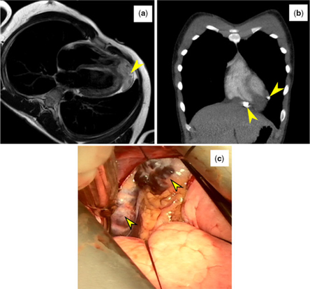
A 17-year-old boy presented with lethargy. An electrocardiogram revealed a prolonged QTc interval with T-wave inversion. An initial echocardiogram was normal, so a cardiac MRI was obtained to evaluate for regional hypertrophy or fibrosis. The cardiac MRI revealed a lobulated, irregular mass in the pericardial space at the apex of the heart with possible myocardial invasion (Fig 1a). The working differential diagnosis included sarcoma, teratoma, venous malformation, fibroma, and lymphoma. Cardiac CT angiogram revealed a minimally enhancing mass containing several calcified foci (Fig 1b). A working diagnosis of a cardiac teratoma was made and surgical resection was planned.
A T2 turbo spin echo in a four-chamber axial plane demonstrates an irregular apical mass with invasion of the right ventricular free wall and apex (1A, yellow arrow). Follow-up imaging via CT angiogram in the coronal plane revealed two discrete calcified foci within the mass (1B, yellow arrows). Gross inspection of the mass following a left thoracotomy unveiled a purple, compressible, and non-pulsatile mass composed of dilated veins with infiltrating epicardial fat and muscle (1C).

Gross inspection via left thoracotomy revealed a spongy, compressible, and non-pulsatile mass attached to the apical right ventricular free wall. It was composed of dilated veins with infiltrating epicardial fat and muscle (Fig 1c). Intra-operative transesophageal echocardiogram showed low-velocity flow. Based on these findings, a diagnosis of a venous malformation was made. Due to the bleeding risk, neither resection nor biopsy was performed. A cardiac MRI obtained 6 months after the original imaging showed no growth. The patient’s electrocardiogram abnormalities resolved spontaneously, and he has remained asymptomatic.
In summary, echocardiograms are considered the first-line modality for diagnosing cardiac venous malformations. However, multi-modality imaging is often necessary to evaluate the structural, physiological, and perfusion properties.
Acknowledgement
The authors acknowledge and thank Dr. Peace C. Madueme for his assistance with preparing and interpreting the images included in this report.
Financial Support
This research received no specific grant from any funding agency, commercial or not-for-profit sectors.
Conflicts of Interest
None.