Introduction
Several techniques have been described for the excision of lesions in the retropharyngeal space. Whilst a transoral approach has been used for smaller lesions, an external cervical approach is currently preferred for the complete excision of larger and more complex lesions.
With the advent of the robotic system, larger tumours within the retropharyngeal space can now be excised transorally. We describe this technique used in a case of a large retropharyngeal schwannoma.
Case report
A 48-year-old plumber underwent a magnetic resonance imaging (MRI) scan for the investigation of unilateral sensorineural hearing loss. Apart from snoring at night, he was otherwise asymptomatic.
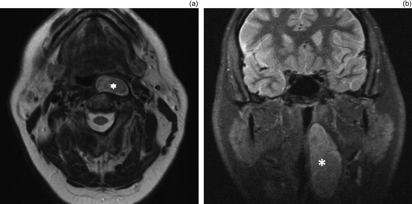
The MRI scan revealed a large incidental solid retropharyngeal mass, measuring 2.7 × 1.2 cm in axial dimensions and over 5.8 cm in craniocaudal length, extending from the level of the post-nasal space to the level of the arytenoids. Appearances were suggestive of a benign lesion, possibly of neurogenic origin. Fine needle aspiration cytology was not diagnostic. This was followed by a period of close observation, with interval MRI scans, after discussion with the patient.
Eighteen months later, the patient became symptomatic, with the sensation of a lump and progressive dysphagia. An MRI scan performed at that time showed that the lesion had grown larger, measuring 3 × 1.9 cm in axial dimensions and 6.7 cm in craniocaudal length. The pre-operative MRI scans are shown in Figure 1. His care was subsequently transferred to the head and neck team.
Pre-operative magnetic resonance imaging scans of the retropharyngeal mass (asterisks), as seen on axial imaging (a) and T2-weighted coronal imaging (b), showing extension from the level of post-nasal space inferiorly beyond the level of the tongue base.

Surgical management with robot-assisted transoral excision was proposed given the size of the retropharyngeal lesion.
Surgical method
The patient was positioned supine under general anaesthetic, with an Olympus® FK-WO Laryngo-Pharyngoscope System (Feyh- Kastenbauer-Weinstein-O'Malley Retractor) in situ. The da Vinci® robotic system (Intuitive Surgical, Sunnyvale, California, USA) was set up.
A paramedian incision was made into the oropharyngeal mucosa using robotic monopolar cautery, and blunt dissection was performed with Maryland dissecting forceps to identify the correct plane. The upper lobe was initially dissected out, and the multiple other lobes were subsequently freed from their attachments. Minor blood vessels were encountered during the procedure and haemostasis was easily achieved with monopolar cautery. The lesion was delivered en bloc with an intact capsule, and the incision was closed primarily with Vicryl 3-0 sutures. The surgical steps are presented in Figure 2.
Surgical steps: (a) and (b) show the location of the paramedian incision in relation to the tongue (‘1’ = Maryland forceps, ‘2’ = 5 mm monopolar diathermy), (c) shows further dissection to identify the correct plane, (d) shows delivery of the upper lobe, and (e) shows delivery of multiple lower lobes.
No untoward incidents or complications were encountered. Total surgical time was 1 hour and 55 minutes, excluding a robot set-up time of 35 minutes.
Post-operative course
The patient resumed an oral diet the following day. He did not require a surgical drain and was discharged home on post-operative day 1. No peri- or post-operative complications were recorded.
At follow up in the out-patient clinic, at six weeks and at three months post-operatively, the patient was asymptomatic, with normal swallowing and resolution of snoring. There was no clinical evidence of recurrence.
Discussion
The robotic system provides the crucial advantages of better three-dimensional high-definition visualisation around the tongue base, and improved dexterity with the use of more surgical instruments. Angled endoscopes and wristed instruments are extremely useful for precise dissection in certain areas that are not easily accessible otherwise.
Smaller retropharyngeal lesions can be removed through transoral approaches. In our case, given the size and craniocaudal extent of the mass, if the robotic system had been unavailable, excision could only have been achieved through a transcervical route. The da Vinci system allowed excision of this large retropharyngeal space lesion through the transoral route. This avoided the need for an external excision and the associated morbidity, which includes a long scar, longer hospital stay and associated pain.
We were most impressed by the rapid resumption of swallowing in our patient, which was the result of minimal disruption to the pharyngeal plexus. The patient had returned to his usual daily living activities within one week post-operatively. He opted to take three months off work as his job involved heavy lifting and working in dusty environments.
Robotic assistance has been used for the transoral excision of retropharyngeal lymph nodes.Reference Byeon, Duvvuri, Kim, Park, Hong and Koh1, Reference Moore, Ebrahimi, Price and Olsen2 However, primary tumours of the retropharyngeal space are rare. This is, to our knowledge, the first report of this technique used in the excision of a large retropharyngeal space mass. A systematic search of Medline, PubMed and Google Scholar databases, individual online journals, conference proceedings, and organisational websites indicated no previous use of this technique.
Tumours in the parapharyngeal space are more common, and robot-assisted excision of parapharyngeal space masses has been successful in avoiding the need for mandibulotomy and transparotid approaches.Reference Mendelsohn3 A recently published case series of pleomorphic adenomas has shown promising outcomes, with no recurrence after a mean follow up of 18.5 months.Reference Chan, Tsang, Eisele and Richmon4
Future research is needed to investigate the clinical outcomes of robot-assisted transoral resection of retropharyngeal space lesions and to educate on patient selection for this technique.
Acknowledgement
We would like to thank Ms Jacqueline Howard for her assistance in our literature search.